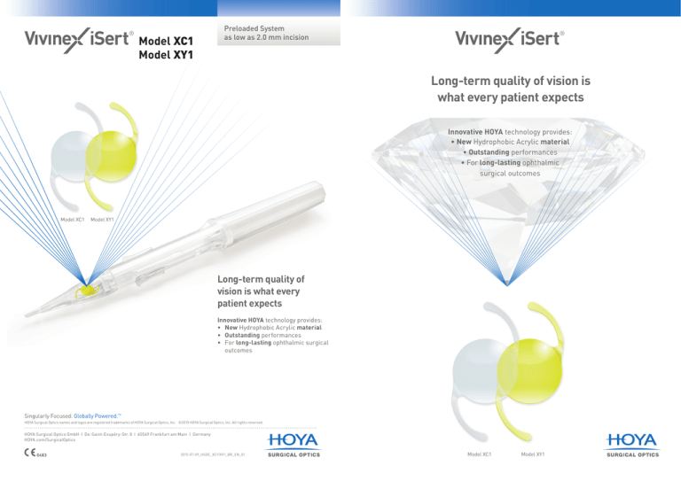
Model XC1
Model XY1
Preloaded System
as low as 2.0 mm incision
Long-term quality of vision is
what every patient expects
Innovative HOYA technology provides:
• New Hydrophobic Acrylic material
• Outstanding performances
• For long-lasting ophthalmic
surgical outcomes
Model XC1
Model XY1
Long-term quality of
vision is what every
patient expects
Innovative HOYA technology provides:
• New Hydrophobic Acrylic material
• Outstanding performances
• For long-lasting ophthalmic surgical outcomes
Singularly Focused. Globally Powered.TM
HOYA Surgical Optics names and logos are registered trademarks of HOYA Surgical Optics, Inc. ©2015 HOYA Surgical Optics, Inc. All rights reserved.
HOYA Surgical Optics GmbH I De-Saint-Exupéry-Str. 8 I 60549 Frankfurt am Main | Germany
HOYA.com/SurgicalOptics
2015-07-09_HSOE_XC1/XY1_BR_EN_01
Model XC1
Model XY1
A NCE
Textured-rough Haptic
• Better grip onto capsular
surface is expected
• To avoid the haptic-tip sticks
to the optic when it’s folded
Improved Square Edge
• Very sharp edges
• Helps to prevent PCO1
Lo
P ER F ORM
According to in vitro tests, the new acrylic polymers
properties of Vivinex™ drastically reduce glistening
ng
Ter
P ER F ORM
m
n
g Ter
m
Cutting-edge IOL* technology provides research-driven
benefits to protect your patients’ “long-term vision quality”
Long-term Transparency
Lo
Quality for Long-term Vision
A NCE
• No glistening was seen based on
in vitro testing (35ºC to 23ºC)4
•The innovative hydrophobic material Vivinex™
is associated with a significant ­decrease in
glistening
Long term visual quality with “ABC Design”
The “ABC Design” of this Aspheric optic maintains high image quality
even if the lens is not centrally aligned with the visual axis.5
HOYA Vivinex
™
Anterior
Surface
Posterior
Surface
1.
2. 3. *
COMPANY 1
HOYA AF-1
Optical Surface Quality3
•High-quality, precise smooth
surface
• Vivinex™ has the similar surface
smoothness and optical quality
as every marketed HOYA IOL
Nishi O, Nishi K, Akura J. Speed of capsular bend formation at the optic edge of acrylic, silicone, and poly(methyl methacrylate) lenses. J Cataract Refract Surg 2002; 28(3):431-437.
Meacock W, et al. The Effect of Texturing the Intraocular Lens Edge on Postoperative Glare Symptoms. Archives of Ophthalmology 2002; Vol 120: 1294-1298.
Data on file
IOL = Intra-Ocular Lens
Unique control
of HOAs to
­reduce effect
of decentration
100%
90%
Cornea SA = +0.27 microns
Pupil = 4 mm
Spatial Frequency = 50 cycles/mm
80%
70%
60%
50%
Sph IOL
40%
30%
Neg Aspheric
20%
HOYA Aspheric
21
Spherical IOL
Power (D)
Optic Edge Texturing Finish
• To reduce Dysphotopsia2
Modulation Transfer Function
Theoretical Eye Model
20
19
HOYA Aspheric
18
Negative Asperic
10%
0%
0 0.20.40.6 0.8
Deviation of IOL Center from Visual Axis (mm)
SA = Spherical aberration
17
01 2 3
Distance from optic center (mm)
Data for a +20.0D IOL
HOA = Higher-order aberrations
4. Data on file: in vitro test achieved according to published method: Marrie van der Mooren et al. “Effects of glistening in intraocular lens”, BIOMEDICAL OPTICS EXPRESS, vol 4, No.8,
P1294-1304(2013).
5. Data on file
PCO** reduction proven in in vivo tests
A NCE
• Strong capsular adhesion reduced the risk of PCO
• Rabbits receiving lenses with proprietary surface treatment showed a low level
of PCO
Right Eye
Year 1
Year 2
Year 3
Patient
A
Case 2
Patient
B
Case 3
Patient
C
Case 4
Control =
hydrophobic acrylic
material
With Surface Treatment =
same acrylic material as
control + surface treatment
6. Hiroyuki Matsushima, et al. Active oxygen processing for acrylic intraocular lenses to prevent.posterior capsule opacification. J Cataract Refract Surg. 2006; 32:1035-1040.
** PCO = Posterior Capsule Opacification
ng
Ter
P ER F ORM
• Effective long-term PCO inhibition
• 30 eyes were enrolled and YAG rate was 3.3% at 3 years post-operative time8
Left Eye
Case 1
Lo
Lo
P ER F ORM
Clinical outcome shows very low PCO rate in
post-operative time7
Images courtesy of Hiroyuki Matsushima, MD, PhD, Department of Ophthalmology,
Dokkyo Medical University, Japan
7. Japanese clinical study carried out in 2010 : internal report
8. Hiroyuki Matsushima, Dokkyo Medical University. Presented at 68th Annual Congress of Japan Clinical Ophthalmology;
November 13, 2014 Kobe Japan
m
n
g Ter
m
in vivo test on rabbit eyes shows that proprietary surface
treatment offers strong PCO reduction6
PCO reduction proven in human eyes
A NCE
P ER F ORM
A NCE
The HOYA surface treatment on the posterior surface and the
new feature of the Vivinex™ iSert® design provides
outstanding performances
2.0 mm
Lo
Ter
Vivinex™ iSert®: The innovative 1-piece acrylic
lens for long term patient satisfaction
ng
Ter
P ER F ORM
Model
VivinexTM iSert® XC1
VivinexTM iSert® XY1
Optic Design
Aspheric “ABC Design“ with
sharp textured optic edge
Optic & Haptic
Materials
Hydrophobic acrylic (Vivinex™)
with UV filtering (Model XC1)
with blue light filtering (Model XY1)
Haptic Design
Textured-rough haptic surface
Dimension
(Optic/OAL)
6.0 mm/13.0 mm
Power
+6.0 to +30.0 D (in 0.5 D increments)
Incision size
as low as 2.0 mm
Textured-rough haptic
Ergonomically designed
parts offering better
procedural control
Anterior
Posterior
6.0 mm
13.0 mm
Slider
Aspheric
“ABC Design”
Precision tip
designed for the
corneal incision
to be as small as
2.0 mm
Optimized sharp
& textured
optic edge
New screw design
with fixed grip
Model XC1
Optimized sharp
& textured
optic edge
Model XY1
Body
Irreversible slider
advancement to prevent
lens dislocation
Nozzle
OVD
infusion
area
• New iSert® offers easy handling and a better surgical comfort
• Very small incision size reduces the risk of surgically-induced astigmatism
OVD
infusion
port
Cover
Release
Tab
Trailing
haptic is
tucked
correctly,
and rod tip
pushes the
optic edge
correctly
Case
Body
Slider
Leading
haptic is
tucked
correctly
Hold the body
with your thumb
Step A
Step B
Step C
Step D
Infuse the OVD into the
injector through the infusion
port with the cannula pointed
in a direction perpendicular
to the body. Fill up the area
indicated by dotted lines with
the OVD and confirm that the
OVD has covered the entire
intraocular lens.
Press the release tabs,
lift up and remove the
cover from the case.
Push the slider slowly until
it stops, holding the body
with your thumb. Remove
the injector from the case.
Carefully insert the nozzle
into the eye through the
incision, keeping bevel
down. Slowly rotate the
screw plunger to inject the
lens into the capsular bag.
m
Easy to insert through an incision as low as
ng
m
The ergonomically-designed iSert® system provides highly
predictable, reproducible IOL delivery through a very
small incision
mm
Lo
Easy to insert through an incision as low as 2.0
A NCE


