Saccharomyces cerevisiae
advertisement

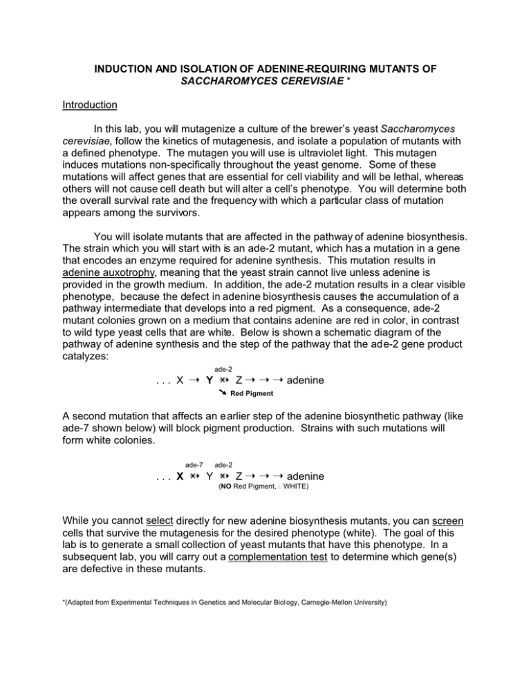
INDUCTION AND ISOLATION OF ADENINE-REQUIRING MUTANTS OF SACCHAROMYCES CEREVISIAE * Introduction In this lab, you will mutagenize a culture of the brewer’s yeast Saccharomyces cerevisiae, follow the kinetics of mutagenesis, and isolate a population of mutants with a defined phenotype. The mutagen you will use is ultraviolet light. This mutagen induces mutations non-specifically throughout the yeast genome. Some of these mutations will affect genes that are essential for cell viability and will be lethal, whereas others will not cause cell death but will alter a cell’s phenotype. You will determine both the overall survival rate and the frequency with which a particular class of mutation appears among the survivors. You will isolate mutants that are affected in the pathway of adenine biosynthesis. The strain which you will start with is an ade-2 mutant, which has a mutation in a gene that encodes an enzyme required for adenine synthesis. This mutation results in adenine auxotrophy, meaning that the yeast strain cannot live unless adenine is provided in the growth medium. In addition, the ade-2 mutation results in a clear visible phenotype, because the defect in adenine biosynthesis causes the accumulation of a pathway intermediate that develops into a red pigment. As a consequence, ade-2 mutant colonies grown on a medium that contains adenine are red in color, in contrast to wild type yeast cells that are white. Below is shown a schematic diagram of the pathway of adenine synthesis and the step of the pathway that the ade-2 gene product catalyzes: ade-2 . . . X ÿ Y ÿ Z ÿ ÿ ÿ adenine ú Red Pigment A second mutation that affects an earlier step of the adenine biosynthetic pathway (like ade-7 shown below) will block pigment production. Strains with such mutations will form white colonies. ade-7 ade-2 . . . X ÿ Y ÿ Z ÿ ÿ ÿ adenine (NO Red Pigment, WHITE) While you cannot select directly for new adenine biosynthesis mutants, you can screen cells that survive the mutagenesis for the desired phenotype (white). The goal of this lab is to generate a small collection of yeast mutants that have this phenotype. In a subsequent lab, you will carry out a complementation test to determine which gene(s) are defective in these mutants. *(Adapted from Experimental Techniques in Genetics and Molecular Biol ogy, Carnegie-Mellon University) Protocol Ultraviolet light will be used to induce mutations in cells which have been plated onto agar plates. The plates must be incubated for at least three days before you score them. The media which you will use is extremely rich and readily supports the growth of bacterial and fungal contaminants. Therefore, you must handle all glassware and media with close attention to sterile technique! Day 1 You will receive an ade-2 culture of yeast with a known cell concentration. Your teaching assistant will tell you what is this starting cell concentration. Dilute the culture to obtain cell suspensions of 105, 104, and 103 cells/ml. Spread plate 0.1 ml of the105 cells/ml culture onto 4 plates and label the bottom of these plates “104 cells”. Spread plate 0.1 ml of the 104 cells/ml culture onto 4 plates and label these plates “103 cells”. Finally, spread plate 0.1 ml of the 103 cells/ml culture onto 4 plates and label these plates “102 cells”. You will expose one of each of these plates to ultraviolet light for different lengths of time (0 seconds as a control,15, 30, and 60 seconds). Before doing so, make sure to label one of each group of plates (“104 cells”, “103 cells”, “102 cells”) with their UV exposure time. Place the 3 plates to be mutagenized for 15 seconds under the UV lamp. Remove the lids of the petri dishes and expose the cells to UV light for 15 seconds. Mutagenize the remaining plates for the designated periods of time (30 and 60 seconds). When you have finished, place all of the plates in the 30oC incubator and incubate for 3 days. Day 2 After 3 days of growth at 30oC, observe the mutagenized plates. They will have red colonies, white colonies, possibly some “sectored” colonies with both red and white sectors, and colonies that are red with white “papillae” (white, raised spots). Red colonies are expressing the original ade-2 mutant phenotype and are not of interest. White colonies are putative double mutants that carry the original ade-2 mutation in addition to a mutation in a gene which affects an earlier step of the adenine biosynthetic pathway (“ade-2; ade-X”). Sectored colonies are ones in which the mutation may have occurred when a cell was dividing (budding), or a spontaneous mutation may have occurred during the growth of the colony. The white portions typically grow faster than the red. Very small white colonies and white papillae are usually “petite” mutants that are affected in mitochondrial function and cannot oxidize organic molecules such as glycerol, ethanol, pyruvate, or lactate. They can only metabolize glucose via glycolysis. Ade-2 mutants that are also “petite” are white and they usually grow slowly and form smaller colonies. Scoring the mutagenesis Count the number of red, white, sectored, and papillated colonies from the plates of each irradiation time point. Plot the percent surviving cells vs. time of irradiation – a typical mutagenesis “kill curve”. On this same graph, plot the percent of white colonies among the survivors vs. time of irradiation. Picking putative ade-2;ade-X double mutants Using sterile toothpicks, pick a total of 50 white colonies onto a YEPD plate, following the pattern provided on the attached ‘master plate’ template. Choose the largest white colonies first. The ade-2;ade-X double mutants should grow faster than the original red ade-2 strain. Avoid picking tiny white colonies which are probably “petites” and not the ade-2; ade-X double mutants you are interested in obtaining. You may also pick some of the white sectors of sectored colonies. Incubate overnight at 30oC. The plates will be transferred to 4oC after this overnight incubation and will be stored there until your next lab session. COMPLEMENTATION ANALYSIS OF ADENINE-REQUIRING MUTANTS OF SACCHAROMYCES CEREVISIAE Introduction In this experiment you will continue your analysis of the white yeast colonies you isolated from the previous experiment. You will determine if this new phenotype is the consequence of additional mutation(s) in known adenine biosynthesis pathway genes. You will make this determination by carrying out a complementation test on your white yeast colonies. To do so, you will cross these strains with a number of known adenine auxotrophic mutants, analyze the phenotype of the diploid cells resulting from the crosses, and determine if your strains carry mutations in these genes. Remember that the starting strain, ade-2, is red because it is blocked at a specific point in the adenine biosynthetic pathway and it accumulates a pathway intermediate that develops into a red pigment. In fact, the ade-2 gene encodes the enzyme that converts a molecule of 5'-phosphoribosyl-5-aminoimidazole (molecule Y, below) to 5'-phosphoribosyl-5aminoimidazole-4-carboxylic acid (molecule Z). A mutation in the ade-2 gene renders its gene product non-functional which causes the accumulation of a byproduct that has a distinct red color. ade-2 . . . X ÿ Y ÿ Z ÿ ÿ ÿ adenine ú Red Pigment A white strain derived from the ade-2 mutant may carry a mutation in a gene whose product is involved in an earlier stage of the adenine biosynthetic pathway (for example, ade-7), which would prevent the formation of molecule “Y” so the red byproduct would not accumulate (see below). ade-7 ade-2 . . . X ÿ Y ÿ Z ÿ ÿ ÿ adenine (NO Red Pigment, WHITE) A complementation test can determine if your white strain carries a mutation in a known gene whose product functions earlier than ade-2 in adenine synthesis. For example, if the mutation that has made a yeast strain white is in the ade-7 gene, and you cross this white mutant to an ade-7 tester strain, the two strains and the resultant diploid strain will have these genotypes: white mutant (ade-7 ; ade-2) X ade-7 tester strain (ade-7 ; +) DIPLOID = ade-7 ; ade-2 ade-7 ; + The phenotype of the diploid would be adenine auxotrophic, because both parents carried the ade-7 mutation. In this case, there is NO COMPLEMENTATION and this tells you that your strain is mutated in ade-7. This result tells you that your mutant and the ade-7 tester strain are allelic, i.e., they have mutations in the same gene. If you were to cross this same white mutant to a different tester strain, e.g., ade-4, the genotypes of the two parents, the cross, and the resulting diploid cells would look like this: white mutant (ade-7 ; ade-2 ; +) X ade-4 tester strain (+ ; + ; ade-4) DIPLOID = ade-7 ; ade-2 ; + + + ade-4 The phenotype of this diploid would be adenine prototrophic, because each parent provides a wild type allele (“+”) in the diploid. In other words, the two strains “complement” each other. This POSITIVE COMPLEMENTATION tells you that the white mutant is NOT mutated in ade-4. Crossing your white mutants to a collection of tester strains with known ade mutations and analyzing the phenotype of resulting diploid cells will tell you if the white phenotype is caused by mutations in one or more of these known genes. A final example: If a different white strain you isolated carries a second mutation in another gene whose product is required for adenine biosynthesis (e.g., ade-4), its genotype, the cross to the ade-7 tester strain, and the resultant diploids would look like this: ade-4 ade-7 ade-2 . . . N ÿ ÿ ÿ X ÿ Y ÿ Z ÿ ÿ ÿ adenine (NO Red Pigment, WHITE) white mutant (+ ; ade-2 ; ade-4) X ade-7 tester strain (ade-7; + ; +) DIPLOID = + ; ade-2 ; ade-4 ade-7 + + The phenotype of the diploid would be adenine prototrophic,indicating POSITIVE COMPLEMENTATION and telling you that your strain is not mutated in ade-7. When you cross this white mutant to the ade-4 tester strain, you will attain a diploid strain in which you will see no complementation, telling you that it is mutated in ade-4 (as with the first ade-7 example above). Please note: To this point, all of the yeast cells you have handled have been plated onto non-selective medium (YEPD) which contains adenine. So, you have not scored directly for adenine auxotrophy. The only phenotype you have followed is colony color. This has allowed you to screen for and isolate putative double mutants. In the complementation test you will carry out, you will plate crosses of your white isolates with tester strains onto selective medium (SC-ade) which lacks adenine, together with a control plate (SC) and score for complementation by assessing growth or lack of growth on the selective (adenine deficient) plate. Protocol Day 1 Observe carefully the master plate you prepared the previous week. Identify 20 strains that have a clear white phenotype. Label the bottoms of 10 YEPD plates on which you will cross these 20 white strains to each of 5 tester strains that carry known adenine auxotrophic mutations: “ade-2 tester/colonies 1 - 10" “ade-2 tester/colonies 11 - 20" “ade-4 tester/colonies 1 - 10" “ade-4 tester/colonies 11 - 20", etc. You will require two plates for the crosses to each tester strain. Drawn below is the template that you will use to set up the plates for the crosses: The short vertical lines numbered 1 - 20 represent small streaks of your 20 white colonies. Transfer each of the 20 strains from the master plate to the YEPD crossing plate with a sterile toothpick. (Your first ten strains will be on one plate, 11-20 on the second of the pair). The broad horizontal line across the middle of the plates represents a streak of a tester strain (in this case, ade-2). Streak a pure red ade-2 colony with a sterile toothpick across the center of both plates. Prepare pairs of plates for crosses of these same 20 white strains to all other tester strains (ade-4, ade-5, etc.). After all of the colonies and all of the tester strains have been streaked out, you are ready to mix cells and allow them to mate overnight and form diploids. To do so, take a small amount of each of your white colonies with a sterile toothpick and smear it around in a small spot between the vertical colony streak and the horizontal tester streak (represented by the grey circle on the diagram). With a second sterile toothpick, take a small amount of tester strain cells and mix them together with the white colony cells in the same spot. Do this for each cross. After completing all crosses, place the plates in the 30oC incubator and incubate overnight. Day 2 With a sterile toothpick, transfer a very small amount of each mating mixture that contains diploid cells onto the surface of an SC and an SC-adenine plate, using the master plate template provided. It is essential that you transfer a very small quantity of each sample so you can distinguish between growth or lack of growth after a 24 hour incubation period. Altogether, you will have 100 small streaks (20 white mutants x 5 tester strains) which will require two of each plate. Make sure that you record carefully which cross is in which position on the SC and SC-adenine plates. Incubate these plates in the 30oC incubator overnight. Day 3 Score the SC and SC-adenine plates for growth (+) or lack of growth (–) of diploid cells and record your data in tabular form, i.e.: White Colony 1 2 . . 20 Cross to ade-2 SC-ade SC Cross to ade-4 SC-ade SC Cross to ade-7 SC-ade SC – – + + + – + + – + + + – + + + + + Note: Growth on selective medium (SC-adenine) is indicative of POSITIVE COMPLEMENTATION. The SC plate is used as a control. It is a complete medium which contains adenine. A mating mix can only be scored as NO COMPLEMENTATION if there is no growth on the selective plate and growth on the non-selective plate. Questions 1. Determine the genotype of the twenty white colonies you chose to analyze. How many are allelic with ade-4, ade-5, etc? 2. If mutations in a certain ade gene appear at a higher frequency than others, what would be a reasonable explanation to account for this result? 3. Did any of your strains complement ade-2? Explain how this could happen. 4. A certain percentage of your white mutants may be petites. What would be a quick and easy way of assaying this?


