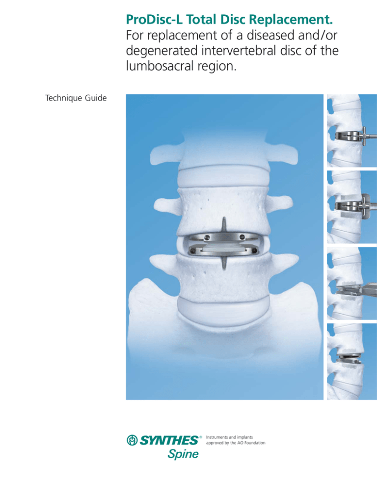
ProDisc-L Total Disc Replacement.
For replacement of a diseased and /or
degenerated intervertebral disc of the
lumbosacral region.
Technique Guide
Instruments and implants
approved by the AO Foundation
Table of Contents
Introduction
Surgical Technique
Product Information
Image intensifier control
Introduction
2
Features
3
Indications for Use
4
Contraindications
4
Patient Exclusion Recommendations
4
MRI Information
4
Preoperative Considerations
6
Patient Positioning
7
Anterior Access and Approach
7
Marking Midline
9
Discectomy, End Plate Preparation
and Remobilization
9
Implantation
11
1. Trial
12
2. Chisel
14
3. Insert Implant
15
Polyethylene Inlay Insertion
17
Final Implant Verification
20
Postoperative Care
20
Implants
21
Instruments
22
Set Lists
25
Synthes Spine
Introduction
The ProDisc-L Total Disc Replacement is intended to replace a
diseased and /or degenerated intervertebral disc of the lumbosacral region in patients with discogenic pain associated
with degenerative disc disease (DDD) at one lumbar spinal
segment level between L3 and S1. Total disc replacement is
intended to significantly reduce discogenic pain and improve
patient function by allowing for the removal of the diseased
disc while restoring the normal disc height.
The ProDisc-L Total Disc Replacement design is based on
a ball and socket principle composed of three implant
components — two cobalt chrome alloy (CoCrMo) end plates
and an ultra-high-molecular-weight polyethylene (UHMWPE)
inlay. The polyethylene inlay includes a radiopaque tantalum
marker. The ProDisc-L Total Disc Replacement end plates have
central keels and small spikes for initial fixation to the
vertebral bodies and a plasma sprayed titanium coating on
all bone-contacting surfaces to promote bony integration
(Figure 1). The UHMWPE / CoCrMo coupling has historically
been used in total joint replacement and has been used in
spinal arthroplasty procedures for two decades.
MRI Information
The ProDisc-L Total Disc Replacement is labeled
MR Conditional, where it has been demonstrated to
pose no known hazards in a specified MR environment
with specified conditions of use. Please refer to page 4
for further information.
2
Synthes Spine ProDisc-L Total Disc Replacement Technique Guide
Figure 1
Features
Ball and socket principle provides a fixed center
of rotation (Figure 2)
– Designed to allow controlled dynamic motion through
the physiological range of motion
– Designed to prevent pure translational motion to
theoretically protect the facets from excessive shear
loading
Modular design accommodates anatomical needs
of individual patients (Figures 3 and 4)
– Medium and large footprints
– 10, 12 and 14 mm heights
– 6° and 11° lordotic angles*
– 12 anatomical combinations
Figure 2
M: 34.5 mm
L: 39 mm
M: 27 mm
L: 30 mm
Central keel facilitates midline placement and provides
secure primary fixation, and titanium plasma sprayed
porous coating helps foster bony integration
The ProDisc-L Total Disc Replacement provides the surgeon
with a motion-preserving system for treating patients with
degenerative disc disease. Successful application and clinical
outcomes of this technology depend on a number of other
critical factors:
– Completion of a company-sponsored training program on
the use of ProDisc-L Total Disc Replacement and associated
instrumentation
– Proper patient selection
– Safe and adequate surgical approach and exposure to
the treated level
– Complete and meticulous discectomy, end plate
preparation, and remobilization of the disc space
– Optimal implant footprint, height, lordosis selection,
and placement
Figure 3
10 mm
12 mm
14 mm
6°
11°
Figure 4
Synthes Spine
3
Indications for Use
The ProDisc-L Total Disc Replacement is indicated for spinal
arthroplasty in skeletally mature patients with degenerative
disc disease (DDD) at one level from L3 to S1. DDD is defined
as discogenic back pain with degeneration of the disc
confirmed by patient history and radiographic studies. These
DDD patients should have no more than grade 1 spondylolisthesis at the involved level. Patients receiving the ProDisc-L
Total Disc Replacement should have failed at least six months
of conservative treatment prior to implantation of the
ProDisc-L Total Disc Replacement.
Contraindications
The ProDisc-L Total Disc Replacement should not be implanted
in patients with the following conditions:
– Active systemic infection or infection localized to the site
of implantation
– Osteopenia or osteoporosis defined as DEXA bone density
measured T-score < -1.0
– Bony lumbar spinal stenosis
– Allergy or sensitivity to implant materials (cobalt, chromium,
molybdenum, polyethylene, titanium, tantalum)
– Isolated radicular compression syndromes, especially due
to disc herniation
– Pars defect
– Involved vertebral end plate dimensionally smaller than
34.5 mm in the medial-lateral and / or 27 mm in the
anterior-posterior directions
– Clinically compromised vertebral bodies at affected level
due to current or past trauma
– Lytic spondylolisthesis or degenerative spondylolisthesis
of grade > 1
Patient exclusion recommendations
Patient selection is extremely important. In selecting patients
for a total disc replacement the following factors can be of
extreme importance to the success of the procedure:
– The patient’s occupation or activity level
– A condition of senility, mental illness, alcoholism,
or drug abuse
– Certain degenerative diseases (e.g., degenerative scoliosis
or ankylosing spondylitis) that may be so advanced at the
time of implantation that the expected useful life of the
device is substantially decreased
4
Synthes Spine ProDisc-L Total Disc Replacement Technique Guide
MRI Information
Synthes ProDisc-L implants are labeled MR Conditional
according to the terminology specified in ASTM F2503-05,
Standard Practice for Marking Medical Devices and Other Items
for Safety in the Magnetic Resonance Environment. Nonclinical
testing of the ProDisc-L demonstrated that the implant is
MR Conditional. A patient with a ProDisc-L implant may be
scanned safely under the following conditions:
– Static magnetic field of 1.5 Tesla and 3.0 Tesla at Normal
Operating Mode or First Level Controlled Mode
– Highest spatial gradient magnetic field of 900 Gauss / cm
or less
– Maximum MR system reported whole body averaged
specific absorption rate (SAR) of 2 W / kg for the Normal
Operating Mode and 4 W / kg for the First Level Controlled
Mode for 15 minutes of scanning
Note: In nonclinical testing, a Synthes ProDisc-L implant of
largest geometrical volume and mass was tested for heating
and results showed a maximum observed heating of 1.8ºC
for 1.5 T and a maximum observable heating of 1.7°C for
3.0 T with a machine reported whole body averaged SAR of
2 W / kg as assessed by calorimetry.
Patients may be safely scanned in the MRI chamber at the
above conditions. Under such conditions, the maximal expected
temperature rise is less than 2°C. To minimize heating, the
scan time should be as short as possible and the SAR as low
as possible. Temperature rise values obtained were based
upon a scan time of 15 minutes.
The above field conditions tested in a 1.5 T and a 3.0 T Philips
Achieva (Philips Healthcare, Software release 2.6.3 SP4) MR
scanner should be compared with those of the user’s MR
system in order to determine if the item can safely be brought
into the user’s MR environment. Synthes MR Conditional
ProDisc-L implants may have the potential to cause artifact in
the diagnostic imaging.
Artifact Information
MR image quality may be compromised if the area of interest
is in the same area or relatively close to the position of the
ProDisc-L implant and it may be necessary to optimize MR
imaging parameters in order to compensate for the presence
of the implant.
A representative implant has been evaluated in the MRI
chamber and worst case artifact information is provided
below. Overall, artifacts created by ProDisc-L implants may
present issues if the MR imaging area of interest is in or
near the area where the implant is located.
– For FFE sequence: Scan duration: 3 min, TR 100 ms,
TE 15 ms, flip angle 15°, worst case artifact will extend
approximately 5.0 cm from the implant
– For SE sequence: Scan duration: 4 min, TR 500 ms,
TE 20 ms, flip angle 70°, worst case artifact will extend
approximately 4.0 cm from the implant
Synthes Spine
5
Preoperative Considerations
Perform a thorough review of patient history, physical exam
and imaging studies to identify possible contraindications to
total disc replacement or midline anterior lumbar approach
and to verify that the lumbar disc in question is a significant
pain generator. It is recommended that you use AP and
lateral radiographs to preoperatively determine implant size
and coordinate the surgical procedure with a spinal-access
trained vascular or general surgeon.
Note: In order to minimize the risk of atraumatic
periprosthetic vertebral fractures, surgeons must consider all co-morbidities, past and present medications,
previous treatments, etc. Upon reviewing all relevant
information the surgeon must determine whether a
bone density scan is prudent. A screening questionnaire for osteoporosis, SCORE (Simple Calculated
Osteoporosis Risk Estimation)1 may be used to screen
patients to determine if a DEXA bone mineral density
measurement is necessary. If DEXA is performed, exclusion from receiving the device should be considered if
the DEXA bone density measured T-score is < -1.0, as
the patient may be osteopenic.
WARNINGS
Correct placement of the device is essential to optimal
performance. Use of the ProDisc-L Total Disc Replacement should only be undertaken after the surgeon has
become thoroughly knowledgeable about spinal
anatomy and biomechanics, has had experience with
anterior approach spinal surgeries, and has had handson training in the use of this device.
In order to achieve proper prosthesis fit and fixation,
patients with involved vertebral end plates dimensionally smaller than 34.5 mm in the medial-lateral and /or
27 mm in the anterior-posterior directions are not
appropriate candidates for ProDisc-L surgery due to
limitations in the prosthesis sizes available (Figure 5).
1 Lydick E, Cook K, Turpin J, et al. “Development and validation of a simple
questionnaire to facilitate identification of women likely to have low bone
density.” Am J Man Care 1998; 4:37 – 48.
6
Synthes Spine ProDisc-L Total Disc Replacement Technique Guide
27 mm
34.5 mm
Figure 5
Surgical Technique
Patient positioning
Insertion of the ProDisc-L implant is dependent on the use of
AP and lateral fluoroscopy throughout the procedure. Patient
positioning should allow for circumferential use of the
C-arm at the operative levels with unobstructed movement
in and out of the sterile field.
Position the patient in a supine, neutral position on a
radiolucent operating table with arms abducted 90°.
Alternatively, the arms may be adducted and crossed over
the chest (Figure 6).
Figure 6
Anterior access and approach
Locate the correct operative disc level and incision location
by taking a lateral fluoroscopic view while holding a straight
metal instrument at the side of the patient. This ensures that
the incision and exposure will allow for direct visualization
into the disc space (Figure 7).
Expose the operative disc level through a standard miniopen retroperitoneal approach.
Perform a transverse skin incision, beginning at midline and
continuing laterally 5 – 6 cm to the left. Incise the anterior
rectus sheath along the same line, extending the dissection
beyond the ends of the skin incision (Figure 8). Elevate the
anterior rectus sheath to allow for full mobilization of the
rectus muscle (Figure 9).
Figure 7
Figure 8
Figure 9
Synthes Spine
7
Surgical Technique continued
Anterior access and approach continued
Retract the rectus muscle towards the midline, and incise the
posterior sheath vertically to the peritoneum. Carefully push
the peritoneum posteriorly at the edge of the fascial incision.
Manually develop a plane between it and the abdominal
wall, into the retroperitoneal space.
Bluntly elevate the peritoneum away from the psoas muscle.
Identify the ureter and lift it away with the peritoneum.
Palpate medially to feel for the disc, vertebral body and iliac
artery. Elevate the peritoneum away in all directions to expose the disc space (Figure 10).
Note: Avoid tearing the peritoneum when elevating it.
Close any tears immediately before proceeding with
the approach.
L3 – L4, L4 – L5
The L3 – L4 and L4 – L5 disc spaces are typically located posterior to the aortic and vena cava bifurcation. Mobilize the left
iliac artery, ligate and cut the iliolumbar vein(s), and then
retract the artery and vein from left to right to provide
adequate exposure of the disc space and vertebral bodies
(Figure 11).
Figure 10
Note: Care must be taken to ensure the left iliac vein
and artery are mobile prior to retracting.
L5 – S1
The L5 – S1 disc space is typically located below the aortic and
vena cava bifurcation. Retract the left and right common iliac
vessels laterally and superiorly to provide adequate exposure
of the disc space and vertebral bodies. Take the middle sacral
vessels to provide clear exposure of the disc space (Figure 12).
Figure 11
Note: Use cautery cautiously to avoid injury to the
superior hypogastric plexus.
Figure 12
8
Synthes Spine ProDisc-L Total Disc Replacement Technique Guide
Marking midline
Instruments
PDL118
Midline Indicator
PDL120
Midline Marker, 8 mm width, 250 mm
Use AP fluoroscopy to identify the midline of the operative
level before the discectomy is initiated (Figure 13). Mark the
midline on the superior and inferior vertebral bodies adjacent
to the operative level so the mark remains visible throughout
the entire procedure. The midline indicator and midline
marker may be used to facilitate this step.
Figure 13
Discectomy, end plate preparation, and remobilization
Instruments
PDL114
Vertebral Body Spreader, angled
PDL116
Bone Elevator, 17 mm width, 337 mm
Note: Performing a complete and meticulous discectomy
and remobilization of the disc space is critical to the
success of the surgery.
The surgeon must remobilize the diseased segment, restore
the disc height, and properly balance the soft tissues prior
to implantation of the ProDisc-L Total Disc Replacement.
Figure 14
The ProDisc-L Total Disc Replacement may maintain
motion, but it cannot create motion.
Create an annulotomy centered on midline and wide enough
to accommodate the ProDisc-L Total Disc Replacement
implant (Figure 14).
Perform a thorough discectomy using the bone elevator and
standard rongeurs, Kerrisons, and curettes, ensuring the
posterolateral corners are freed of disc material (Figure 15).
Figure 15
Synthes Spine
9
Surgical Technique continued
Discectomy, end plate preparation, and remobilization
continued
Under fluoroscopic control, insert the vertebral body
spreader to the posterior margin of the vertebral bodies
to gradually remobilize the motion segment (Figure 16).
Placement of the tips to the posterior margin will minimize
the risk of end plate fracture. Place the spreader on one side
to facilitate the discectomy on the contralateral side, and
then repeat for the other side.
Vertebral body spreader
Remove the cartilaginous end plates to bleeding bone, taking
care to not compromise the integrity of the bony end plates.
The posterior annulus should be completely exposed, and
resected as necessary to expose the posterior longitudinal
ligament (PLL) and to remobilize the segment.
If posterior remobilization cannot be achieved, the PLL may
need to be released from the posterior vertebral body with a
curved curette, transected, or completely resected (Figure 17).
Two vertebral body spreaders can be used to obtain
balanced remobilization.
Note: Ensure that a complete discectomy is performed
and the integrity of the bony end plates is preserved to
provide a firm base for mechanical stability and to
reduce the potential for device subsidence. Fluoroscopy
must be used to ensure the tips of the vertebral body
spreader are resting on the posterior margin prior to
distracting, to minimize the risk of vertebral end plate
fracture. Remodeling of the end plates is only recommended to remove significant posterior osteophytes
that may interfere with prosthesis insertion. Remobilization and restoration of the disc height must be fully
achieved prior to implantation of the ProDisc-L Total
Disc Replacement. Parallel distraction is critical for
restoration of disc height.
Figure 16
Figure 17
10
Synthes Spine ProDisc-L Total Disc Replacement Technique Guide
Implantation
The ProDisc-L Total Disc Replacement contains 12 trials that
correspond to the 12 possible ProDisc-L Total Disc Replacement implant sizes. Trials are placed into the disc space
intraoperatively to determine the appropriate implant footprint, lordotic angle and disc height.
The trial is used as a jig to control the direction and depth of
the chisel cuts in the vertebral bodies. The central keels on
the ProDisc-L Total Disc Replacement end plates will follow
the path of the chisel cuts to its final position. Lastly, the
end plates are distracted and the polyethylene inlay is inserted
and locked into the inferior end plate.
Implantation of the ProDisc-L Total Disc Replacement implant
is performed in three steps:
1
2
3
Trial
Chisel
Insert implant
Synthes
11
Surgical Technique continued
1
Trial
Instruments
PDL102
Slotted Mallet
PDL202
Handle, for Trial Implant
PDL206
Screwdriver, for Adjustable Stop
PDL208
Adjustable Stop, for Trial Implants
PDL222 –
PDL256
Trial Implants
Figure 18
Use the screwdriver to assemble the stop into the trial.
Ensure the stop is fully seated in the trial.
Connect the handle to the shaft of the trial by pulling back
on the flange. The handle locks onto the shaft of the trial
implant and can be oriented in one of four positions.
Insert the trial into the intervertebral disc space, centered on
the midline mark and aligned with the sagittal plane of the
vertebral body (Figure 18).
Trial
12
Synthes Spine ProDisc-L Total Disc Replacement Technique Guide
Handle
Under lateral fluoroscopic control, advance the trial to the posterior margin
of the vertebral bodies with the slotted
mallet. The stop can be backed out to
allow the trial to be positioned more
posteriorly (Figure 19). Each full counterclockwise rotation of the stop allows the
trial to be advanced 1 mm posterior.
Select the largest footprint to maximize
coverage of the vertebral bodies, the
disc height to match that of a normal
adjacent disc space, and the lordosis
angle that matches the anatomy.
Note: Ensure that the adjustable
stop instrument is fully seated to
the trial body prior to inserting the
trial device. The optimal position of
the trial is at the posterior margin
of the vertebral bodies, and centered on the midline. Ensure that
the largest footprint is selected to
minimize the potential for implant
subsidence. Correct sizing, placement and lordotic angle are critical
to ensure optimal performance.
Figure 19
Synthes Spine
13
Surgical Technique continued
2
Chisel
Instruments
PDL102
Slotted Mallet
PDL322–
PDL326
Chisels
Remove the handle from the trial.
Slide the chisel onto the shaft of the trial. Under lateral
fluoroscopic control, advance the chisel into the vertebral
bodies with the slotted mallet until the chisel is fully seated
on the trial (Figure 20).
The chisel should be fully seated on the trial; this will ensure
that the chisel depth is adequate.
Figure 20
The chisel and trial are left in place until the ProDisc-L Total
Disc Replacement implant is ready for insertion.
Note: Ensure that the stop on the trial is fully seated
against the vertebral body prior to chiseling. Chiseling
must be performed under lateral fluoroscopic control.
The depth of the chisel cuts within both vertebral
bodies must be adequate prior to ProDisc-L Total Disc
Replacement implant insertion. The chisel should be
fully inserted on the trial until the “stop” on the trial
is reached; this will ensure that the chisel depth is
adequate.
It may be necessary to perform two chisel cuts to ensure
adequate chisel depth on each vertebral body. The first chisel
cut should follow the angle of the inferior vertebral body,
then, the chisel should be cephalized so the second cut
follows the angle of the superior vertebral body (Figure 21).
Figure 21
14
Synthes Spine ProDisc-L Total Disc Replacement Technique Guide
3
Insert implant
Inserter
Instrument
PDL102
Slotted Mallet
PDL402–
PDL404
Inserters
The ProDisc-L Total Disc Replacement implant end plates are
inserted in a collapsed position and then distracted for polyethylene inlay insertion. This modular technique facilitates
end plate insertion and provides efficient distraction for
polyethylene inlay assembly.
End plate insertion
Press the release button on the back of the inserter and
rotate the inferior arms outward. Assemble the inferior end
plate onto the pins of the inferior arms. Press the release
button and rotate the arms inward to lock the inferior end
plate onto the pins (Figure 22).
Figure 22
Load the superior end plate onto the pins on the superior
arms of the inserter (Figure 23).
Nest and hold the two end plates together by firmly gripping
the inserter arms (Figure 24).
Note: Ensure inferior arms of inserter are correctly
locked prior to end plate insertion.
Figure 23
Figure 24
Synthes Spine
15
Surgical Technique continued
3
Insert implant continued
Remove the chisel and trial. Ensure the disc space is clear of
any disc or bony debris.
Align the keels of the ProDisc-L Total Disc Replacement
implant with the chisel cuts. Under fluoroscopic control, use
the slotted mallet to insert the ProDisc-L Total Disc Replacement end plates to the posterior margin of the vertebral
bodies (Figure 25).
Note: Visually confirm that the anterior edge of the
prosthesis is within the anterior edge of the vertebral
body.
16
Synthes Spine ProDisc-L Total Disc Replacement Technique Guide
Figure 25
Polyethylene inlay insertion
Instrument
PDL422–
PDL426
Distractors
Distractor
Insert the polyethylene inlay into the grooves in the inferior
arms of inserter with the “Dome Up and Dome Up”
(Figure 26).
Note: Rounded
profile faces
anterior
Note: Ensure that the polyethylene inlay is placed in
the proper direction by confirming that the rounded
profile is facing anterior.
Dome up
Advance the polyethylene inlay to the first ball detent
(Figure 27).
Dome up
Figure 26
Ball detent
Figure 27
Synthes Spine
17
Surgical Technique continued
Polyethylene inlay insertion continued
Assemble the distractor to the inserter
(Figure 28). Under fluoroscopic control,
use the thumbscrew to fully advance
the distractor (Figure 29). Verify that
the posterior edge of the end plates
have separated from each other (Figure
30); this ensures adequate clearance for
the insertion of the polyethylene inlay.
Note: Do not attempt to force and
lock the polyethylene inlay if the
end plates have not separated.
Additional discectomy and
remobilization may be required if
the end plates do not separate.
Figure 28
Figure 30
18
Synthes Spine ProDisc-L Total Disc Replacement Technique Guide
Figure 29
Instrument
PDL432–
PDL434
Inlay Pushers
Inlay pusher
Insert the inlay pusher into the same grooves in the inferior
arms. Manually push and lock the polyethylene inlay into the
inferior end plate (Figure 31). Remove the inlay pusher.
Visually confirm the polyethylene inlay is locked into the
inferior end plate (Figure 32). A nerve hook may be used to
verify that NO STEP and NO GAP are present.
Remove the distractor from the inserter.
Note:
Ensure that the polyethylene inlay is securely locked
within the inferior plate component. If the
polyethylene inlay is not securely locked, anterior
displacement of the polyethylene inlay will occur.
Visually confirm the polyethylene inlay is locked into
the inferior end plate by using a nerve hook to verify
that NO STEP and NO GAP are present at the anterior
edge of the end plate (Figure 33).
Figure 31
The tantalum marker does not ensure whether or not
the inlay is fully seated in the inferior plate. It is still
necessary to check visually and manually (e.g. “NO
STEP” and “NO GAP”) the seating of the inlay.
Press the release button of the inserter and rotate the inferior
arms outward to unlock the inserter. Gently remove the
inserter from the ProDisc-L Total Disc Replacement implant.
STEP
“NO STEP”
GAP
“NO GAP”
Figure 32
Figure 33
Synthes Spine
19
Surgical Technique continued
Final implant verification
Verify final implant position with lateral
and AP imaging (Figure 34).
The surgical wound is closed in routine
fashion appropriate for the surgical exposure utilized.
Postoperative care
Following surgery, patients can begin
ambulating on postoperative days 1 – 3
with supervised use of a walker and a
simple corset when out of bed. Isometric leg exercises are recommended for
the first two weeks postoperatively,
with the subsequent initiation of outpatient physical therapy.
Figure 34
Patients should be instructed to avoid
excessive bending or lifting for the first
two weeks postoperatively, and can
begin driving, light bending, and lifting
from 2 to 6 weeks postoperatively,
gradually resuming normal activities
beginning at 6 weeks postoperatively.
20
Synthes Spine ProDisc-L Total Disc Replacement Technique Guide
Implants
ProDisc-L Implant Components sold separately
Superior Endplates, sterile
PDL-M-SP06S Medium, 6°
PDL-M-SP11S Medium, 11°
PDL-L-SP06S Large, 6°
PDL-L-SP11S Large, 11°
Polyethylene Inlays with Tantalum
Marker, sterile
PDL-M-PT10S Medium, 10 mm
PDL-M-PT12S Medium, 12 mm
PDL-M-PT14S Medium, 14 mm
PDL-L-PT10S Large, 10 mm
PDL-L-PT12S Large, 12 mm
PDL-L-PT14S Large, 14 mm
Inferior Endplates, sterile
PDL-M-IP00S Medium
PDL-L-IP00S
Large
ProDisc-L Implant Component
Reference Table
27 mm
30 mm
34.5 mm
Superior Endplates
Polyethylene Inlays
Inferior Endplates
39 mm
Medium
Large
6°
PDL-M-SP06S
PDL-L-SP06S
11°
PDL-M-SP11S
PDL-L-SP11S
10 mm
PDL-M-PT10S
PDL-L-PT10S
12 mm
PDL-M-PT12S
PDL-L-PT12S
14 mm
PDL-M-PT14S
PDL-L-PT14S
PDL-M-IP00S
PDL-L-IP00S
Synthes Spine
21
Instruments
PDL102
Slotted Mallet
PDL114
Vertebral Body Spreader, angled
PDL116
Bone Elevator, 17 mm width, 337 mm
PDL118
Midline Indicator
PDL120
Midline Marker, 8 mm width, 250 mm
PDL202
Handle, for Trial Implant
PDL206
Screwdriver, for Adjustable Stop
22
Synthes Spine ProDisc-L Total Disc Replacement Technique Guide
PDL222
PDL224
PDL226
Trial Implant, medium, 6°, 10 mm
Trial Implant, medium, 6°, 12 mm
Trial Implant, medium, 6°, 14 mm
PDL232
PDL234
PDL236
Trial Implant, medium, 11°, 10 mm
Trial Implant, medium, 11°, 12 mm
Trial Implant, medium, 11°, 14 mm
PDL242
PDL244
PDL246
Trial Implant, large, 6°, 10 mm
Trial Implant, large, 6°, 12 mm
Trial Implant, large, 6°, 14 mm
PDL252
PDL254
PDL256
Trial Implant, large, 11°, 10 mm
Trial Implant, large, 11°, 12 mm
Trial Implant, large, 11°, 14 mm
Synthes Spine
23
Instruments continued
PDL208
Adjustable Stop, for Trial Implants
PDL322
PDL324
PDL326
Chisel, 10 mm
Chisel, 12 mm
Chisel, 14 mm
PDL402
PDL404
Inserter, medium
Inserter, large
PDL422
PDL424
PDL426
Distractor, 10 mm
Distractor, 12 mm
Distractor, 14 mm
PDL432
PDL434
Inlay Pusher, medium
Inlay Pusher, large
PDL442
Lever
24
Synthes Spine ProDisc-L Total Disc Replacement Technique Guide
ProDisc-L Instrument Set (PDL1000)
Graphic Cases
690.225
Graphic Case, for ProDisc-L Instruments (1)
690.224
Graphic Case, for ProDisc-L Instruments (2)
Instruments
PDL102
PDL114
PDL116
PDL118
PDL120
PDL202
PDL206
PDL208
PDL322
PDL324
PDL326
PDL402
PDL404
PDL422
PDL424
PDL426
PDL432
PDL434
PDL442
Slotted Mallet
Vertebral Body Spreader, angled, 2 ea.
Bone Elevator, 17 mm width, 337 mm
Midline Indicator, 2 ea.
Midline Marker, 8 mm width, 250 mm
Handle, for Trial Implant, 2 ea.
Screwdriver, for Adjustable Stop
Adjustable Stop, for Trial Implants, 12 ea.
Chisel, 10 mm
Chisel, 12 mm
Chisel, 14 mm
Inserter, medium, 2 ea.
Inserter, large, 2 ea.
Distractor, 10 mm
Distractor, 12 mm
Distractor, 14 mm
Inlay Pusher, medium
Inlay Pusher, large
Lever
690.225
690.224
Note: For additional information, please refer to package insert.
For detailed cleaning and sterilization instructions, please refer to
http://us.synthes.com/Medical + Community / Cleaning + and + Sterilization.htm
or to the below listed inserts, which will be included in the shipping container:
– Processing Synthes Reusable Medical Devices — Instruments, Instrument Trays
and Graphic Cases — DJ1305
Synthes Spine
25
ProDisc-L Instrument Set (PDL1000) continued
Trial Implants
PDL222
PDL224
PDL226
PDL232
PDL234
PDL236
PDL242
PDL244
PDL246
PDL252
PDL254
PDL256
26
Trial Implants
Trial Implant, Medium, 6°, 10 mm
Trial Implant, Medium, 6°, 12 mm
Trial Implant, Medium, 6°, 14 mm
Trial Implant, Medium, 11°, 10 mm
Trial Implant, Medium, 11°, 12 mm
Trial Implant, Medium, 11°, 14 mm
Trial Implant, Large, 6°, 10 mm
Trial Implant, Large, 6°, 12 mm
Trial Implant, Large, 6°, 14 mm
Trial Implant, Large, 11°, 10 mm
Trial Implant, Large, 11°, 12 mm
Trial Implant, Large, 11°, 14 mm
Synthes Spine ProDisc-L Total Disc Replacement Technique Guide
Synthes Spine
1302 Wrights Lane East
West Chester, PA 19380
Telephone: (610) 719-5000
To order: (800) 523-0322
Fax: (610) 251-9056
© 2006 Synthes, Inc. or its affiliates. All rights reserved.
www.synthesprodisc.com
www.synthes.com
ProDisc and Synthes are trademarks of Synthes, Inc. or its affiliates.
Printed in U.S.A. 6/11 J6206-D


