32
Review
TRENDS in Neurosciences Vol.25 No.1 January 2002
Generating neuronal diversity in the
retina: one for nearly all
Till Marquardt and Peter Gruss
Visual perception of our environment essentially depends on the correct
assembly of seven principal cell types into the functional architecture of the
neuroretina. During retinogenesis these cell types derive from a common
population of multipotent retinal progenitor cells (RPCs) residing in the inner
layer of the optic cup. In contrast to other well studied regions of the
developing CNS, retinal cell diversification is apparently not achieved by spatial
prepatterning into distinct progenitor domains, but rather by the sequential
production of cell types in a defined histogenetic order. Several lines of
evidence suggest that this observation reflects substantial intrinsic changes in
the retinogenic potential of RPCs. Recent advances, however, point at the
existence of a common molecular framework underlying the retinogenic
potential of RPCs throughout retinal neurogenesis.
Rods
Bipolar
Müller glia
Ganglion
Horizontal
Cones
Amacrine
11 13
16 Birth
Embryonic
3
6
9 11 Days
Postnatal
Retinogenesis
Cell differentiation in the vertebrate retina is
initiated in the inner layer of the central optic cup and
progresses concentrically in a wave-like fashion until
reaching the peripheral edges of the retina [1].
Another characteristic feature of vertebrate
retinogenesis is the relatively fixed chronological
sequence after which the different retinal cell types
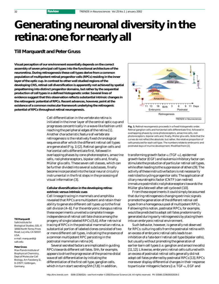
are generated (Fig. 1) [2]. Retinal ganglion cells and
horizontal cells differentiate first, followed in
overlapping phases by cone-photoreceptors, amacrine
cells, rod-photoreceptors, bipolar cells and, finally,
Müller glia cells. These seven cell classes, which can
be further divided into several subclasses, finally
become incorporated into the local neural circuitry
instrumental in the first steps in the processing of
visual information [3].
Cellular diversification in the developing retina:
extrinsic versus intrinsic cues
Till Marquardt
Salk Institute for
Biological Studies, GEL-P,
10010 North Torrey Pines
Road, La Jolla, CA 92037,
USA.
e-mail: marquardt@
salk.edu
Peter Gruss
Max-Planck-Institute of
Biophysical Chemistry,
Dept of Molecular Cell
Biology, Am Fassberg 11,
D-37077, Göttingen,
Germany.
Cell lineage tracing in mammals and amphibia
revealed that RPCs are multipotent and retain their
ability to generate different cell types up to the final
cell division [4–6]. For the embryonic Xenopus retina
these experiments unveiled a complete lineageindependence of retinal cell fate choice among the
progeny of single labeled RPCs [5,6]. After retroviral
tracing of RPCs in the postnatal mammalian retina, a
substantial portion of labeled clones consisted of two
or more different cell types, indicating the presence of
a common multipotent RPC persisting in the
postnatal mammalian retina [4].
Several secreted factors are implicated in guiding
RPCs towards different cell fates. Shh, for example,
appears to drive the progression of the proximo-distal
wave of cell differentiation by initiating the
differentiation of the first cell type, ganglion cells,
which in turn start secreting Shh [7,8]. In addition,
http://tins.trends.com
TRENDS in Neurosciences
Fig. 1. Retinal neurogenesis proceeds in a fixed histogenetic order.
Retinal ganglion cells and horizontal cells differentiate first, followed in
overlapping phases by cone-photoreceptors, amacrine cells, rodphotoreceptors, bipolar cells and, finally, Müller glia cells. Note that the
curves do not reflect the absolute, but rather, the relative proportion of
cells produced for each cell type. The numbers relate to embryonic and
postnatal days of murine development. Modified from [2].
transforming growth factor-α (TGF-α), epidermal
growth factor (EGF) and leukemia inhibitory factor can
stimulate the production of particular retinal cell types,
while often leading to the suppression of others [9]. The
activity of these instructive factors is not necessarily
restricted to cycling progenitor cells: The application of
ciliary neurotrophic factor (CNTF) can redirect
immature postmitotic rod photoreceptors towards the
Müller glia fate well after cell cycle exit [10].
From these experiments it could simply be assumed
that during retinogenesis changing extrinsic signals
promote the generation of the different retinal cell
types from a homogenous pool of multipotent RPCs.
Following this notion, postnatal RPCs, for example,
would be predicted to adopt cell fates predominantly
generated during early retinogenesis by placing them
into an embryonic retinal environment.
Such behavior, however, is generally not observed
for RPCs: culturing cells from the postnatal retina with
an excess of embryonic retinal cells leads to an
inhibition of a ‘late-born’ retinal cell type (bipolar cells),
but usually without promoting the generation of
earlier-born cell types (i.e. ganglion and amacrine cells)
[11,12]. Likewise, embryonic retinal cells cultured with
an excess of postnatal retinal cells generally did not
adopt cell fates preferred by postnatal RPCs [13]. RPCs
moreover display differential changes in their response
to particular mitogenic factors [i.e. TGF-α, EGF and
0166-2236/02/$ – see front matter © 2002 Elsevier Science Ltd. All rights reserved. PII: S0166-2236(00)02028-2
Review
TRENDS in Neurosciences Vol.25 No.1 January 2002
(a)
E12
rpe
E15
nr
le
PN8
onl
nbl
inl
gcl
gcl
Key:
Rx1
(b)
Chx10
rpe
onl
Pax6 and Six3
inl
E12
E17
PN8
Pax6, Six3,
RPCs
Rx1, Chx10 and Hes1
Adult
TRENDS in Neurosciences
Fig. 2. Transcription factor expression during murine retinogenesis. (a) Pax6 is localized to virtually all
mitotic retinal progenitor cells (RPCs) throughout retinogenesis, as revealed by immunohistochemistry
with antibodies against Pax6 (red) and proliferating cell nuclear antigen (PCNA; green). Pax6 and PCNA
colocalize in RPCs (yellow), whereas Pax6 expression is maintained in postmitotic ganglion, horizontal
and amacrine cells. The residual RPC population in the postnatal day 8 (PN8) retina is still Pax6+
(arrowheads). (b) A set of transcription factors coexpressed initially in all RPCs becomes segregated in
expression with the increasing proportion of postmitotic cells in the developing retina [43,44,79–81]. It is
still unclear whether these factors actually colocalize in all RPCs. Arrows and circular arrows denote
approximately patterns of cell migration and mitotic activity, respectively. Abbreviations: E10–17,
embryonic day 10–17; gcl, ganglion cell layer; inl, inner nuclear layer; le, lens; nbl, neuroblast layer; onl,
outer nuclear layer; rpe, retinal pigment epithelium.
fibroblast growth factor (FGF)] with progression of
retinogenesis [14].
Besides the action of extrinsic signals influencing
cell fate, cell autonomous mechanisms must therefore
operate in mediating changes in the intrinsic
responsiveness of RPCs to particular extracellular
signals. To account for these observations it was
proposed that during the successive stages of
retinogenesis RPCs switch between different
competence states [15,16]. In molecular terms,
however, it still remains unclear what defines these
intrinsic changes that broadly appear to affect the
Pax6+
Extrinsic
signals
bHLH+
Pax6+
Pax6+
Additional
signals?
Cell cycle exit
RPC pool
Delta
Delta
bHLH
X
Notch
bHLH
Delta
Terminal
differentiation Differentiation
factors
Differentiation
TRENDS in Neurosciences
Fig. 3. The activity of Pax6 in all retinal progenitor cells (RPCs) is a prerequisite for the activation of
retinogenic bHLH transcription factors (such as Math5, Ngn2 and Mash1) [33], possibly triggered by
extrinsic signals, such as Shh or epidermal growth factor (EGF) [7,17]. Their activation, presumably,
underlies the transition from uncommitted to lineage-restricted RPCs. Inset: activation of Notch
receptor by high levels of Delta ligand present on the surface of the adjacent RPC leads to the
suppression of Delta and bHLH factor expression [61]. The resulting lateral inhibition consequently
assures that the activation of bHLH factors occurs only in a subset of RPCs. The action of bHLH factors
is thought to accelerate cell cycle exit [23], possibly assisted by additional signals. In addition, bHLH
factors, such as Math5, are implicated in activating terminal differentiation factors, such as Brn3b, in
postmitotic precursor cells [30,31].
http://tins.trends.com
RPC pool. A clue is provided by the observation that
the level of EGF receptor (EGF-R) expression
increases in RPCs from late embryonic to early
postnatal stages, underlying a shift in the
responsiveness to EGF [17,18]. A possible mechanism
therefore involves changes in the expression level of
cell surface receptors, which thereby mediate
intrinsic differences in the response of RPCs when
challenged by the same extrinsic signals.
Rx1, Chx10 and Hes1
gcl
E10
33
Complexity of the retinal progenitor cell pool
Another issue which has to be taken into account
when interpreting these findings is the heterogeneity
of the RPCs pool during retinogenesis, with the
apparent coexistence of non-overlapping RPC
subpopulations with distinct preferences for the
range of cell types generated [9]. One such lineagerestricted RPC population that preferentially gave
rise to amacrine cells and, later, photoreceptor cells,
was identified on the basis of selective expression of
particular epitopes [19].
A set of transcription factors, most prominently of
the basic helix–loop–helix (bHLH) class, are prime
candidates to mediate such cell fate biases of RPC
sub-populations [20,21]. Neuronal bHLH
transcription factors are generally thought to promote
the acquisition of pan-neuronal characteristics
[20,22,23]. However, in recent years evidence
accumulated that proneural bHLH factors are also
involved in more specific aspects of neurogenesis, such
as the specification of particular neuronal fates
[12,20]. In neural crest stem cells, for instance, Mash1
mediates intrinsic changes in the competence to
respond to BMP signaling [24], whereas the activity of
Olig2 promotes motor neuron and oligodendrocytic
fates in the ventral spinal cord [25,26]. In the
developing retina, several bHLH factors are localized
to subsets of RPCs and where shown to direct such
cells to particular fates (Figs 2,3) [27–30].
The activity of Math5 in a sub-population of RPCs
essentially leads to the activation of the POU domain
transcription factor Brn3b, thereby driving these
progenitor cells towards the ganglion cell fate (Fig. 3)
[30–32]. Intriguingly, Mash1 and Ngn2 become
activated in two strictly nonoverlapping RPC
populations that both appear to give rise to bipolar
and photoreceptor cells [27,28,33] (Marquardt et al.,
unpublished).
To integrate these observations with the changes in
competence of the RPC pool it was suggested that
during retinogenesis lineage-restricted RPCs might
shift from one competence state to the other, although it
remains elusive whether such a switch can indeed occur
[16,34]. Since overexpression of retinal bHLH factors
leads to a strong and cell-autonomous bias towards
particular cell fates [29,35], a switch in competence or
cell fate bias consequently should be reflected by a shift
in the expression profile of such factors.
Such a switch in cell fate bias and/or molecular
profile of a particular RPC subpopulation was so far
34
Fig. 4. Potential repressive
interactions between bHLH
transcription factors in
subsets of lineagerestricted RPCs help to
establish the correct
proportion of cell types. In
wild-type RPCs, Math5
promotes the acquisition
of ganglion cell fate by
activation of Brn3b [30,31],
while at the same time
inhibiting amacrine cell
fate [30] and conephotoreceptor fate [38]
(not shown). NeuroD in
turn promotes amacrine
cell differentiation and
concomitantly suppresses
Müller glia (MCs) and
bipolar cell (BPCs) fate [35].
In the absence of Math5
the RPCs normally defined
by the presence of Math5
now preferentially adopt
the amacrine cell fate,
possibly by derepression
of NeuroD [33]. In Pax6deficient RPCs, retinogenic
factors other than NeuroD
fail to be activated, leading
to the channeling of the
RPCs towards the
amacrine cell fate.
Review
TRENDS in Neurosciences Vol.25 No.1 January 2002
Wild-type
RPCs
Math5-deficient
RPCs
NeuroD+
Other
cell fates
Pax6-deficient
RPCs
Other
cell fates
Math5+
NeuroD
Math5
?
BPCs MCs
NeuroD Brn3b
Amacrine cells
Ganglion cells
NeuroD
X
Amacrine cells
Math5
X
?
NeuroD
NeuroD
Amacrine cells
TRENDS in Neurosciences
only observed in the experimental situation. The
inactivation of Ngn2 leads to the up-regulation of
Mash1 in the formerly Ngn2+ Mash1− progenitor cells
in the developing neocortex and retina, in this case
leading to an apparent functional compensation
[36,37] (Marquardt et al., unpublished). Inactivation
of NeuroD, on the other hand, leads to a severe
reduction in the number of amacrine cells,
accompanied by a marked increase in the number of
bipolar and Müller glia cells, while NeuroD
overexpression essentially leads to the opposite
outcome [35]. Likewise, the failure to generate
ganglion cells in Math5 null mutants is accompanied
by a marked increase in the number of amacrine and
cone photoreceptor cells [30,38]. These results
indicate that besides directly guiding cells towards
particular fates, potential repressive interactions
among retinogenic bHLH factors might control the
correct numbers of the different cell types generated
in the neuroretina (Fig. 3).
In this respect such cell-intrinsic mechanisms
potentially complement the control of relative cell
numbers via extrinsic signaling. The production of
retinal ganglion cells, for instance, appears to be
controlled by negative feedback signaling by newly
post-mitotic ganglion cells, which in part seems to be
mediated by NGF signaling [39,40].
Pax6 at the link between early eye development and
retinal cell fate determination
In all vertebrate species analyzed so far, a similar set
of pivotal transcription factors, most prominently
Pax6, Rx1, Six3/6 and Lhx2, which act in initiating
vertebrate eye development, continues to be present
during the ensuing steps of retinal neurogenesis
(Fig. 4b) [41–47]. In the developing mouse retina,
http://tins.trends.com
Pax6 is expressed in virtually all mitotic RPCs during
all stages of retinogenesis, including postnatal stages
shortly before the last retinal cells become postmitotic
(Fig. 4a; Marquardt and Gruss, unpublished). Forced
expression of the Pax family transcription factor
Pax6, as well as the homeodomain transcription
factors Six3, Six6/Optx2 and Rx1/rax in fish and frog
embryos promotes the formation of ectopic retinal
tissue [42,45–47].
The coexpression of such factors appears to be a
defining feature of RPCs and it is well imaginable
that the combinatorial action of these transcription
factors controls the range of cell fates generated from
RPCs. This scenario would be similar to the situation
in the developing caudal CNS, where the generation
of specific neuronal subtypes at particular positions
along the dorsoventral axis is defined by the
coexpression of specific sets of paired- and
homeodomain transcription factors in the progenitor
cells of the ventricular zone [48].
Null mutations in the genes encoding Pax6, Lhx2
and Rx1, however, lead to an early arrest or, as in the
case of Rx1, to a failure to initiate optic vesicle
formation, resulting in a complete absence of
functional eye structures [42,49,50]. It therefore
remained obscure to what extent the activity of these
factors actually contributes to the retinogenic
potential of RPCs.
This constraint was overcome by conditional
inactivation of the gene encoding Pax6, specifically in
the RPCs of the distal optic cup, just prior to the onset
of cell differentiation [33,51]. At first glance, Pax6
deficient RPCs merely displayed reduced mitotic
activity, but maintained principal retinal identity and
with a delay, began to differentiate into neurons.
However, the Pax6 deficient RPCs displayed a
Review
TRENDS in Neurosciences Vol.25 No.1 January 2002
complete restriction towards the generation of only
one of the seven principal cell fates normally available
to RPCs, resulting in the exclusive differentiation of
amacrine interneurons (Fig. 3) [33].
The observation that a single factor, active in all
RPCs throughout retinogenesis, is essential for the
formation of nearly all retinal cell types, affecting
early and late born types alike, suggests the existence
of a common molecular framework underlying RPCs
during all stages of retinogenesis (Fig. 4). In this
respect the intrinsic changes in the retinogenic
potential of RPCs at particular stages of retinal
development might merely be superimposed on a
more ‘primitive’ RPC state. These intrinsic
differences apparently cannot easily be overcome
under certain culture conditions [11–13]. Recently,
however, the possibility of partially relieving these
constraints in vitro and promoting the production of
early born retinal cell types by late RPCs has been
reported [52].
The intrinsic changes in the retinogenic potential
which appear to affect the whole RPC pool might arise
from a shift in the relative expression level of
transcription factors expressed by all RPCs. For
example, the relative level of Pax6 activity in RPCs
becomes markedly lower during late embryogenesis
(Fig. 4a; Marquardt and Gruss, unpublished). Such
changes in the profile of transcription factor activity
might in turn affect the expression level of cell surface
receptors, thereby mediating differences in the
response of RPCs elicited by the same extrinsic
signals.
How can such shifts in the level of cellautonomously acting factors be achieved in the first
place? A likely scenario would be that such changes
might be mediated by an accumulative effect of
signals to which RPCs were exposed in the course of
retinogenesis. In this respect, the intrinsic changes in
the retinogenic potential of RPCs might be driven
forward by alterations in the signaling environment,
which in turn result from the continuous increase in
the number of postmitotic cells of different types.
Retinogenic bHLH transcription factors at the link
between Pax6 and retinal cell fate determination
How does Pax6 operate in mediating the retinogenic
potential of RPCs? The bHLH factors Ngn2, Mash1
and Math5 all fail to be activated in Pax6 deficient
RPCs (Fig. 3) [33]. Moreover, these factors appear to
constitute direct targets of Pax6 mediated
transcriptional activation [22,33,53]. Pax6 might
control the availability of the full range of cell fates to
RPCs essentially by mediating the activation of such
retinogenic transcription factors (Figs 2,3).
Another retinogenic bHLH factor, NeuroD, in
contrast turned out to be activated independently of
Pax6 (Fig. 3). Most importantly, NeuroD is strongly
implicated in mediating amacrine cell differentiation
[35]. However, since Pax6 is expressed in all RPCs
(Fig. 4a) and by virtually all postmitotic amacrine
http://tins.trends.com
35
cells [33] (Marquardt and Gruss, unpublished), the
observed behavior of Pax6 deficient RPCs is not likely
to be due to a direct derepression of amacrine cell fate.
In this context, it is of considerable interest to recall
the marked increase in the number of amacrine cells
in the retina of Math5 deficient mice. By mediating
the activation of Math5 in a subset of RPCs, which in
turn suppresses amacrine cell fate (Fig. 3), Pax6
might indirectly control the number of RPCs biased
towards the amacrine cell fate.
Another striking observation is that the
retinoblastomas that form in the retina of
Rb/p107-deficient chimeric mice exclusively comprise
cells with amacrine cell marker characteristics [54].
Because Rb, independently of its function in cell cycle
control, is known to interact with transcription
factors in the control of cell differentiation (most
notably the myogenic bHLH factor MyoD) [55,56],
this observation hints at the intriguing possibility
that Pax6 and Rb might act cooperatively during
retinogenesis.
From multipotency to lineage-restriction
To explain the observed Pax6 dependent activation of
bHLH factors in particular subsets of RPCs, in an
otherwise homogeneously Pax6+ RPC pool, the most
parsimonious interpretation has been that these
findings reflect a transition from an uncommitted
(possibly stem-cell-like) towards a lineage-restricted
RPC state [33]. In this respect the Pax6+ population
defines the most ‘primitive’ and the bHLH+Pax6+
population demarcates the lineage-restricted state
(Fig. 2).
This situation would be analogous to the observed
complex population of neural stem cells and distinct
lineage-restricted progenitor cell populations
coexisting in the ventricular zone of other regions of
the developing CNS [9,34,57,58]. Indeed, small
numbers of multipotent RPCs with stem cell
characteristics (i.e. passagability and neurosphere
formation) can be retrieved from the late embryonic
retina, while the majority of RPCs apparently
undergo immediate differentiation when cultured
in vitro [52,59].
The activity of pivotal retinal factors like Pax6 in
all RPCs potentially imposes a ‘retinal identity’ to the
response of the progenitor cells upon encountering
quite widely utilized signaling molecules like Shh or
EGF. However, as holds true for other regions of the
developing CNS, it still remains to be demonstrated
whether such instructive signals promote the
generation of lineage-restricted progenitors from
stem cells or if they stimulate direct differentiation to
particular cell fates [60].
How can distinct progenitor cell sub-populations
arise in a previously homogenous RPC pool?
Neurogenic bHLH factors are in general thought to
be subject to lateral inhibition mediated by
Notch/Delta signaling [61], which presumably
underlies their mosaic-like expression pattern in the
36
Review
TRENDS in Neurosciences Vol.25 No.1 January 2002
developing retina and elsewhere (Fig. 2)
[33,37,62,63]. Since neurogenic bHLH factors can
drive neural progenitor cells out of the cell cycle
[23,25], this mechanism potentially prevents the
premature depletion of the RPC pool, as has been
observed after inactivation of the Notch effector Hes1
[64]. The concomitant action of Notch/Delta
mediated lateral inhibition and repressive
interactions among the activated bHLH factors could
indicate that the induction of a given retinogenic
factor occurs only in a subset of RPCs (Fig. 2). The
interplay of these mechanisms thereby results in a
heterogeneous progenitor cell pool with distinct RPC
populations possessing different retinogenic
potentials (Fig. 3), which ultimately underlie the
generation of the different retinal cell types in their
appropriate numbers.
Some unresolved issues
Acknowledgements
We thank the members of
the P.G. and Michael
Kessel laboratories for
support and helpful
discussions. We are
particularly grateful to
Anastassia Stoykova,
Nicole Andrejewski and
Ruth Ashery-Padan for
discussions and critical
reading of the
manuscript. The studies
from which this review is
derived were supported
by an EU grant (B104CT96-0042) and by the
Max-Planck-Gesellschaft.
An important issue which remains unclear concerns
the precise lineage relationships between biased RPC
subpopulations. In particular it has to be elucidated
how fixed or plastic the restrictions towards
particular cell fates are for certain molecularly
definable RPC subpopulations. Furthermore, it
remains unclear how the action of secreted
instructive factors such as Shh or TGF-α are linked to
the expression of retinogenic factors like Math5 or
NeuroD in subsets of RPCs. In this respect, although
factors like Math5 appear directly to promote the
determination of particular cell fates, it remains to be
addressed whether other retinogenic bHLH factors
impose a bias on RPCs via changing their competence
to respond to particular extrinsic signals.
The transcription factors Pax6, Rx1 and Chx10,
which are initially coexpressed in RPCs, display an
ominous segregation of their expression domains
with increasing proportion of postmitotic cells in the
retina (Fig. 4b). It remains to be elucidated whether
the continued presence of these factors in particular
lineages serves later roles in terminal differentiation
and consolidation of cell identity, alongside factors
like Crx1 and Brn3b [65–67]. At the same time the
rapid down-regulation of these factors upon cell cycle
exit might be a prerequisite for the correct
specification of other cell lineages. Overexpression of
Pax6, for example, was reported to lead to severe
reduction of the photoreceptor containing outer
References
1 Prada, C. et al. (1991) Spatial and temporal
patterns of neurogenesis in the chick retina.
Europ. J. Neurosci. 3, 559–569
2 Young, R.W. (1985) Cell differentiation in the
retina of the mouse. Anat. Rec. 212, 199–205
3 Dowling, J.E. (1987) The Retina: An Approachable
Part of the Brain, Belknap Press of Harvard
University Press
4 Turner, D.L. and Cepko, C.L. (1987) A common
progenitor for neurons and glia persists in rat
retina late in development. Nature 328, 131–136
5 Holt, C.E. et al. (1988) Cellular determination in
the Xenopus retina is independent of lineage and
http://tins.trends.com
nuclear layer [68], where Pax6 is rapidly downregulated during normal retinogenesis (Fig. 4a).
Recently retinal stem cells could be isolated from
the pigmented ciliary margin of the adult mouse and
human retina [69]. Here it will be highly significant to
analyze the contribution of pivotal retinal factors like
Pax6, Six3/6 and Rx1 in mediating the retinogenic
potential of these stem cells. Subsequently, evidence
was provided that mild injury can induce a certain
regenerative potential exerted by Müller glia cells in
the adult avian retina [70]. The production of new
cells by Müller glia was preceded by the concomitant
up-regulation of Chx10 and Pax6, which normally are
only coexpressed in RPCs (Fig. 4). Evidence has
furthermore been provided for the existence of a rare
population of stem cells in the inner nuclear layer of
the adult teleost retina (possibly Müller glia), which
expresses Pax6 [71]. Hence, these results further
emphasize a role for the combined action of these
transcription factors in mediating the retinogenic
potential of RPCs. The surprising retinogenic
potential of Müller glia cells is remarkably analogous
to the recently uncovered neurogenic function of the
related radial glia cell of the cerebral cortex [72], for
the specification of which Pax6 was shown to play an
essential role [73].
Another issue is whether the factors acting in all
RPCs are linked to the topographic organization of
the optic cup. Patterning of the retina across the
dorsoventral and nasotemporal axes appears to
precede the onset of retinogenesis: the winged helix
transcription factors BF1 and BF2, for example,
already start to be expressed in the nasal and
temporal optic vesicle, respectively [74]. Recently
some light has been shed on how such factors
influence the projection properties of retinal ganglion
cells, by directing the activation of particular axon
guidance molecules [75–77]. However, it remains
unclear whether such factors can subtly influence the
retinogenic potential of RPCs to establish differences
in cellular subtype composition across the two
principal retinal axes [78]. In this respect the
gradient expression of pivotal factors like Pax6 across
the proximodistal axis (Andrejewski et al.,
unpublished) might provide a link to the gradients of
cellular composition and topographic cues that
ultimately underlie the correct representation of
visual space in our brain.
birth date. Neuron 1, 15–26
6 Wetts, R. and Fraser, S.E. (1988) Multipotent
precursors can give rise to all major cell types of
the frog retina. Science 239, 1142–1145
7 Neumann, C.J. and Nuesslein-Volhard, C.
(2000) Patterning of the zebrafish retina by a
wave of sonic hedgehog activity. Science 289,
2137–2139
8 Zhang, X.M. and Yang, X.J. (2001) Regulation of
retinal ganglion cell production by Sonic
hedgehog. Development 128, 943–957
9 Lillien, L. (1998) Neural progenitors and stem
cells: mechanisms of progenitor heterogeneity.
Curr. Opin. Neurobiol. 8, 37–44
10 Ezzeddine, Z.D. et al. (1997) Postmitotic cells
fated to become rod photoreceptors can be
respecified by CNTF treatment of the retina.
Development 124, 1055–1067
11 Belliveau, M.J. et al. (2000) Late retinal
progenitor cells show intrinsic limitations in the
production of cell types and the kinetics of opsin
synthesis. J. Neurosci. 20, 2247–2254
12 Cepko, C.L. (1999) The roles of intrinsic and
extrinsic cues and bHLH genes in the
determination of retinal cell fates. Curr. Opin.
Neurobiol. 9, 37–46
13 Belliveau, M.J. and Cepko, C.L. (1999) Extrinsic
and intrinsic factors control the genesis of
Review
14
15
16
17
18
19
20
21
22
23
24
25
26
27
28
29
30
31
32
amacrine and cone cells in the rat retina.
Development 126, 555–566
Lillien, L. and Cepko, C. (1992) Control of
proliferation in the retina: temporal changes in
responsiveness to FGF and TGF-α. Development
115, 253–266
Cepko, C.L. et al. (1996) Cell fate determination in
the vertebrate retina. Proc. Natl. Acad. Sci.
U. S. A. 93, 589–595
Livesey, F.J. and Cepko, C.L. (2001) Vertebrate
neural cell-fate determination: lessons from the
retina. Nat. Rev. Neurosci. 2, 109–118
Lillien, L. (1995) Changes in retinal cell fate
induced by overexpression of EGF receptor.
Nature 377, 158–162
Lillien, L. and Wancio, D. (1998) Changes in
epidermal growth factor receptor expression and
competence to generate glia regulate timing and
choice of differentiation in the retina. Mol. Cell
Neurosci. 10, 296–308
Alexiades, M.R. and Cepko, C.L. (1997) Subsets of
retinal progenitors display temporally regulated
and distinct biases in the fates of their progeny.
Development 124, 1119–1131
Guillemot, F. (1999) Vertebrate bHLH genes and
the determination of neuronal fates. Exp. Cell
Res. 253, 357–364
Perron, M. and Harris, W.A. (2000)
Determination of vertebrate retinal progenitor
cell fate by the Notch pathway and basic
helix–loop–helix transcription factors. Cell Mol.
Life Sci. 57, 215–223
Scardigli, R. et al. (2001) Cross regulation
between Neurogenin2 and pathways specifying
neuronal identity in the spinal cord. Neuron 31,
203–217
Farah, M.H. et al. (2000) Generation of neurons
by transient expression of neural bHLH proteins
in mammalian cells. Development 127, 693–702
Lo, L. et al. (1997) MASH1 maintains competence
for BMP2-induced neuronal differentiation in
post-migratory neural crest cells. Curr. Biol. 7,
440–450
Novitch, B.G. et al. (2001) Coordinate regulation
of motor neuron subtype identity and panneuronal properties by the bHLH repressor Olig2.
Neuron 31, 773–789
Zhou, Q. et al. (2001) The bHLH transcription
factor Olig2 promotes oligodendrocyte formation
in collaboration with Nkx2.2. Neuron 31, 791–807
Tomita, K. et al. (1996) Mash1 promotes neuronal
differentiation in the retina. Genes Cells 1,
765–774
Perron, M. et al. (1999) X-ngnr-1 and Xath3
promote ectopic expression of sensory neuron
markers in the neurula ectoderm and have
distinct inducing properties in the retina. Proc.
Natl. Acad. Sci. U. S. A. 96, 14996–15001
Tomita, K. et al. (2000) Mammalian achaete-scute
and atonal homologs regulate neuronal versus
glial fate determination in the central nervous
system. EMBO J. 19, 5460–5472
Wang, S.W. et al. (2001) Requirement for math5 in
the development of retinal ganglion cells. Genes
Dev. 15, 24–29
Liu, W. et al. (2001) The Ath5 proneural genes
function upstream of Brn3 POU domain
transcription factor genes to promote retinal
ganglion cell development. Proc. Natl. Acad. Sci.
U. S. A. 98, 1649–1654
Kay, J.M. et al. (2001) Retinal ganglion cell
genesis requires lakritz, a zebrafish atonal
homolog. Neuron 30, 725–736
http://tins.trends.com
TRENDS in Neurosciences Vol.25 No.1 January 2002
33 Marquardt, T. et al. (2001) Pax6 is required for the
multipotent state of retinal progenitor cells. Cell
105, 43–55
34 Gage, F.H. (2000) Mammalian neural stem cells.
Science 287, 1433–1438
35 Morrow, E.M. et al. (1999) NeuroD regulates
multiple functions in the developing neural retina
in rodent. Development 126, 23–36
36 Fode, C. et al. (2000) A role for neural
determination genes in specifying the
dorsoventral identity of telencephalic neurons.
Genes Dev. 14, 67–80
37 Nieto, M. et al. (2001) Neural bHLH genes control
the neuronal versus glial fate decision in cortical
progenitors. Neuron 29, 401–413
38 Brown, N.L. et al. (2001) Math5 is required for
retinal ganglion cell and optic nerve formation.
Development 128, 2497–2508
39 Waid, D.K. and McLoon, S.C. (1998) Ganglion
cells influence the fate of dividing retinal cells in
culture. Development 125, 1059–1066
40 Gonzales-Hoyuela, M. et al. (2001) The
autoregulation of retinal ganglion cell number.
Development 128, 117–124
41 Walther, C. and Gruss, P. (1991) Pax-6, a murine
paired box gene, is expressed in the developing
CNS. Development 113, 1435–1449
42 Mathers, P.H. et al. (1997) The Rx homeobox gene
is essential for vertebrate eye development.
Nature 387, 603–607
43 Oliver, G. et al. (1995) Six3, a murine homologue
of the sine oculis gene, demarcates the most
anterior border of the developing neural plate and
is expressed during eye development.
Development 121, 4045–4055
44 Jean, D. et al. (1999) Six6 (Optx2) is a novel
murine Six3-related homeobox gene that
demarcates the presumptive
pituitary–hypothalamic axis and the ventral optic
stalk. Mech. Dev. 84, 31–40
45 Loosli, F. et al. (1999) Six3 overexpression intiates
the formation of ectopic retina. Genes Dev. 13,
649–654
46 Chow, R.L. et al. (1999) Pax6 induces ectopic eyes
in a vertebrate. Development 126, 4213–4222
47 Zuber, M.E. et al. (1999) Giant eyes in Xenopus
laevis after overexpression of Xoptx2. Cell 98,
341–352
48 Jessell, T.M. (2000) Neuronal specification in the
spinal cord: inductive signals and transcriptional
codes. Nat. Rev. Genet. 1, 20–29
49 Grindley, J.C. et al. (1995) The role of Pax-6 in eye
and nasal development. Development 121,
1433–1442
50 Porter, F.D. et al. (1997) Lhx2, a LIM homeobox
gene, is required for eye, forebrain, and definitive
erythrocyte development. Development 124,
2935–2944
51 Ashery-Padan, R. et al. (2000) Pax6 activity in the
lens primordium is required for lens placode
formation and the correct placement of a single
retina in the eye. Genes Dev. 14, 2701–2711
52 Ahmad, I. et al. (1999) In vitro analysis of a
mammalian retinal progenitor that gives rise to
neurons and glia. Brain Res. 831, 1–10
53 Stoykova, A. et al. (2000) Pax6 modulates the
dorsoventral patterning of the mammalian
telencephalon. J. Neurosci. 20, 8042–8050
54 Robanus-Maandag, E. et al. (1998) p107 is a
suppressor of retinoblastoma development in
pRb-deficient mice. Genes Dev. 12, 1599–1609
55 Toma, J.G. et al. (2000) Evidence that
helix–loop–helix proteins collaborate with
37
56
57
58
59
60
61
62
63
64
65
66
67
68
69
70
71
72
73
74
75
retinoblastoma tumor suppressor protein to
regulate cortical neurogenesis. J. Neurosci. 20,
7648–7656
Sellers, W.R. and Kaelin, W.G. (1996) RB as a
modulator of transcription. Biochim. Biophys.
Acta 1288, M1–M5
Mayer-Proschel, M. et al. (1997) Isolation of
lineage-restricted neuronal precursors from
multipotent neuroepithelial stem cells. Neuron
19, 773–785
Anderson, D.J. (2001) Stem cells and pattern
formation in the nervous system: the possible
versus the actual. Neuron 30, 19–35
Jensen, A.M. and Raff, M.C. (1997) Continuous
observation of multipotential retinal progenitor
cells in clonal density culture. Dev. Biol. 188,
267–279
Anderson, D.J. et al. (2001) Can stem cells cross
lineage boundaries? Nat. Med. 7, 393–395
Artavanis-Tsakonas, S. et al. (1999) Notch
signaling: cell fate control and signal integration
in development. Science 284, 770–776
Kuroda, K. et al. (1999) Delta-induced Notch
signaling mediated by RBP-J inhibits MyoD
expression and myogenesis. J. Biol. Chem. 274,
7238–7244
Fode, C. et al. (2000) A role for neural
determination genes in specifying the
dorsoventral identity of telencephalic neurons.
Genes Dev. 14, 67–80
Tomita, K. et al. (1996) Mammalian hairy and
enhancer of split homolog 1 regulates
differentiation of retinal neurons and is essential
for eye morphogenesis. Neuron 16, 723–734
Livesey, F.J. et al. (2000) Microarray analysis of
the transcriptional network controlled by the
photoreceptor homeobox gene Crx. Curr. Biol. 10,
301–310
Xiang. M. (1996) Requirement for Brn-3b in early
differentiation of postmitotic retinal ganglion cell
precursors. Dev. Biol. 197, 155–169
Gan, L. et al. (1999) POU domain factor Brn-3b is
essential for retinal ganglion cell differentiation
and survival but not for initial cell fate
specification. Dev. Biol. 210, 469–480
Schedl, A. et al. (1996) Influence of PAX6 gene
dosage on development: overexpression causes
severe eye abnormalities. Cell 86, 71–82
Tropepe, V. et al. (2000) Retinal stem cells in
the adult mammalian eye. Science 287,
2032–2036
Fischer, A.J. and Reh, T.A. (2001) Müller glia are a
potential source of neural regeneration in the
postnatal chicken retina. Nat. Neurosci. 4,
247–252
Otteson, D.C. et al. (2001) Putative stem cells and
the lineage of rod photoreceptors in the mature
retina of the goldfish. Dev. Biol. 232, 62–76
Malatesta, P. et al. (2000) Isolation of radial glial
cells by fluorescent-activated cell sorting
reveals a neuronal lineage. Development 127,
5253–5263
Gotz, M. et al. (1998) Pax6 controls radial glia
differentiation in the cerebral cortex. Neuron 21,
1031–1044
Hatini, V. et al. (1994) Expression of winged helix
genes, BF-1 and BF-2, define adjacent domains
within the developing forebrain and retina.
J. Neurobiol. 25, 1293–1309
Yuasa, J. et al. (1996) Visual projection map
specified by topographic expression of
transcription factors in the retina. Nature 382,
632–635
38
Review
TRENDS in Neurosciences Vol.25 No.1 January 2002
76 Schulte, D. et al. (1999) Misexpression of the
Emx-related homeobox genes cVax and mVax2
ventralizes the retina and perturbs the
retinotectal map. Neuron 24, 541–553
77 Schulte, D. and Cepko, C.L. (2000) Two homeobox
genes define the domain of EphA3 expression in the
developing chick retina. Development 127, 5033–5045
78 Szel, A. et al. (1996) Distribution of cone
photoreceptors in the mammalian retina. Microsc.
Res. Tech. 35, 445–462
79 Belecky-Adams, T. et al. (1997) Pax-6, Prox 1, and
Chx10 homeobox gene expression correlates with
phenotypic fate of retinal precursor cells. Invest.
Ophthalmol. Vis. Sci. 38, 1293–1303
80 Furukawa, T. et al. (1997) rax, a novel paired-type
homeobox gene, shows expression in the anterior
neural fold and developing retina. Proc. Natl.
Acad. Sci. U. S. A. 94, 3088–3093
81 Perron, M. et al. (1998) The genetic sequence of
retinal development in the ciliary margin of the
Xenopus eye. Dev. Biol. 199, 185–200
Neuronal injury in bacterial meningitis:
mechanisms and implications for
therapy
Roland Nau and Wolfgang Brück
In bacterial meningitis, long-term neurological sequelae and death are caused
jointly by several factors: (1) the systemic inflammatory response of the host,
leading to leukocyte extravasation into the subarachnoid space, vasculitis, brain
edema and secondary ischemia; (2) stimulation of resident microglia within the
CNS by bacterial compounds; and (3) possible direct toxicity of bacterial
compounds on neurons. Neuronal injury is mediated by the release of reactive
oxygen intermediates, proteases, cytokines and excitatory amino acids, and is
executed by the activation of transcription factors, caspases and other
proteases. In experimental meningitis, dexamethasone as an adjunct to
antibiotic treatment leads to an aggravation of neuronal damage in the
hippocampal formation, suggesting that corticosteroids might not be the ideal
adjunctive therapy. Several approaches that interfere selectively with the
mechanisms of neuronal injury are effective in animal models, including the use
of nonbacteriolytic protein synthesis-inhibiting antibiotics, antioxidants and
inhibitors of transcription factors, matrix metalloproteinases, and caspases.
Roland Nau
Dept of Neurology,
University of Göttingen,
University Hospital,
Robert-Koch-Str. 40,
D-37075 Göttingen,
Germany.
e-mail: rnau@gwdg.de
Wolfgang Brück
Dept of Neuropathology,
Humboldt-University
Berlin, Charité,
Augustenburger Platz 1,
D-13353 Berlin, Germany.
Bacterial meningitis is still associated with a high
mortality and incidence of neurological sequelae,
including cognitive impairment in at least one-third
of survivors. Approximately 600 000 cases of
meningitis occur worldwide every year, with 180 000
deaths and 75 000 cases of severe hearing
impairment [1–3]. In the last four decades, mortality
from community-acquired bacterial meningitis has
remained unchanged (5–10% in children and ~25% in
adults), in spite of improved diagnostic techniques,
the introduction of new antibacterials, adjunctive
therapies and progress in intensive care [3].
Of the various adjunctive therapeutic approaches
effective in animal experiments, only dexamethasone
has been widely used in clinical practice. When given
before the first antibiotic dose in children with
Haemophilus influenzae meningitis, dexamethasone
reduces hearing impairment and overall neurological
sequelae [4]. Surprisingly, dexamethasone aggravates
neuronal injury in the hippocampal formation in a
rabbit model of Streptococcus pneumoniae meningitis
[5]. It is unknown whether dexamethasone also
http://tins.trends.com
increases hippocampal damage in experimental
meningitis caused by Gram-negative bacteria. This
could have major implications for the clinical use of
dexamethasone in human meningitis.
Entry of bacteria into the subarachnoid space
Most organisms causing community-acquired
meningitis colonize the mucosal membranes of the
nasopharynx (e.g. Neisseria meningitidis,
S. pneumoniae and H. influenzae) and
gastrointestinal tract (e.g. Listeria monocytogenes).
Pneumococci bind to the polymeric immunoglobulin
receptors to cross the nasopharyngeal epithelium [6].
Meningococcal pili adhere to the CD46 and CD66
receptors of nonciliated mucosal cells of the
nasopharynx and cross the epithelium through
phagocytic vacuoles [7].
Bacteria enter the CNS via the bloodstream or focal
infections in the vicinity of the CNS (Fig. 1).
Escherichia coli enters brain endothelial cells by
interaction of bacterial proteins (e.g. outer membrane
protein A) with endothelial receptors [8]. By binding to
the receptor for platelet-activating factor, pneumococci
can enter and cross cerebral microvascular endothelia
by transcytosis in a manner dependent on the
presence of pneumococcal choline-binding protein A
[9]. Within the cerebrospinal fluid (CSF), bacteria
multiply, lyse spontaneously and release
proinflammatory and toxic compounds by autolysis
and secretion [3,10,11]. Understanding interactions
between bacteria and cells of the blood–brain and
blood–CSF barrier will allow the development of new
strategies to prevent meningitis by blocking bacterial
adherence to cerebral endothelia [8,9].
Leukocyte migration into the CNS
Host defense mechanisms in the subarachnoid space
are insufficient to eliminate encapsulated bacteria.
0166-2236/02/$ – see front matter © 2002 Elsevier Science Ltd. All rights reserved. PII: S0166-2236(00)02024-5


