specification of color: chromaticity
advertisement
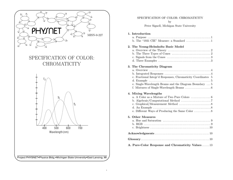
SPECIFICATION OF COLOR: CHROMATICITY by Peter Signell, Michigan State University 1. Introduction a. Purpose . . . . . . . . . . . . . . . . . . . . . . . . . . . . . . . . . . . . . . . . . . . . . . . . . 1 b. The “1931 CIE” Measure: a Standard . . . . . . . . . . . . . . . . . . . 1 MISN-0-227 2. The Young-Helmholtz Basic Model a. Overview of the Theory . . . . . . . . . . . . . . . . . . . . . . . . . . . . . . . . . 2 b. The Three Types of Cones . . . . . . . . . . . . . . . . . . . . . . . . . . . . . . 2 c. Signals from the Cones . . . . . . . . . . . . . . . . . . . . . . . . . . . . . . . . . . 3 d. Three Examples . . . . . . . . . . . . . . . . . . . . . . . . . . . . . . . . . . . . . . . . . 3 SPECIFICATION OF COLOR: CHROMATICITY 3. The Chromaticity Diagram a. Overview . . . . . . . . . . . . . . . . . . . . . . . . . . . . . . . . . . . . . . . . . . . . . . . . 4 b. Integrated Responses . . . . . . . . . . . . . . . . . . . . . . . . . . . . . . . . . . . . 4 c. Fractional Integr’d Responses, Chromaticity Coordinates 5 d. Example . . . . . . . . . . . . . . . . . . . . . . . . . . . . . . . . . . . . . . . . . . . . . . . . 5 e. Single-Wavelength Beams and the Diagram Boundary . . . 5 f. Mixtures of Single-Wavelength Beams . . . . . . . . . . . . . . . . . . . . 6 zl 4. Mixing Wavelengths a. A Color as a Mixture of Two Pure Colors . . . . . . . . . . . . . . . 6 b. Algebraic/Computational Method . . . . . . . . . . . . . . . . . . . . . . . 7 c. Graphical/Measurement Method . . . . . . . . . . . . . . . . . . . . . . . . .8 d. An Example . . . . . . . . . . . . . . . . . . . . . . . . . . . . . . . . . . . . . . . . . . . . .8 e. Different Ways of Producing the Same Color . . . . . . . . . . . . . 8 yl xl 400 500 600 Wavelength (nm) 5. Other Measures a. Hue and Saturation . . . . . . . . . . . . . . . . . . . . . . . . . . . . . . . . . . . . . 9 b. RGB . . . . . . . . . . . . . . . . . . . . . . . . . . . . . . . . . . . . . . . . . . . . . . . . . . . . 9 c. Brightness . . . . . . . . . . . . . . . . . . . . . . . . . . . . . . . . . . . . . . . . . . . . . . 10 700 Acknowledgments . . . . . . . . . . . . . . . . . . . . . . . . . . . . . . . . . . . . . . . . . . 10 Glossary . . . . . . . . . . . . . . . . . . . . . . . . . . . . . . . . . . . . . . . . . . . . . . . . . . . . . 10 A. Pure-Color Response and Chromaticity Values . . . . . . 13 Project PHYSNET · Physics Bldg. · Michigan State University · East Lansing, MI 1 ID Sheet: MISN-0-227 THIS IS A DEVELOPMENTAL-STAGE PUBLICATION OF PROJECT PHYSNET Title: Specification of Color: Chromaticity Author: Peter Signell, Department of Physics and Astronomy, Michigan State University, East Lansing, MI Version: 3/15/2002 Evaluation: Stage 0 Length: 1 hr; 24 pages Input Skills: 1. Vocabulary: wavelength (MISN-0-201), spectrum (MISN-0-212). Output Skills (Knowledge): K1. Vocabulary: monochromatic, hue, saturation, brightness, cones, chromaticity coordinates, integrated response values, fractional integrated response values, tri-stimulus response values, Chromaticity Diagram, RGB, white, black, spectral components. K2. Describe how you could generate the Chromaticity Diagram, complete with color labels scattered around it, using only the 1931 CIE Tri-stimulus values, graph paper, a calculator, and two variablewavelength light beams. K3. Explain how two light beams can have different spectral components and yet have exactly the same color. K4. Explain how a color TV set produces colors and why the set cannot produce all the colors that can be seen by the eye. The goal of our project is to assist a network of educators and scientists in transferring physics from one person to another. We support manuscript processing and distribution, along with communication and information systems. We also work with employers to identify basic scientific skills as well as physics topics that are needed in science and technology. A number of our publications are aimed at assisting users in acquiring such skills. Our publications are designed: (i) to be updated quickly in response to field tests and new scientific developments; (ii) to be used in both classroom and professional settings; (iii) to show the prerequisite dependencies existing among the various chunks of physics knowledge and skill, as a guide both to mental organization and to use of the materials; and (iv) to be adapted quickly to specific user needs ranging from single-skill instruction to complete custom textbooks. New authors, reviewers and field testers are welcome. PROJECT STAFF Andrew Schnepp Eugene Kales Peter Signell Webmaster Graphics Project Director Output Skills (Rule Application): R1. Given a beam of light of a specified wavelength, use a Tri-Stimulus Table to determine the beam’s chromaticity coordinates. R2. Given the chromaticity coordinates of a color, determine its hue and saturation, and vice versa. R3. Given a color that is to be produced by mixing two fully saturated beams, determine the proper fraction of each beam in the mixture, and vice versa (given the component beams, determine the resulting color). See this module’s Local Guide. ADVISORY COMMITTEE D. Alan Bromley E. Leonard Jossem A. A. Strassenburg Yale University The Ohio State University S. U. N. Y., Stony Brook Views expressed in a module are those of the module author(s) and are not necessarily those of other project participants. c 2002, Peter Signell for Project PHYSNET, Physics-Astronomy Bldg., ° Mich. State Univ., E. Lansing, MI 48824; (517) 355-3784. For our liberal use policies see: External Resources (Required): 1. A ruler, preferably a clear one. http://www.physnet.org/home/modules/license.html. 3 4 MISN-0-227 1 SPECIFICATION OF COLOR: CHROMATICITY by Peter Signell, Michigan State University 1. Introduction 1a. Purpose. In this module we show you how to make use of an international standard for the quantitative description of color. According to this standard, each color is specified by a unique combination of three numbers: two numbers specifying a color’s “chromaticity” and one number specifying its “brightness.” In this module we deal mainly with a color’s chromaticity and only briefly with its brightness. A modern Tri-Stimulus Colorimeter containing a microprocessor can be used to do the whole job of color measurement: light of the desired color enters the device, there is a short period of measurement and computation, then its readout displays the three numbers. Here we show you the physics that is basic to the processes going on in the device and we show you how to interpret the results. In addition, the understandings developed along the way will provide a basis for developing an understanding of the colors that we perceive for various objects under various conditions.1 The subject of color specification is of considerable importance to industry in both designing the perceived colors of objects under various lighting conditions and in maintaining quality control, over extended periods of time, for paints, textiles, plastics, foods, etc. 1b. The “1931 CIE” Measure: a Standard. The “1931 CIE” measure is a basic industry standard for specifying a color. The measure was fixed by an international congress (the “CIE”) in 1931, with the intent of mimicking the processing that takes place in the eye-brain system of a “standard observer.” 1 See “Colors from Spectral Distributions” (MISN-0-229), which extends the treatment of the present module to cover light containing all visible wavelengths, the kind of light normally entering the human eye. Then “Color Effects from Light Source and Reflection” (MISN-0-265) follows changes in the spectral distribution of a ray of light as it emerges from its source, travels to an object, reflects off the object, travels to the eye, then produces in the brain a perception of “the color of the object.” Finally, “Land’s Observations on Color Perception” (MISN-0-228) shows the striking effects that the content of an entire scene can have on the colors perceived for various objects in the scene. 5 MISN-0-227 2 Since 1931 a lot more has been learned about how the eye-brain systems of most people produce color perception and this is currently an active field of research. Current theories of color perception greatly refine and expand on the ideas present in the 1931 CIE standard, but the resulting revisions to the numbers used in the standard have only been small and only need to be taken into account where great precision is required. Thus the 1931 standard is still widely used in specifying colors and it makes a good beginning point for study of the eye-brain color perception system. 2. The Young-Helmholtz Basic Model 2a. Overview of the Theory. The Young-Helmholtz theory, which is a main basis for the 1931 CIE standard, is an “additive three-component” description.2 From many experiments Young deduced that there are three types of receptors in the eye, and that each type of receptor sends separate information to the brain for building a perception of color. Helmholtz later made an important addition by saying that each type of receptor responds to a wide range of wavelengths. Young gave a color name to each of the three types of receptors, but we will simply call them x, y, and z (this is a universal designation and professionals generally consider Young’s color names to be interchangeable with these labels). 2b. The Three Types of Cones. The retina3 of a human eye contains about seven million color-detecting nerves, called cones, each of which belongs to one of three distinct cone types. All cones of the same type respond similarly to light from a particular source. However, cones of differing type respond differently to light from the same source (except for the color “white,” as we shall discuss later). Thus any “x” cone responds similarly to any other “x” cone but differently from a “y” cone or a “z” cone. The responses of the “x,” “y,” and “z” cones to light of individual wavelengths are plotted in Fig. 1 and are listed in Table A-1, Appendix A, this module. These responses are called the “CIE 1931 TriStimulus Values” and are written xλ , yλ , and zλ . The λ subscripts show that they are functions of wavelength.4 Notice that in Fig. 1 the x cones 2 Note that this is not a three-color description. eye’s “retina” is a surface, in the back of the eye, composed of nerve endings. Light absorbed on a retinal nerve ending causes a chemical reaction that starts a signal down that particular nerve, heading toward the brain, whereupon the nerve “resets” and is ready to fire again. 4 In producing the responses shown in Fig. 1 and Table A-1, the light incident on the cones is kept at the same intensity at all wavelengths so the responses can be said to all 3 The 6 MISN-0-227 3 1.8 relative response 1.6 1.4 yl 1.0 xl 0.8 0.6 0.4 0.2 0.0 400 500 600 4 of 600 nm.7 When the retina of your eye absorbs that light, the x cones are highly stimulated, the y cones are moderately stimulated, and the z cones are negligibly stimulated (see Fig. 1). We say there is a large x-cone response, a modest y one, and a negligible z one. The x cones send out large signals, the y ones modest signals, and the z ones negligible ones. As a result of this combination of signals from the cones, the brain constructs a perception of the color “yellow-orange.” zl 1.2 MISN-0-227 700 Wavelength (nm) Figure 1. The 1931 CIE presumed response curves (“tristimulus values”) for the three types of retinal cones. are generally more sensitive on the right side (“red”)of the graph, the y cones in the middle (“green”) part, and the z cones on the left (“blue”). Note also that the “x” cones have two peaks of sensitivity. 2c. Signals from the Cones. When light falls on cones in the retina of the eye, each cone sends a message that communicates the intensity of its response. This signal goes out over an optic nerve5 to a preprocessing center back of the eye. The preprocessing center combines the signals from three adjacent x, y, and z cones and sends three signals, containing the information in a somewhat altered form, on to the brain. The brain, in turn, converts the three numbers to a perception of colored light at a point corresponding to the location, in the eye, of the three adjacent cones. The collection of all such sets of three signals from all over the back of the eye allow the brain to construct a complete colored scene.6 2d. Three Examples. As a first example, consider what happens when you look at a light source that emits light at the single wavelength have the same cone stimulus. If equal-intensity light at all these wavelengths are mixed, the resultant color is said to be “idealized white light.” There is no natural source that produces such light, but overhead sunlight in the tropics is a modest approximation. 5 The eye’s “optic nerve” is a bundle of nerve cells leaving the retina of the eye, heading toward the brain. These cells transport the visual information received at the retina. 6 For more information see Color: A Guide to Basic Facts and Concepts, Burnham, Hanes, and Bartleson, John Wiley and Sons, 1963, pp. 56. For access, see this module’s Local Guide. 7 As a second example, suppose you look at a light source that emits light at the single wavelength of 570 nm, down from the 600 nm used in the previous example. Now the x and y cones send out equal-strength signals but the z cones send out only a small signal. As a result of this combination of signals from the cones, the brain produces a perception of the color “yellow.” Finally, suppose the light entering the eye is such that all three cone types send out equal-strength signals. As a result of this combination of signals from the cones, the brain produces a perception of the color “white” (or “grey” if the brightness is low).8 3. The Chromaticity Diagram 3a. Overview. The Tri-Stimulus Values can be used to make a diagram that numerically specifies two non-brightness parts of a color’s description: this is called the CIE Chromaticity Diagram. We will first discuss the procedure for measuring the various quantities that describe the response of the eye to specific light beams. Then we will construct the periphery of the Chromaticity Diagram by plotting the positions (on the diagram) of single-wavelength light beams. Next we will plot the positions of colors corresponding to mixtures of single-wavelength light beams. Finally, we will use the diagram to relate colors to the color-technology terms “hue,” “saturation” and “brightness.” 3b. Integrated Responses. If a light beam having a single wavelength falls on, say, an x cone, that cone responds, as shown in Fig. 1, for that type of cone and that wavelength. If we now add, to the beam, a second ray having the same intensity as the first, but a different wavelength, the response of the cone will increase and the amount of increase will be just the amount shown in Fig. 1 for the second wavelength. In general, 7 Such a single-wavelength light beam is said to be “monochromatic” and is routinely called a “monochromatic beam.” 8 See “The Measure of Perceived Color” (MISN-0-229), a systematic presentation. 8 MISN-0-227 5 light has a continuous distribution of wavelengths and thus each type of cone has an “Integrated Response” to the light. The three Integrated Responses, one for each type of cone, are denoted X, Y , and Z. The apparent brightness of a light beam is just the total integrated response, X + Y + Z. 3c. Fractional Integr’d Responses, Chromaticity Coordinates. The apparent brightness of a light beam is normally eliminated from the discussion by dividing each of the three integrated responses by the sum of the three. The resulting fractional integrated responses are labeled x, y, and z: x = X/(X +Y +Z), y = Y /(X +Y +Z), z = Z/(X +Y +Z). Then if x = 0.3 for some light beam, then we can say that 30% of the response of the eye to that beam is from x cones. Now of course x + y + z = 1, so knowing any two of x, y, and z immediately gives us the third. Thus a knowledge of all three is superfluous. When describing the color of a light beam entering the eye, the convention is to just specify x and y and these are called the “chromaticity coordinates” of the color. 3d. Example. Suppose that, for a particular light beam entering the eye, the integrated cone responses have the values X = 40, Y = 80, and Z = 120. Then the total response is X + Y + Z = 240 and the fractional responses are x = 40/240 = 0.167, y = 80/240 = 0.333, and z = 120/240 = 0.500. ¤ Show that, for the beam quoted above, the “chromaticity coordinates” for our Example color are: x = 0.167, y = 0.333. 3e. Single-Wavelength Beams and the Diagram Boundary. To obtain the chromaticity coordinates of a single-wavelength light beam, we need merely calculate the fractional values x and y for that beam. For example, one sees from Fig. 1 and Table A-1 (Appendix A, this module), that a beam of 540 nm light has the integrated response values X = 0.290, Y = 0.954, Z = 0.020. For this beam X +Y +Z = 1.264 so x = 0.229 and y = 0.755. These latter two numbers are given in the last two columns of the table, but to greater accuracy. If you physically look at a 540 nm beam you will see that its color is green, so you could plot the point x = 0.229, y = 0.755 on the diagram and label it “540 nm” and “green.” After doing this for many different singlewavelength light beams you would have constructed the curved boundary in the Chromaticity Diagram, as shown in Fig. 2. There are two distinct parts to the boundary: the curved part where each different point corresponds to a different wavelength, and the straight 9 MISN-0-227 6 part at the bottom where each different point corresponds to a different combination of the violet and red end-point wavelengths. ¤ Show that the Chromaticity coordinates in Fig. 2 for a 600 nm gold light beam are as given in the Table A-1. 3f. Mixtures of Single-Wavelength Beams. Given the single wavelengths on the boundary of the Chromaticity Diagram, one can move on to colors that correspond to mixtures of such pure wavelengths. Any beam of visible light can be broken up into its single-wavelength components, so all colors must be either on the periphery or in the interior of the Chromaticity Diagram. For example, mixing equal intensities of 625 nm red and 470 nm violet light beams produces a perception of a light purple beam so we could locate the point that is half way between the 625 nm and 470 nm points on the Chromaticity Diagram and write in the word “light purple.” Similarly, a mixture having equal components of 489 nm blue light and 595 nm orange light produces a perception of a “white” light beam so we locate the point that is half way between the 489 nm and 595 nm points on the Diagram and label it “white.” Of course the color “pink” turns out to be between the colors “red” and “white” on the diagram. 4. Mixing Wavelengths 4a. A Color as a Mixture of Two Pure Colors. Given a position on the Chromaticity Diagram, corresponding to a particular color, one can measure on the diagram and determine a combination of two purewavelength light beams which will produce that color. ¤ Show that a straight line from the 450 nm violet point to the 550 nm green point passes through the x = 0.277, y = 0.580 point in Fig. 2, a shade of green. We will hereafter refer to this as the (0.277,0.580) point on the Diagram. Since the point is closer to the 550 nm end of the line, that must be the dominant partner in this “450 nm plus 550 nm” combination. To find the actual fractions of each member of the combination, one can either do a calculation based on the values in the Table A-1, or one can measure distances among the three points on the Chromaticity Diagram. Of course the point corresponding to the target color must lie on the straight line connecting the two proposed components, or the target color cannot be produced by any combination of those two components. The equivalence of the calculational and measurement methods can be easily 10 MISN-0-227 7 520 0.8 dark green 510 ¤ Suppose you interchange the ray labels #1 and #2 and then redo the calculation of Eqns. (1). Show that in all such cases you will get exactly the same values for x and y that you got before the interchange. Help: [S-4] Given this result, does it matter which ray you label #1 and which one you label #2 in order to do the calculation? 540 550 green 560 0.6 TV 570 500 4c. Graphical/Measurement Method. To use graphical measurements to find the combination of two rays that produces a given color, first draw the straight line that connects the three points on the Chromaticity Diagram, then measure the distances between the points. Call d1 the distance from the point for component #1 to the point for the target point, and d2 the distance from the target point to the point for component #2. Then the fraction of component #2 in the mixture is: 580 y gold warm white cool white pink 0.4 daylight 490 equal energy point deep blue 480 0.2 0.0 590 600 610 620 TV 640 red 780 f2 = d1 /(d1 + d2 ), blue 470 460 450 violet 380 0.2 (2) where this equation comes from solution of Eq. (1) for f2 plus a little trig work. Help: [S-3] purple TV 0.4 0.6 4d. An Example. In Sect. 4a it was proposed that a 450 nm light ray and a 550 nm light ray be combined to produce the color at point (0.277,0.580) on the Chromaticity Diagram. 0.8 x 0.0 8 also in Table A-1. You need only solve one of the two equations labeled Eq. (1): the two are redundant if the three points actually do lie on a straight line as assumed in Eq. (1). For an example, see Sect. 4d. pure wavelength locus (wavelength in nm) 530 MISN-0-227 ¤ Solve this problem by the algebraic method, showing that the solution is 83% 550 nm light, 17% 450 nm light. Help: [S-1] Figure 2. The “1931 CIE” Chromaticity Diagram shown (see below). 4b. Algebraic/Computational Method. To find the combination of two rays that produces a given color, (x,y), assume that the combination has a fraction “f2 ” of component ray #2 with color (x2 ,y2 ), and hence a fraction 1 − f2 of component ray #1 with color (x1 ,y1 ). Then you can write for the color (x,y): x = (1 − f2 )x1 + f2 x2 y = (1 − f2 )y1 + f2 y2 (1) ¤ Solve the problem again, now by the graphical method. Show that the answer so obtained is the same as that found in the algebraic method, within the accuracy of graphical work. 4e. Different Ways of Producing the Same Color. Any color that is in the interior of the Chromaticity Diagram (not on its boundary) can be produced in an infinite number of different ways. For the example, there are an infinite number of straight lines that can be drawn through the point at (0.277,0.580), all at different angles. For each of these lines one can determine the percentages of the two boundary components that will produce the color at (0.277,0.580). where you should remember that f2 is defined as the fraction in the mixture that is color #2 and that (x2 ,y2 ) is the position of color #2 on the Chromaticity Diagram. If it is a pure wavelength color, its (x2 ,y2 ) are ¤ Show that a (60% 510 nm, 40% 610 nm) combination will produce the same color as a (16% 450 nm, 84% 550 nm) combination (within the 2digit accuracy of the quoted percentages). 11 12 MISN-0-227 9 5. Other Measures 5a. Hue and Saturation. Instead of x and y, the two chromaticity coordinates, one can specify position on the Chromaticity Diagram by considering the color to be a mixture of “white” and a color on the boundary of the Diagram. The color on the boundary is called the target color’s “hue” while the fraction of the boundary color in the mixture is called the color’s “saturation.” Thus both light and dark pink would be described as having a red hue, but the light color would be said to be less saturated than the dark one. As an example, consider a color described as “86% saturated 573 nm light.” On the Chromaticity Diagram, the color of this light would be 86% of the way from the “white” point at (.333,.333) to the 573 nm point on the boundary. Another way of saying it is that the color could be produced by mixing 573 nm yellow-green light and white light, with the yellow-green light being 86% of the mixture. To get that color’s x and y values, its Chromaticity Coordinates, you simply draw, right on a copy of the diagram, a straight line that goes from the “white” point to the 573 nm point on the boundary. Finally, locate the target color as the point that is 86% of the way from the white point to the boundary. ¤ Show that, for 86% saturated 573 nm light, x = 0.45, y = 0.51. A slight complication arises when the line drawn from the “white” point to the target point, and on to the boundary, hits the boundary at a point on the straight line section along the bottom of the Diagram. There the boundary points do not correspond to pure wavelengths but rather to mixtures of pure-wavelength light. One solution, good for anywhere on the boundary, is to specify the hue as the polar angle of the line that goes from “white” to the point in question and then on to the boundary.9 For example, a 0◦ hue is “red,” a 90◦ hue is “green,” a 180◦ hue is “blue,” and a 270◦ hue is “purple.” ¤ Show that the color at (0.277,0.580) can be described as 61% saturated 541 nm light. 5b. RGB. The RGB method of specifying a color is useful when dealing with colors on a TV screen or on a computer’s color monitor. Color monitors (including the screens in ordinary color TV sets) have three phosphor dots next to each other at each location on the screen, and each MISN-0-227 10 dot gives out a certain intensity of light at any particular instant. The dots are sufficiently small so the human eye does not resolve them individually, but only responds to the three as a group. The colors produced individually by each of the three types of phosphor dots are labeled “TV” on Fig. 2, and they are known by the names “Red,” “Green,” and “Blue.” Collectively, the three are referred to by the letters RGB. All colors produced by a TV set or color monitor are within the triangle formed by connecting the three RGB points marked “TV” on Fig. 2. Any color within the triangle can be specified by the relative amounts of R, G, and B light which will produce that color. Any color outside the triangle cannot be produced by a TV set or color monitor using those standard phosphors. In practical terms, one can determine the amounts of R, G, and B necessary to produce a particular color by first combining any two of R, G, and B, then combining the resulting color with the other one of the R, G, and B set. 5c. Brightness. As one varies the total intensity of a light beam, its chromaticity coordinates stay fixed but its “brightness” changes. One can do this for all the colors on the Diagram at once by watching the Diagram as its brightness is changed continuously. The diagrams would all have identical shapes and boundaries but the labels we place on the diagram would vary from diagram to diagram. Thus, for example, as intensity decreased the white point on the Diagram would turn from white to light grey to dark grey and finally to black. The latter color, black, is the total absence of light, so it corresponds to zero intensity and a totally black diagram. Color charts are available that show the progression of color changes perceived as brightness alone is changed. Acknowledgments We thank K. Franklin and J. Kovacs for their contributions to a precursor of this module. Professor Jim Linneman made a useful suggestion. Preparation of this module was supported in part by the National Science Foundation, Division of Science Education Development and Research, through Grant #SED 74-20088 to Michigan State University. Glossary 9 This method is used in the color-specification part of the computer programming c Carnegie-Mellon University). language CT (° • black: this is the “color” sensation produced by the absence of light. 13 14 MISN-0-227 11 • brightness (of a particular color): overall energy intensity of a particular light; e.g., “60 watts/square meter.” Brightness can be varied independently of hue and saturation. • chromaticity coordinates: the fractional integrated responses of the x and y cones to a particular light beam. The numbers describe the “color” of a light beam but omit variations in perceived color produced by variations in intensity. • cones: one of two types of nerve cells in the retina of the eye that act as light receptors. There are three varieties of cones, each with a different sensitivity to various wavelengths of the visible spectrum. The cones are responsible for color vision. MISN-0-227 12 is measured. The bin light-energy values are plotted at their respective wavelength center-points and a smooth curve is drawn. The narrower the bins, the greater the accuracy of the resulting curve. A bin width greater than 10 nm will generally give only very crude results. • tri-stimulus response curves/values: numbers that represent the responses, as functions of wavelength, of the three types of cones of a “standard observer,” when those cones are successively subjected to equal intensities at each visible wavelength. • white: The color sensation produced by light that has chromaticity coordinates (0.333,0.333). This color also includes all shades of gray, obtained by keeping the “color” fixed and varying the intensity. • hue (of a particular color): the single-wavelength color that can be mixed with white to produce the color being specified; e.g., “380 nm violet” is a “hue.” Hue can be varied independently of saturation and brightness. • monochromatic: an adjective applied to a light beam, indicating that the beam contains only a single wavelength. In reality, any light beam has a range of wavelengths but that range can often be made so small that it can be considered to be a single wavelength for the purpose at hand. A device that emits essentially monochromatic light whose wavelength can be changed at will is called a “monochromator.” • RGB: the three color beams used to excite the phosphors in television and computer screens; so-called because the three beams excite screen phosphors whose colors are red, green, and blue, respectively. Such screens can only produce colors whose chromaticity coordinates are within the chromaticity-diagram triangle defined by the chromaticity coordinates of the three screen phosphors. • saturation (of a particular color): the fractional amount of the hue, in the mixture of hue plus white, needed to produce the color being specified; e.g., “15% saturated 650 nm red” is a mixture of 15 parts 650 nm red to 85 parts white: it is perceived in the brain as “light pink.” Saturation can be varied independently of hue and brightness. • spectral components: the single-wavelength light beams that are the constituents of a multi-component beam. In practice, to analyze a spectrum that is continuous, we break it down into very narrow wavelength bands, called “bins,” and the amount of light energy in each bin 15 16 MISN-0-227 13 MISN-0-227 LG-1 A. Pure-Color Response and Chromaticity Values λ(nm) 400 410 420 430 440 450 460 470 480 490 500 510 520 530 540 550 560 570 580 590 600 610 620 630 640 650 660 670 680 690 700 tri-stimulus values xλ yλ zλ 0.014 0.000 0.068 0.044 0.001 0.207 0.134 0.004 0.646 0.284 0.012 1.386 0.348 0.023 1.747 0.336 0.038 1.772 0.291 0.060 1.669 0.195 0.091 1.288 0.096 0.139 0.813 0.032 0.208 0.465 0.005 0.323 0.272 0.009 0.503 0.158 0.063 0.710 0.078 0.166 0.862 0.042 0.290 0.954 0.020 0.433 0.995 0.009 0.595 0.995 0.004 0.762 0.952 0.002 0.916 0.870 0.002 1.026 0.757 0.001 1.062 0.631 0.001 1.003 0.503 0.000 0.854 0.381 0.000 0.642 0.265 0.000 0.448 0.175 0.000 0.284 0.107 0.000 0.165 0.061 0.000 0.087 0.032 0.000 0.047 0.017 0.000 0.023 0.008 0.000 0.011 0.004 0.000 LOCAL GUIDE singlewavelength chromaticity coordinates x y 0.1733 0.0048 0.1726 0.0048 0.1714 0.0051 0.1689 0.0069 0.1644 0.0109 0.1566 0.0177 0.1440 0.0297 0.1241 0.0578 0.0913 0.1327 0.0454 0.2950 0.0082 0.5384 0.0139 0.7502 0.0743 0.8338 0.1547 0.8059 0.2296 0.7543 0.3016 0.6923 0.3731 0.6245 0.4441 0.5547 0.5125 0.4866 0.5752 0.4242 0.6270 0.3725 0.6658 0.3340 0.6915 0.3083 0.7079 0.2920 0.7190 0.2809 0.7260 0.2740 0.7300 0.2700 0.7320 0.2680 0.7334 0.2666 0.7344 0.2656 0.7347 0.2653 Note: The graphical method described in Section 4c may be required on the exam you take so you should bring a ruler (preferably a clear one) to the Exam Room. The book listed in this module’s ID Sheet is on reserve for you in the Physics-Astronomy Library, Room 230 in the Physics-Astronomy Building. Tell the person at the desk that you want a book that is on reserve for CBI (a BOOK, not a reading). Then tell the person the name of the book you want. 17 18 MISN-0-227 PS-1 MISN-0-227 PROBLEM SUPPLEMENT AS-1 SPECIAL ASSISTANCE SUPPLEMENT ¤ Warning: this symbol marks problems scattered throughout the text. Those problems are part of this module’s problem set: do all of them properly before attempting the problems below. Note: The problems below also occur on this module’s Model Exam. 1. Starting with the 1931 CIE Tri-Stimulus Values in Appendix A, determine the Chromaticity Coordinates of: (a) 100% saturated 500 nm light and (b) 100% saturated 600 nm light. 2. Using the results of Problem 1, determine the Chromaticity Coordinates of a 2:1 mixture of 500 nm and 600 nm light. S-1 Student: Go back to this module’s text. Start at the beginning of the module and go through the text, understanding each paragraph before going on to the next. Work each “try-it” as you come to it, writing out all the steps explicitly on paper. If you get stuck on a “try-it,” bring what you have done, up to the point where you became stuck, to a Consultant and get help on getting going again. If you finish all of the try-it’s and still can’t work this problem, bring your explicitly workedout try-it’s and your work on this problem, up to the point where you became stuck, to a Consultant. S-2 Brief Answers: (from TX-4d, PS-Problem 1, (a) and (b)) Consultant: do not help this student unless all of the relevant work indicated below is shown to you in the manner described. (from PS-Problem 2) First, work on Problem 1, above, until you get the right answers. Write out every step you used in solving the try-it’s and the problem. Do the same for this problem. If you still can’t get this problem, bring all of your work to a Consultant for help. 1. a. (0.008,0.538) Help: [S-1] b. (0.627,0.372) Help: [S-1] 2. x = 0.21, y = 0.48. Help: [S-2] S-3 (from TX-4a,4c) To derive Eq. 2 from Eq. 1, first formally solve Eq. 1 for f . Then, on a sketch imitating part of the Chromaticity Diagram, draw a line connecting point (x1 , y1 ) and point (x2 , y2 ) and going through the target point (x, y). Draw straight lines connecting points (x1 , y1 ), (x2 , y2 ), and (x2 , y1 ), making a triangle. Then connect points (x, y) and (x, y1 ). Now look at the sketch and see that the ratio d1 /d equals the solution of Eq. 1 that you just found for f . S-4 (from TX-4b) For (1 − f1 ) substitute its equivalent, f2 . Then notice that, if you everywhere exchange the labels #1 and #2, the equations themselves do not change. 19 20 MISN-0-227 ME-1 MISN-0-227 MODEL EXAM ME-2 520 0.8 dark green 510 1. See Output Skills K1-K4 on this module’s ID Sheet. One or more of these skills, or none, may be on the actual exam. pure wavelength locus (wavelength in nm) 530 540 550 2. Starting with the 1931 CIE Tri-Stimulus Values, determine the Chromaticity Coordinates of: (a) 100% saturated 500 nm light and (b) 100% saturated 600 nm light. green 560 0.6 TV 570 500 3. Using the results of Problem 2, determine the Chromaticity Coordinates of a 2:1 mixture of 500 nm and 600 nm light. λ(nm) 400 420 440 460 480 500 520 540 560 580 600 620 640 660 680 700 580 y gold warm white cool white tri-stimulus values xλ yλ zλ 0.014 0.000 0.068 0.134 0.004 0.646 0.348 0.023 1.747 0.291 0.060 1.669 0.096 0.139 0.813 0.005 0.323 0.272 0.063 0.710 0.078 0.290 0.954 0.020 0.595 0.995 0.004 0.916 0.870 0.002 1.062 0.631 0.001 0.854 0.381 0.000 0.448 0.175 0.000 0.165 0.061 0.000 0.047 0.017 0.000 0.011 0.004 0.000 590 600 610 620 TV 640 red 780 pink 0.4 daylight 490 equal energy point deep blue 480 0.2 blue 470 460 450 purple TV 0.0 0.0 violet 380 0.2 0.4 0.6 0.8 x Brief Answers: 1. See this module’s text. 2. See this module’s Problem Supplement, problem 1. 3. See this module’s Problem Supplement, problem 2. 21 22 23 24
