Neurobiology: Nerve cell interactions
TEB
Principle
To use a nerve function model to study the following aspects of nerve cell interactions with
other nerve cells or with motoric and sensoric cells:
• RENSHAW INHIBITION
• Motoneuron signals with recurrent inhibition by Renshaw cell
• Motoneuron signals without recurrent inhibition
• Functional characteristics of Renshaw inhibition
• LATERAL INHIBITION AND CONTRAST IMPROVEMENT
• Lateral inhibition
• Contrast improvement
• NEURONAL PRINCIPLES OF CLASSIC CONDITIONING
• Conditioned reflex
• Reversed stimulus succession does not bring about a conditioned reflex
Equipment
1
1
1
Neurobiology Lab
Additional nerve cell
PC, Windows® 95 or higher
65963-11
65963-10
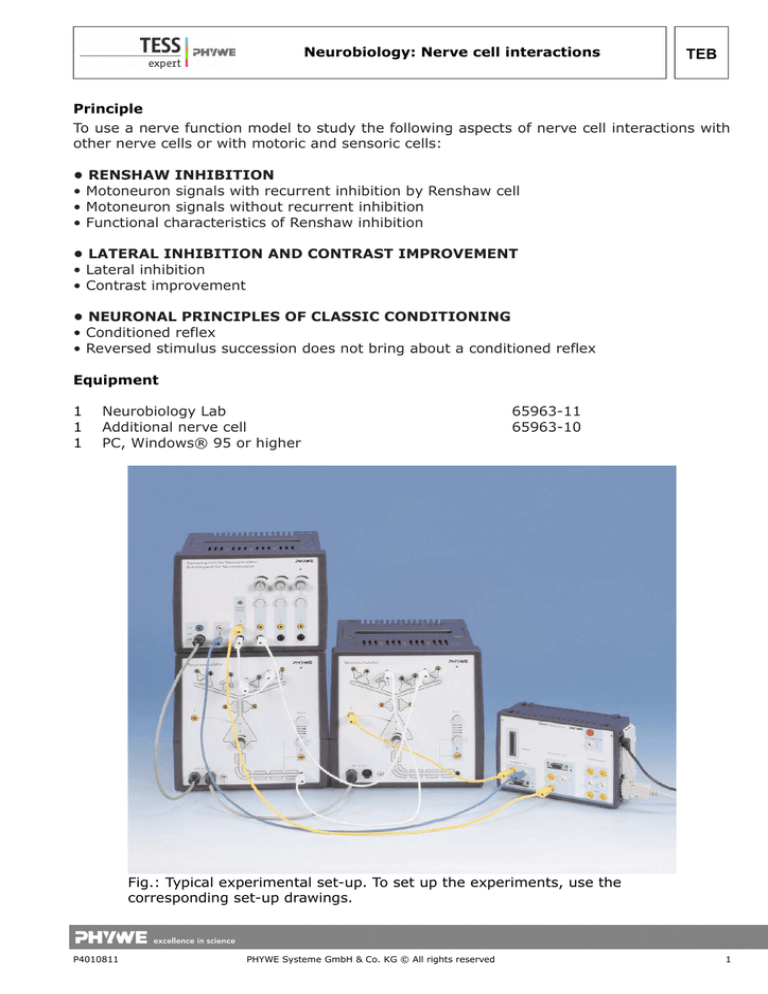
Fig.: Typical experimental set-up. To set up the experiments, use the
corresponding set-up drawings.
P4010811
PHYWE Systeme GmbH & Co. KG © All rights reserved
1
TEB
Neurobiology: Nerve cell interactions
Set-up and procedure
For all experiments, use the software settings as shown in Fig. 2.
RENSHAW INHIBITION (FIG. 1)
Internet search keywords: Renshaw inhibition, Renshaw cell, motoneuron, motoric neuron,
hyper-excitation.
The Renshaw cell, a special neuron, protects a motoneuron and the muscle fiber, which is
driven by the motoneuron, from hyper-excitation. Its circuit causes negative feedback of
the excitory stimulation of the motoric neuron. The circuit can be realized with two
Neurosimulator modules, whereby one is the motoneuron and one the Renshaw cell. The
stimulus to the motoneuron is amplified by spatial summation.
The Renshaw cell is also found in the spinal cord of vertebrates.
Fig. 1: Experimental set-up
2
PHYWE Systeme GmbH & Co. KG © All rights reserved
P4010811
Neurobiology: Nerve cell interactions
TEB
Fig. 2
a) Motoneuron signals with recurrent inhibition by Renshaw cell (Fig. 3)
Fig. 3
b) Motoneuron signals without recurrent inhibition (Fig. 4)
(inhibitory synapses deactivated):
P4010811
PHYWE Systeme GmbH & Co. KG © All rights reserved
3
TEB
Neurobiology: Nerve cell interactions
Fig. 4
c) Functional characteristics of Renshaw inhibition (Fig. 5)
No inhibition at low stimulus intensities, however, distinct inhibitory effect at higher
intensities.
Fig. 5
LATERAL INHIBITION AND CONTRAST IMPROVEMENT (FIG. 6)
Lateral inhibition is a useful function of the nervous system in brain and sensory organs. It
serves to intensify differences in the stimulation of neighbouring elements and so to
increase the resolving power. E.g., it causes an improvement in the contrast between
adjacent areas in the visual system.
4
PHYWE Systeme GmbH & Co. KG © All rights reserved
P4010811
Neurobiology: Nerve cell interactions
TEB
a) Lateral inhibition
In this measurement, mutual inhibition of both Neurosimulator modules is measured.
Before the measurement, with the thresholds at 0, the stimulation levels of both modules
need to be calibrated so that they are identical.
Disconnect the connections between both modules by removing the white cables from each
of the efferent axons. Then, in the measuring mode, however, before starting the
measurement, while pressing both stimulation buttons, turn one of the two stimulation
knobs so that the digital displays of both analog channels are identical (i.e. depolarization
of both Neurosimulators is equally strong):
Fig. 6: Experimental set-up
P4010811
PHYWE Systeme GmbH & Co. KG © All rights reserved
5
TEB
Neurobiology: Nerve cell interactions
During the measurement the cables must be replugged to show lateral inhibition:
Start measurement and press each stimulation button for about one second. Then replug
both cables and press both buttons at the same time to get the result shown in Fig. 7.
The graph shows that depolarization is less pronounced for both Neurosimulators when
laterally inhibited.
Ratio of depolarization (see Table 1):
Fig. 7
b) Contrast improvement (see Fig. 8)
One of the two stimuli is reduced and the measurement is repeated.
The measurements can be repeated with different excitation intensities to determine
inhibition factors. It can be shown that the ratio of the exhibition intensities of the two
channels – the contrast – can increase significantly (here: 1.40 vs. 1.17) (see Table 2):
Fig 8
6
PHYWE Systeme GmbH & Co. KG © All rights reserved
P4010811
Neurobiology: Nerve cell interactions
Table 1
Uninhibited 1 Uninhibited 2
-5.2 V
-5.2 V
TEB
Ratio
Inhibited 1
Inhibited 2
Ratio
Inhib. factor
1
-5.75 V
-5.75 V
1
1
Ratio
Inhibited 1
Inhibited 2
Ratio
Inhib. factor
Table 2
Uninhibited 1 Uninhibited 2
-6.4 V
-5.2 V
1.23
-7.3 V
-5.2 V
1.40
1.17
…
…
…
…
…
…
…
NEURONAL PRINCIPLES OF CLASSIC CONDITIONING (FIG. 9)
Pavlov's dog experiment is no doubt the most well-known example of classic conditioning.
The sound of a bell becomes associated with the smell of food and salivary excretion
follows as reaction. The perception of the smell of food and the salivary secretion that
results from it is called an (inborn) unconditioned reflex. The reaction to the sound of the
bell that is associated with the unconditioned reflex as a result of learning is called a
conditioned reflex. In all experiments on classic conditioning, it is absolutely necessary that
the stimulus for the conditioned reflex lies before that for the unconditioned reflex in time.
Should this succession in time be reversed, then conditioning is impossible.
In the first experiment the experiment is shown with the proper time sequence of
conditioning. In the second experiment it is shown that proper sequencing is essential for
the conditioned reflex to be brought about.
P4010811
PHYWE Systeme GmbH & Co. KG © All rights reserved
7
TEB
Neurobiology: Nerve cell interactions
Fig 9: Experimental set-up
a) Conditioned reflex (see Fig. 10)
Set-up: Stimulus knob of channel 1 at maximum.
Two neurosimulators simulate the interneuron-associative neuron pair. In the experiment,
the associative neuron is conditioned to increase its level of intracellular potential
(depolarization) without involvement by the interneuron.
Experiment: Covering the photosensor simulates the bell in the classical Pavlov
experiment, pressing the stimulus button simulates the presentation of food.
8
PHYWE Systeme GmbH & Co. KG © All rights reserved
P4010811
Neurobiology: Nerve cell interactions
TEB
Fig. 10
To verify that the level of depolarization is low for the associative neuron before
conditioning, the photosensor is covered once (without pressing the button for "meat").
After that, repeat the following procedure about 20 times: cover the photosensor for half a
second, immediately after that press the button. Depolarization of the associative neuron
increases gradually. Eventually, only the photosensor is covered, and the conditioned reflex
is manifest.
b) Reversed stimulus succession does not bring about a conditioned reflex (see Fig. 11)
First, to unlearn the conditioned reflex, press the reset button of the associative neuron. To
verify that the level of depolarization is low for the associative neuron, the photosensor is
covered once. Then, repeat the same procedure as in the experiment about the conditioned
reflex above, but this time press the button first, then cover the photo sensor.
Fig. 11
P4010811
PHYWE Systeme GmbH & Co. KG © All rights reserved
9


