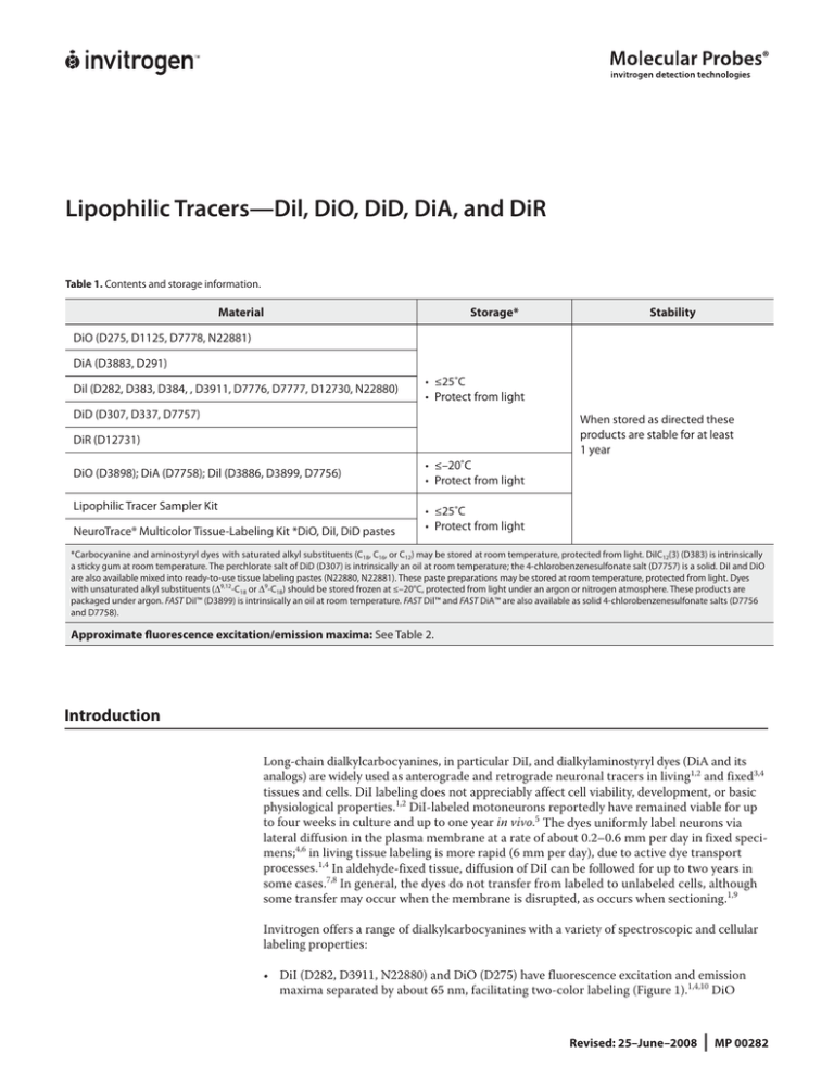
Lipophilic Tracers—Dil, DiO, DiD, DiA, and DiR
Table 1. Contents and storage information.
Material
Storage*
Stability
DiO (D275, D1125, D7778, N22881)
DiA (D3883, D291)
Dil (D282, D383, D384, , D3911, D7776, D7777, D12730, N22880)
• ≤25˚C
• Protect from light
DiD (D307, D337, D7757)
When stored as directed these
products are stable for at least
1 year
DiR (D12731)
DiO (D3898); DiA (D7758); Dil (D3886, D3899, D7756)
Lipophilic Tracer Sampler Kit
NeuroTrace® Multicolor Tissue-Labeling Kit *DiO, DiI, DiD pastes
• ≤–20˚C
• Protect from light
• ≤25˚C
• Protect from light
*Carbocyanine and aminostyryl dyes with saturated alkyl substituents (C18, C16, or C12) may be stored at room temperature, protected from light. DiIC12(3) (D383) is intrinsically
a sticky gum at room temperature. The perchlorate salt of DiD (D307) is intrinsically an oil at room temperature; the 4-chlorobenzenesulfonate salt (D7757) is a solid. DiI and DiO
are also available mixed into ready-to-use tissue labeling pastes (N22880, N22881). These paste preparations may be stored at room temperature, protected from light. Dyes
with unsaturated alkyl substituents (Δ9,12-C18 or Δ9-C18) should be stored frozen at ≤–20°C, protected from light under an argon or nitrogen atmosphere. These products are
packaged under argon. FAST DiI™ (D3899) is intrinsically an oil at room temperature. FAST Dil™ and FAST DiA™ are also available as solid 4-chlorobenzenesulfonate salts (D7756
and D7758).
Approximate fluorescence excitation/emission maxima: See Table 2.
Introduction
Long-chain dialkylcarbocyanines, in particular DiI, and dialkylaminostyryl dyes (DiA and its
analogs) are widely used as anterograde and retrograde neuronal tracers in living1,2 and fixed3,4
tissues and cells. DiI labeling does not appreciably affect cell viability, development, or basic
physiological properties.1,2 DiI-labeled motoneurons reportedly have remained viable for up
to four weeks in culture and up to one year in vivo.5 The dyes uniformly label neurons via
lateral diffusion in the plasma membrane at a rate of about 0.2–0.6 mm per day in fixed specimens;4,6 in living tissue labeling is more rapid (6 mm per day), due to active dye transport
processes.1,4 In aldehyde-fixed tissue, diffusion of DiI can be followed for up to two years in
some cases.7,8 In general, the dyes do not transfer from labeled to unlabeled cells, although
some transfer may occur when the membrane is disrupted, as occurs when sectioning.1,9
Invitrogen offers a range of dialkylcarbocyanines with a variety of spectroscopic and cellular
labeling properties:
• DiI (D282, D3911, N22880) and DiO (D275) have fluorescence excitation and emission
maxima separated by about 65 nm, facilitating two-color labeling (Figure 1).1,4,10 DiO
Revised: 25–June–2008
|
MP 00282
•
•
•
•
staining is usually less intense than that of DiI, and occasionally fails completely in fixed
tissues.6,11
DiD (D307, D7757) is an analog of DiI with markedly red-shifted fluorescence excitation
and emission spectra (Figure 1). This characteristic is useful in avoiding autofluorescence
and phototoxic effects,12 as well as for two-color labeling.13,14 DiR (D12731) has excitation
and emission maxima in the near infrared region, where many tissues are optically transparent.
DiIC12(3) (D383), DiIC16(3) (D384), and DiOC16(3) (D1125) have shorter alkyl substituents
(C12 or C16) than DiI and DiO (C18). These slightly less lipophilic probes have been found
by some to incorporate into membranes more easily than the DiI and DiO.15
FAST DiI™ (D3899, D7756) and FAST DiO™ (D3898) have diunsaturated Δ 9,12-C18 alkyl
substituents in place of the saturated C18 tails of DiI and DiO, resulting in accelerated
diffusion within membranes.16 A monounsaturated Δ9-C18 analog of DiI is aso available
(Δ9-DiI; D3886).
Sulfonated derivatives of DiI and DiO, and CM-DiI (a thiol-reactive DiI derivative) produce staining that persists after fixation and permeabilization treatments. These tracers
are described in instruction manual MP 06999.
In addition to neuronal tracing, lipophilic carbocyanines have many other applications including:
•
•
•
•
•
Detection of cell-cell fusion17-19 and adhesion 20
Tracing cell migration during development and after transplantation 21-23
Lipid diffusion in membranes by FRAP (Fluorescence Recovery After Photobleaching) 24,25
Cytotoxicity assays 26,27
Labeling of lipoproteins 28,29
The lipophilic aminostyryl dyes DiA (D3883), and 4-Di-10-ASP (D291) are also often used for
neuronal tracing.30,31 DiA has been used as a second-color neuronal tracer in conjunction
with DiI.6,32,33 Fixed tissue has been successfully labeled with DiA in some cases where DiO
staining failed.6 FAST DiA™ (D7758), a diunsaturated Δ9,12-C18 DiA analog designed for accelerated diffusion within membranes is also available from Invitrogen.
Spectral Characteristics
The spectral properties of the dialkylcarbocyanines are largely independent of the lengths of
the alkyl chains, but are instead determined by the heteroatoms in the terminal ring systems
and the length of the connecting bridge. They have extremely high extinction coefficients,
moderate fluorescence quantum yields, and short excited state lifetimes in lipid environments
(~1 ns).34 They are insoluble in water, but their fluorescence is readily detected when incorporated into membranes. A summary of spectral properties is shown in Table 2, together with
appropriate filter sets for fluorescence microscopy applications.
Figure 1. Normalized fluorescence emission spectra of DiO, DiI, DiD, and DiR bound to phospholipid bilayer
membranes.
Lipophilic Tracers—Dil, DiO, DiD, DiA, and DiR
|
2
Figure 2. Fluorescence excitation and emission spectra of DiA bound to phospholipid bilayer membranes.
Aminostyryl dyes are insoluble in aqueous environments but their fluorescence is easily detected upon insertion into membranes or when diluted into organic solvents. DiA’s excitation and emission maxima measured in DOPC phospholipid bilayers are about 460 nm and
580 nm, respectively (Figure 2). As is typical for aminostyryl dyes, there is a significant wavelength shift relative to spectra measured in solvents such as methanol (Table 2). The emission
spectrum of DiA is very broad, allowing it to be detected as green, orange, or even red fluorescence depending on the optical filter used.
Experimental Applications
Preparing Stock Solutions
Prepare stock solutions of lipophilic tracers in dimethylformamide (DMF), dimethylsulfoxide
(DMSO), or ethanol at 1 to 2.5 mg/mL. DMF is preferable to ethanol as a solvent for DiO. Stock
solutions can be stored for at least six months without deterioration under the same conditions as
the undissolved product.
Dissolving DiO in DMF may require sonication and heating to 50°C, and the dye may precipitate
from this solution after several hours. To obtain a stable concentrated solution of DiO, perform the
following procedure.35
1.1 Dissolve solid DiO in chloroform to prepare a 50 mg/mL DiO/chloroform stock solution. Mix
one volume of the DiO/chloroform stock solution with an equal volume of octadecylamine.
Ratios of DiO/chloroform stock solution and octadecylamine of up to 1:10 have also worked.
1.2 Heat the solution to 50°C, then allow it to precipitate by placing it on ice with two volumes of
methanol.
1.3 Pellet the precipitate by centrifugation for 5 minutes at 13,000 rpm in a microcentrifuge. Carefully remove the supernatant and discard it.
1.4 Dry the pellet using a vacuum microcentrifuge or by allowing the solvent to evaporate overnight through a fine hole poked in the lid of the tube’s cap.
1.5 Dissolve the resulting powder in DMF at 5% (w/v) with sonication, heating to 50°C. Clear the
solution by centrifuging for 10 minutes at 13,000 rpm in a microcentrifuge.
Staining Methods
Methods for cellular labeling with dialkylcarbocyanine and dialkylaminostyryl tracers based on
published procedures and recommendations from our customers are summarized below.
Lipophilic Tracers—Dil, DiO, DiD, DiA, and DiR
|
3
Direct application of dye crystals. Dye crystals can be applied directly to intact or cut neurons
for retrograde or anterograde labeling.4,6,31,36 We offer specially prepared large crystals of DiI
(D3911) for this purpose. FAST Dil™ (D7756) is a crystal but has the appearance of tar. In some
cases, direct application of crystals produces more consistent labeling than injection of a dye solution.36 Crystals are typically applied to a micropipette tip for delivery to the desired labeling site.
Water-setting glue 5 or pieces of gelatin sponge (Gelfoam, Pfizer, Kalamazoo, MI) 31 may be used
to hold the crystals in place during the time required for transport of the dye along the neuronal
pathway. For labeling relatively large areas, a piece of nylon filter or Gelfoam may be soaked with
concentrated dye solution and inserted into a natural or dissected cavity.3
NeuroTrace® tissue labeling pastes. NeuroTrace® tissue labeling pastes consist of DiI, DiO, or
DiD mixed into an inert, water-resistant gel. The pastes are ready to use as supplied and can be applied directly to live or fixed tissue specimens using the tip of a needle. This method of application
improves the penetration of the dye into bundled neurons, labeling axons both on and below the
surface. In similar situations, direct application of dye crystals or microinjection of concentrated
solutions will only label neurons on the surface. This labeling method has also been found to increase
the rate of dye transport by 50–80%.37
Loading of tracers supplied as oils. FAST DiI™ (D3899), Δ9-DiI (D3886), and DiD (D307) are
packaged as a sticky oil. The sticky oil residue may be warmed slightly and applied directly to tissue samples with forceps. The tissue should then be warmed to ~40°C to facilitate transport of the
dye. FAST DiI™, FAST DiA™, and DiD are also available in solid form (D7756, D7758, D7757) for
direct application to cells or tissue.
Loading by injection. Pressure microinjection of a small bolus of concentrated dye solution is
an alternative to direct application of crystalline dye for retrograde and anterograde neuronal
tracing.1,13 A 2.5 mg/mL (0.25% w/v) solution of dye in DMF is typically used. Sonication, centrifugation, or filtration (5 μm pore size) of the concentrated dye solution prior to injection is recommended to remove undissolved dye crystals that might clog the pipette tip. Iontophoretic injection
of DiI (5 mg/mL in ethanol) produces precise labeling of small groups of 2–30 cells for lineage tracing studies.21
Staining fixed and mounted tissue. Specimens for labeling with dialkylcarbocyanine and dialkylaminostyryl tracers (usually by direct application of dye crystals) are typically fixed in 4% paraformaldehyde in 0.1 M phosphate buffer, pH 7.4 at ambient temperature.38 Other fixatives, particularly
glutaraldehyde, tend to produce unacceptably high levels of background fluorescence.3,4 Storage
during the time required for diffusive staining of neuronal pathways (typically several weeks) can
be at 4°C or ambient temperature. Permeabilizing reagents, detergents, and high concentrations of
organic solvents usually result in loss of staining.3 Tissues stained with DiI and other lipophilic carbocyanines can be sectioned by cryostat or vibratome methods.38 It is often reported that cryostat
sectioning severely degrades the resolution of DiI labeling, but a recent report describes the use
of polyethylene glycol (PEG) for this purpose.39 Avoid mounting media containing glycerol, which
can extract membrane-bound dyes.
Labeling cell suspensions or adherent cells. Vybrant® DiI, DiO, and DiD cell-labeling solutions
can be added directly to normal culture media to uniformly label suspended or attached culture
cells. Cell suspensions or adherent cells on coverslips are incubated with the loading solution for
5 minutes to 2 hours at 37°C. After loading, the cells are spun down, rinsed, and resuspended in
fresh medium. For adherent cells, labeling in culture while attached results in improved viability
compared to labeling after dissociation.10 Methods for labeling cultured cells using the Vybrant®
DiI, DiO, and DiD cell-labeling solutions are described in instruction manual MP 22885.
Labeling intracellular membranes. DiI and DiIC16(3) can be used to selectively label the endoplasmic reticulum (ER) in living sea urchin and ascidian eggs. A saturated solution of dye in soybean oil is pressure microinjected into the egg and diffusively labels the entire ER network within
about 30 minutes.39
Lipophilic Tracers—Dil, DiO, DiD, DiA, and DiR
|
4
Combination with Other Labeling Techniques
DiI labeling can be used in combination with immunofluorescent or immunoperoxidase labeling
for detailed neuroanatomical studies.15,38,40-42 The primary limitation is that conventional permeabilization reagents such as Triton® X-100 cannot be used to enhance antibody penetration.41
An optimized protocol developed by Lukas and co-workers38 uses 0.3% Tween 20. DiI-sensitized
photoconversion of diaminobenzidine (DAB) to an electron-dense precipitate allows long-term
preservation of DiI labeling, as well as correlation of fluorescence imaging with transmitted light
and electron microscopy.3,7 Staining may be enhanced by Giesma counterstaining.43 Typically,
tissue sections are pre-incubated in a 2 mg/mL solution of DAB in 0.1 M Tris buffer, pH 8.2 for
approximately 60 minutes at 4°C in the dark. Sections are then irradiated for 1–2 hours using the
same excitation filter used for fluorescence microscopy, changing the DAB solution every 10–20
minutes.3,43 Use of higher-magnification objectives (e.g., 20X) decreases the photoconverted area
of the specimen but produces better image resolution and sensitivity and also reduces the irradiation time.43
References
1. J Cell Biol 103, 171 (1986); 2. Trends in Neurosci 12, 333 (1989); 3. Methods Cell Biol 38, 325 (1993); 4. Development 101, 697 (1987); 5. J Comp
Neurol 302, 729 (1990); 6. Neuron 11, 801 (1993); 7. J Histochem Cytochem 38, 725 (1990); 8. Exp Neurol 102, 92 (1988); 9. J Neurosci Methods
88, 27 (1999); 10. Histochemistry 97, 329 (1992); 11. J Neurosci Methods 36, 17 (1991); 12. J. Histochem Cytochem 32, 608 (1984); 13. J Neurosci
15, 549 (1995); 14. J Cell Biol 126, 519 (1994); 15. J Physiol 517, 533 (1999); 16. J Neurosci Methods 103, 3 (2000); 17. J Cell Biol 135, 63 (1996);
18. Biophys J 71, 479 (1996); 19. Cytometry 21, 160 (1995); 20. J Cell Biol 133, 381 (1996); 21. Meth Cell Biol 51, 147 (1996); 22. Histochemistry
97, 329 (1992); 23. J Neurosci Methods 36, 17 (1991); 24. Biophys J 68, 766 (1995); 25. J Cell Biol 103, 1745 (1986); 26. J Immunol Methods 185,
209 (1995); 27. J Immunol Methods 166, 45 (1993); 28. Cytometry 20, 290 (1995); 29. J Cell Biol 90, 595 (1981); 30. Brain Res 581, 339 (1992);
31. J Neuroscience 15, 1057 (1995); 32. J Neurosci 15, 990 (1995); 33. J Neurosci 12, 3494 (1992); 34. Biochemistry 24, 5176 (1985); 35. Personal
communication from F. Bonhoeffer and T. Trowe; 36. J Comp Neurol 261, 155 (1987); 37. Personal communication from H. Richard Koerber, University of Pittsburgh, School of Medicine; 38. J. Histochem Cytochem 46, 901 (1998); 39. J Cell Biol 114, 929 (1991); 40. J Histochem Cytochem 38,
735 (1990); 41. J Neurosci Methods 42, 45 (1992); 42. J Neurosci Methods 46, 251 (1993); 43. J Neurosci Methods 64, 47 (1996).
Table 2. Spectral characteristics of lipophilic carbocyanine and aminostyryl tracers.
Tracer
Catalog Numbers
Ex * (nm)
Em * (nm)
484
501
Optical Filters †
Omega
XF100, XF23
Chroma
DiO
D275, D1125, D3898, D7778,
N22881
41001, 31001
DiA
D3883, D291, D7758
456
590
XF21
31024
DiI
D282, D383, D384, D3886,
D3899, D7756, D7776, D7777,
D12730, D3911, N22880
549
565
XF108, XF32
41002, 31002
DiD
D307, D337, D7757
644
665
XF110, XF47
41008, 31023
DiR
D12731
750
780 ‡
XF112
41009
* Fluorescence excitation (Ex) and emission (Em) maxima for membrane-bound dye. † Catalog numbers for recommended bandpass filter sets
for fluorescence microscopy. Omega filters are supplied by Omega Optical Inc. (www.omegafilters.com). Chroma filters are supplied by Chroma
Technology Corp. (www.chroma.com).
‡ Fluorescence emission of this dye is invisible to the human eye and must be detected using a CCD camera or other infrared-sensitive detector.
Lipophilic Tracers—Dil, DiO, DiD, DiA, and DiR
|
5
Product List Current prices may be obtained from our website or from our Customer Service Department.
Cat. no.
D291
D383
D3883
D1125
D7778
D384
D7758
D3898
D7756
D3899
D275
D7776
D7777
D12730
D282
D3911
D7757
D307
D337
D12731
D3886
L7781
N22880
N22881
N22884
Product Name
Unit Size
4-(4-(didecylamino)styryl)-N-methylpyridinium iodide (4-Di-10-ASP) ...........................................................................................................................
25 mg
1,1′-didodecyl-3,3,3′,3′-tetramethylindocarbocyanine perchlorate (DiIC12(3)) ...........................................................................................................
100 mg
4-(4-(dihexadecylamino)styryl)-N-methylpyridinium iodide (DiA; 4-Di-16-ASP) ........................................................................................................
25 mg
3,3′-dihexadecyloxacarbocyanine perchlorate (DiOC16(3)) ................................................................................................................................................
25 mg
3,3’-dioctadecyl-5,5’- di(4-sulfophenyl)oxacarbocyanine, sodium salt (SP-DiOC18(3)) .............................................................................................
5 mg
1,1′-dihexadecyl-3,3,3′,3′-tetramethylindocarbocyanine perchlorate (DiIC16(3)).......................................................................................................
100 mg
4-(4-(dilinoleylamino)styryl)-N -methylpyridinium 4-chlorobenzenesulfonate (FAST DiA™ solid; DiΔ9,12-C18ASP, CBS) ...............................
5 mg
3,3′-dilinoleyloxacarbocyanine perchlorate (FAST DiO™ solid; DiOΔ9,12-C18(3), ClO4) ...............................................................................................
5 mg
1,1′-dilinoleyl-3,3,3′,3′-tetramethylindocarbocyanine, 4-chlorobenzenesulfonate (FAST DiI™ solid; DiIΔ9,12-C18(3), CBS) ..........................
5 mg
1,1′-dilinoleyl-3,3,3′,3′-tetramethylindocarbocyanine perchlorate (FAST DiI™ oil; DiIΔ9,12-C18(3), ClO4).............................................................
5 mg
3,3′-dioctadecyloxacarbocyanine perchlorate (‘DiO′; DiOC18(3)) .....................................................................................................................................
100 mg
1,1′-dioctadecyl-3,3,3′,3′-tetramethylindocarbocyanine-5,5′-disulfonic acid (DiIC18(3)-DS) .................................................................................
5 mg
1,1’-dioctadecyl-6,6’- di(4-sulfophenyl)-3,3, 3’,3’-tetramethylindocarbocyanine (SP-DiIC18(3)) .............................................................................
5 mg
1,1’-dioctadecyl-3,3, 3’,3’-tetramethylindodicarbocyanine- 5,5’-disulfonic acid (DiIC18(5)-DS () ...........................................................................
5 mg
1,1′-dioctadecyl-3,3,3′,3′-tetramethylindocarbocyanine perchlorate (‘DiI′; DiIC18(3))..............................................................................................
100 mg
1,1′-dioctadecyl-3,3,3′,3′-tetramethylindocarbocyanine perchlorate (‘DiI′; DiIC18(3)) *crystalline* ...................................................................
25 mg
1,1′-dioctadecyl-3,3,3′,3′-tetramethylindodicarbocyanine, 4-chlorobenzenesulfonate salt (‘DiD′ solid; DiIC18(5) solid) ............................
10 mg
1,1′-dioctadecyl-3,3,3′,3′-tetramethylindodicarbocyanine perchlorate (‘DiD′ oil; DiIC18(5) oil) ............................................................................
25 mg
4,4’-diisothiocyanatostilbene- 2,2’-disulfonic acid, disodium salt (DIDS).......................................................................................................................
100 mg
1,1′-dioctadecyl-3,3,3′,3′-tetramethylindotricarbocyanine iodide (‘DiR′; DiIC18(7)) ..................................................................................................
10 mg
1,1′-dioleyl-3,3,3′,3′-tetramethylindocarbocyanine methanesulfonate (Δ9-DiI) .........................................................................................................
25 mg
Lipophilic Tracer Sampler Kit...........................................................................................................................................................................................................
1 kit
NeuroTrace® DiI tissue-labeling paste ..........................................................................................................................................................................................
500 mg
NeuroTrace® DiO tissue-labeling paste ........................................................................................................................................................................................
500 mg
NeuroTrace® Multicolor Tissue-Labeling Kit *DiO, DiI, DiD pastes, 500 mg each*.......................................................................................................
1 kit
Contact Information
Molecular Probes, Inc.
29851 Willow Creek Road
Eugene, OR 97402
Phone: (541) 465-8300
Fax: (541) 335-0504
Customer Service:
6:00 am to 4:30 pm (Pacific Time)
Phone: (541) 335-0338
Fax: (541) 335-0305
probesorder@invitrogen.com
Toll-Free Ordering for USA:
Order Phone: (800) 438-2209
Order Fax: (800) 438-0228
Technical Service:
8:00 am to 4:00 pm (Pacific Time)
Phone: (541) 335-0353
Toll-Free (800) 438-2209
Fax: (541) 335-0238
probestech@invitrogen.com
Invitrogen European Headquarters
Invitrogen, Ltd.
3 Fountain Drive
Inchinnan Business Park
Paisley PA4 9RF, UK
Phone: +44 (0) 141 814 6100
Fax: +44 (0) 141 814 6260
Email: euroinfo@invitrogen.com
Technical Services: eurotech@invitrogen.com
Further information on Molecular Probes products, including product bibliographies, is available from your local distributor or directly
from Molecular Probes. Customers in Europe, Africa and the Middle East should contact our office in Paisley, United Kingdom. All others
should contact our Technical Service Department in Eugene, Oregon.
Molecular Probes products are high-quality reagents and materials intended for research purposes only. These products must be used
by, or directly under the supervision of, a technically qualified individual experienced in handling potentially hazardous chemicals. Please
read the Material Safety Data Sheet provided for each product; other regulatory considerations may apply.
Limited Use Label License No. 223: Labeling and Detection Technology
The purchase of this product conveys to the buyer the non-transferable right to use the purchased amount of the product and components of the product in research conducted by the buyer (whether the buyer is an academic or for-profit entity). The buyer cannot sell or
otherwise transfer (a) this product (b) its components or (c) materials made using this product or its components to a third party or otherwise use this product or its components or materials made using this product or its components for Commercial Purposes. The buyer
may transfer information or materials made through the use of this product to a scientific collaborator, provided that such transfer is not
for any Commercial Purpose, and that such collaborator agrees in writing (a) to not transfer such materials to any third party, and (b) to
use such transferred materials and/or information solely for research and not for Commercial Purposes. Commercial Purposes means any
activity by a party for consideration and may include, but is not limited to: (1) use of the product or its components in manufacturing; (2)
use of the product or its components to provide a service, information, or data; (3) use of the product or its components for therapeutic,
diagnostic or prophylactic purposes; or (4) resale of the product or its components, whether or not such product or its components are
resold for use in research. Invitrogen Corporation will not assert a claim against the buyer of infringement of the above patents based
upon the manufacture, use or sale of a therapeutic, clinical diagnostic, vaccine or prophylactic product developed in research by the
buyer in which this product or its components was employed, provided that neither this product nor any of its components was used
in the manufacture of such product. If the purchaser is not willing to accept the limitations of this limited use statement, Invitrogen is
willing to accept return of the product with a full refund. For information on purchasing a license to this product for purposes other than
research, contact Molecular Probes, Inc., Business Development, 29851 Willow Creek Road, Eugene, OR 97402, Tel: (541) 465-8300. Fax:
(541) 335-0354.
Several Molecular Probes products and product applications are covered by U.S. and foreign patents and patents pending. All names containing the designation ® are registered with the U.S. Patent and Trademark Office.
Copyright 2008, Molecular Probes, Inc. All rights reserved. This information is subject to change without notice.
For country-specific contact information,
visit www.invitrogen.com.
Lipophilic Tracers—Dil, DiO, DiD, DiA, and DiR
|
6


