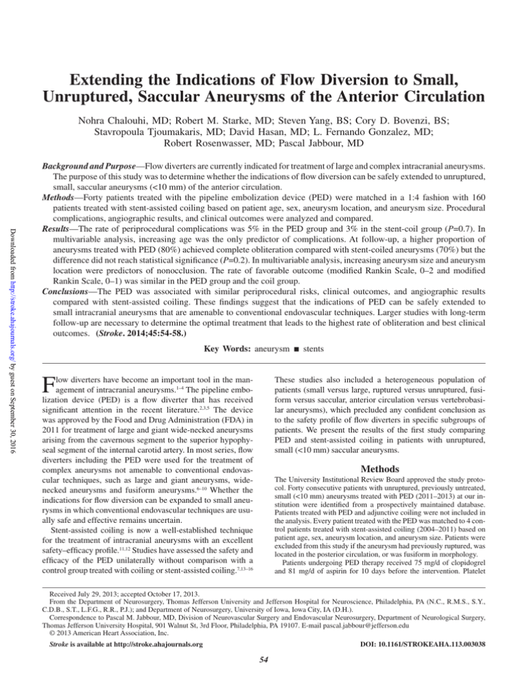
Extending the Indications of Flow Diversion to Small,
Unruptured, Saccular Aneurysms of the Anterior Circulation
Nohra Chalouhi, MD; Robert M. Starke, MD; Steven Yang, BS; Cory D. Bovenzi, BS;
Stavropoula Tjoumakaris, MD; David Hasan, MD; L. Fernando Gonzalez, MD;
Robert Rosenwasser, MD; Pascal Jabbour, MD
Downloaded from http://stroke.ahajournals.org/ by guest on September 30, 2016
Background and Purpose—Flow diverters are currently indicated for treatment of large and complex intracranial aneurysms.
The purpose of this study was to determine whether the indications of flow diversion can be safely extended to unruptured,
small, saccular aneurysms (<10 mm) of the anterior circulation.
Methods—Forty patients treated with the pipeline embolization device (PED) were matched in a 1:4 fashion with 160
patients treated with stent-assisted coiling based on patient age, sex, aneurysm location, and aneurysm size. Procedural
complications, angiographic results, and clinical outcomes were analyzed and compared.
Results—The rate of periprocedural complications was 5% in the PED group and 3% in the stent-coil group (P=0.7). In
multivariable analysis, increasing age was the only predictor of complications. At follow-up, a higher proportion of
aneurysms treated with PED (80%) achieved complete obliteration compared with stent-coiled aneurysms (70%) but the
difference did not reach statistical significance (P=0.2). In multivariable analysis, increasing aneurysm size and aneurysm
location were predictors of nonocclusion. The rate of favorable outcome (modified Rankin Scale, 0–2 and modified
Rankin Scale, 0–1) was similar in the PED group and the coil group.
Conclusions—The PED was associated with similar periprocedural risks, clinical outcomes, and angiographic results
compared with stent-assisted coiling. These findings suggest that the indications of PED can be safely extended to
small intracranial aneurysms that are amenable to conventional endovascular techniques. Larger studies with long-term
follow-up are necessary to determine the optimal treatment that leads to the highest rate of obliteration and best clinical
outcomes. (Stroke. 2014;45:54-58.)
Key Words: aneurysm ◼ stents
F
low diverters have become an important tool in the management of intracranial aneurysms.1–4 The pipeline embolization device (PED) is a flow diverter that has received
significant attention in the recent literature.2,3,5 The device
was approved by the Food and Drug Administration (FDA) in
2011 for treatment of large and giant wide-necked aneurysms
arising from the cavernous segment to the superior hypophyseal segment of the internal carotid artery. In most series, flow
diverters including the PED were used for the treatment of
complex aneurysms not amenable to conventional endovascular techniques, such as large and giant aneurysms, widenecked aneurysms and fusiform aneurysms.6–10 Whether the
indications for flow diversion can be expanded to small aneurysms in which conventional endovascular techniques are usually safe and effective remains uncertain.
Stent-assisted coiling is now a well-established technique
for the treatment of intracranial aneurysms with an excellent
safety–efficacy profile.11,12 Studies have assessed the safety and
efficacy of the PED unilaterally without comparison with a
control group treated with coiling or stent-assisted coiling.7,13–16
These studies also included a heterogeneous population of
patients (small versus large, ruptured versus unruptured, fusiform versus saccular, anterior circulation versus vertebrobasilar aneurysms), which precluded any confident conclusion as
to the safety profile of flow diverters in specific subgroups of
patients. We present the results of the first study comparing
PED and stent-assisted coiling in patients with unruptured,
small (<10 mm) saccular aneurysms.
Methods
The University Institutional Review Board approved the study protocol. Forty consecutive patients with unruptured, previously untreated,
small (<10 mm) aneurysms treated with PED (2011–2013) at our institution were identified from a prospectively maintained database.
Patients treated with PED and adjunctive coiling were not included in
the analysis. Every patient treated with the PED was matched to 4 control patients treated with stent-assisted coiling (2004–2011) based on
patient age, sex, aneurysm location, and aneurysm size. Patients were
excluded from this study if the aneurysm had previously ruptured, was
located in the posterior circulation, or was fusiform in morphology.
Patients undergoing PED therapy received 75 mg/d of clopidogrel
and 81 mg/d of aspirin for 10 days before the intervention. Platelet
Received July 29, 2013; accepted October 17, 2013.
From the Department of Neurosurgery, Thomas Jefferson University and Jefferson Hospital for Neuroscience, Philadelphia, PA (N.C., R.M.S., S.Y.,
C.D.B., S.T., L.F.G., R.R., P.J.); and Department of Neurosurgery, University of Iowa, Iowa City, IA (D.H.).
Correspondence to Pascal M. Jabbour, MD, Division of Neurovascular Surgery and Endovascular Neurosurgery, Department of Neurological Surgery,
Thomas Jefferson University Hospital, 901 Walnut St, 3rd Floor, Philadelphia, PA 19107. E-mail pascal.jabbour@jefferson.edu
© 2013 American Heart Association, Inc.
Stroke is available at http://stroke.ahajournals.org
DOI: 10.1161/STROKEAHA.113.003038
54
Chalouhi et al Flow Diversion 55
Downloaded from http://stroke.ahajournals.org/ by guest on September 30, 2016
function tests were routinely performed using aspirin assay and P2Y12
assay (VerifyNow; Accumetrics, San Diego, CA) to ascertain that the
level of platelet inhibition was between 30% and 90%. Patients with inhibition <30% were reloaded and the assay rechecked. Poor responders
to clopidogrel were then switched to prasugrel (brand name Effient, Eli
Lilly and Company, Indianapolis, IN). Patients with inhibition >90%
were admitted to the hospital, their procedure was canceled, and Plavix
was held until platelet inhibition level fell <90%. An initial 100 U/kg
of heparin bolus was administered and activated clotting time was
maintained at 2× the patient’s baseline intraoperatively. Heparin was
discontinued but not reversed at the conclusion of the procedure. Dual
antiplatelet therapy was continued for ≥6 months after the procedure.
Procedures were performed under general endotracheal anesthesia and
continuous neurophysiologic monitoring, including electroencephalography and somatosensory-evoked potentials. PEDs were deployed
through a Marksman microcatheter (ev3, Irvine, CA) using a triaxial
guide-catheter system. The number of stents deployed was left to the
operator’s discretion but, in general, when stasis was seen in the aneurysm dome no further devices were deployed. Recently, we have been
using only a single device for most aneurysms. The expansion of the
PED was documented under fluoroscopy or with additional DynaCT/
Xpert CT angiography at the operator’s discretion. Inadequate vessel
wall apposition was remedied with Gateway balloon (Boston Scientific,
Fremont, CA) angioplasty when needed. Placement of additional PEDs
was considered at follow-up if the aneurysm remained unchanged or
did not sufficiently decrease in size, despite treatment.
Our protocol and technique for stent-assisted coiling have been detailed previously.11 Briefly, when the use of a stent was anticipated,
patients were pretreated with daily 81 mg of aspirin and 75 mg of clopidogrel for 10 days before the procedure. Dual antiplatelet therapy was
continued for 2 months after the intervention. Coiling was interrupted
when the aneurysm was completely occluded or when no additional
coils could be deployed. Stent-assisted coiling was typically performed
using the microcatheter jailing technique in which the stent is deployed
after the aneurysm is microcatheterized but before coil deployment.
The outcomes of 40 PED patients and 160 stent-coil patients
matched for patient age, sex, aneurysm location, and aneurysm size
were compared. Medical charts were reviewed retrospectively to determine patient demographics, aneurysm characteristics, procedural
specifics, and procedural complications. Only procedural complications with clinical repercussions are reported. Angiographic follow-up
(digital subtraction angiography or magnetic resonance angiography)
was scheduled at 3 to 6 months, 1 year, 2 years, and 5 years after
treatment. Aneurysm obliteration rates were determined as percentages and transformed into a dichotomic variable: complete obliteration (100%) and incomplete obliteration (<100%). Regardless of the
need for further intervention, any filling at the neck or the dome of the
aneurysm was considered <100% occlusion and classified as incomplete obliteration. Clinical outcomes at the last available follow-up
were collected from follow-up notes of the attending physician and
classified using the modified Rankin Scale (mRS).
Statistical Analysis
Data are presented as mean and range for continuous variables and as
frequency for categorical variables. Matched analysis was performed
as appropriate. Univariate conditional (matched) analysis was used
to test covariates predictive of the following dependent variables:
treatment complications, follow-up obliteration, and clinical outcome
(mRS, 0–2 versus 3–6 and mRS, 0–1 versus 2–6). Interaction and confounding were assessed through stratification and relevant expansion
covariates. Factors predictive in univariate analysis (P<0.20)17 were
entered into a multivariate conditional logistic regression analysis. P
values of ≤0.05 were considered statistically significant. Statistical
analysis was performed with Stata 10.0 (College Station, TX).
proportion of female patients was 85% in both groups. Mean
aneurysm size was 6.2±2.4 mm in the PED group and 6.0±1.6
mm in the stent-coil group (P=0.3). The proportion of aneurysms >6 mm was similar in PED (60%) and stent-coil patients
(57%; P=0.8). Aneurysm locations (Table 1) were matched
between the 2 groups.
Aneurysm Treatment
PED deployment was successful in all 40 patients. The number of PEDs used was 1.3±0.4 per aneurysm. A single PED
was used in 26 (65%) aneurysms and 2 PEDs in 14 (35%)
aneurysms. Balloon angioplasty was performed for optimal
PED expansion in 1 (2.5%) patient.
In the stent-coil group (n=160), initial Raymond scores
were I (complete occlusion) in 71 (44%) patients, II (residual neck) in 56 (35%) patients, and III (dome filling) in 33
(20.6%) patients.
Procedural Complications
Procedure-related complications occurred in 2 (5%) patients
(1 ischemic event and 1 distal hemorrhage) in the PED group
versus 5 (3%) patients (4 ischemic events and intraoperative rupture) in the stent-coil group (P=0.7). There was no
procedure-related mortality in either group. No patient had
a symptomatic side-branch occlusion after PED therapy. The
following factors were tested for as predictors of complications: age, sex, aneurysm location, aneurysm size, aneurysm
morphology, and type of treatment. In univariate analysis,
older age (≥65 years; odds ratio [OR], 3.7; 95% confidence
interval [CI], 0.8–17.7; P=0.09) predicted procedural complications. In multivariate analysis, there was a trend for older
age (≥65 years) to predict complications (OR, 3.8; 95% CI,
0.7–17.7; P=0.09). The type of treatment was not a predictor
of complications even after controlling for age.
Angiographic Outcome
Angiographic follow-up was available for 39 (97.5%) patients
treated with PED and 147 (92%) patients treated with stentassisted coiling. Median angiographic follow-up time was
7 months in the PED group and 15 months in the stent-coil
group (P<0.001). At the latest follow-up, a higher proportion
of aneurysms treated with PED (80%; n=31) achieved complete obliteration (100%) compared with coiled aneurysms
(70%; n=103) but the difference fell short of statistical significance (P=0.2; Table 2). In the stent-coil group (n=160),
Raymond scores at the latest follow-up were I (complete
occlusion) in 103 (70%) patients, II (residual neck) in 17
(11.5%) patients, and III (dome filling) in 28 (19%) patients.
Table 1. Aneurysm Locations
Aneurysm Location
PED (%)
Stent-Coil (%)
Carotid ophthalmic/paraclinoid
37 (92.5)
145 (90.7)
1 (2.5)
5 (3.1)
Carotid cavernous
Results
Posterior communicating
1 (2.5)
5 (3.1)
Baseline Characteristics
Middle cerebral artery
1 (2.5)
5 (3.1)
Mean patient age was similar in the PED group (52.1±13.7
years) and the stent-coil group (52.6±11.4 years; P=0.8). The
Total
40
160
PED indicates pipeline embolization device.
56 Stroke January 2014
Downloaded from http://stroke.ahajournals.org/ by guest on September 30, 2016
The following factors were tested for as predictors of occlusion: age, sex, aneurysm location, aneurysm size, aneurysm
morphology, type of treatment, complications, and follow-up
time. In univariable analysis, factors predicting nonocclusion
were increasing aneurysm size (OR, 1.23; 95% CI, 1.01–1.49;
P=0.04) and carotid cavernous-posterior communicating
artery-middle cerebral artery aneurysm location (ie, aneurysm
locations with rates of complete occlusion <70%; P=0.01). In
multivariable analysis, increasing aneurysm size (OR, 5; 95%
CI, 1.4–14; P=0.01) and carotid cavernous-posterior communicating artery-middle cerebral artery aneurysm location (OR,
5; 95% CI, 1.4–14; P=0.01) remained statistically significant
independent predictors of nonocclusion.
Retreatment was necessary in 4 (10%) patients in the PED
group and 13 (9%; P=0.8) patients in the coil group. It should
be noted that retreatment was undertaken for recurrences in
all 13 patients in the stent-coil group, whereas none of the 4
patients in the PED group had a recurrence. In fact, aneurysm
size decreased to some extent in 3 of the 4 PED patients but
the decision was made to place additional devices to accelerate
and increase the likelihood of further aneurysm thrombosis.
Clinical Outcome
Clinical follow-up was available for 39 (97.5%) patients in
the PED group and 148 (93%) patients in the stent-coil group.
Median follow-up time was 7 months in the PED group and 17
months in the stent-coil group (P<0.001). The proportion of
patients with mRS 0 to 2 was 100% (39/39) in the PED group
and 98% in the stent-coil group (99%; 146/148; P=0.9). The
proportion of patients with mRS 0 to 1 was 95% (37/39) in the
PED group and 96% in the stent-coil group (96%; 142/148;
P=0.9). The following factors were tested for as predictors of
outcome: age, sex, aneurysm location, aneurysm size, aneurysm morphology, type of treatment, and complications. In
univariable analysis, increasing aneurysm size (OR, 5.9;
95% CI, 0.6–5.2; P=0.1) predicted a poor clinical outcome
(mRS>1). In multivariable analysis, no factor was a significant predictor of poor clinical outcome.
Discussion
The only flow diverter currently approved by the FDA is the PED.
Other flow diverters include Silk (Balt, Montmorency, France),
Surpass (Stryker, Fremont, CA), and FRED (Microvention,
Tustin, CA).18 The Silk stent has been extensively used outside
the United States, and the Surpass has recently shown promising results in a small series from Europe.4,19,20
Initially reserved for complex, giant, and fusiform aneurysms, flow diverters are currently increasingly used in the
management of small and less complex aneurysms at some
Table 2. Rates of Aneurysm Occlusion
Complete Aneurysm Occlusion
≤6 mo
7 to 12 mo
PED
21/27 (77%)
17/21 (81%)
6/8 (75%)
Stent-assisted
coiling
21/30 (70%)
56/70 (80%)
40/53 (75%)
PED indicates pipeline embolization device.
>12 mo
institutions. Many interventionalists, however, remain wary
of this approach and continue to prefer traditional endovascular strategies, especially for small aneurysms. A recent
meta-analysis on flow diverters by Brinjikji et al,21 including
1451 patients with 1654 aneurysms, found procedure-related
morbidity and mortality rates of 5% and 4%, respectively. The
authors concluded that the risk of procedure-related morbidity
and mortality with flow diverters is not negligible and should
be taken into account when considering the best therapeutic
option for intracranial aneurysms. Another meta-analysis of
15 studies that compiled 897 patients with 1018 aneurysms
found an early mortality rate of 2.8%, a late mortality rate
of 1.3%, and an overall neurological morbidity rate of 9.9%.
The authors of the meta-analysis also found that available data
supporting the use of flow diverters were heterogeneous and
prone to publication biases, concluding that the use of flow
diverters in patients eligible to more conventional treatments
should be restricted to controlled clinical trials.
However, several studies have demonstrated convincingly
that the PED carries a high safety and efficacy profile. In a large
Turkish series of 191 patients treated with the PED, Saatci et al7
reported a 6-month occlusion rate of 91% with an impressive
permanent morbidity rate of only 1%. A recent well-designed
multicenter international trial reported a success rate of 99%,
an occlusion rate of 74%, and a major ipsilateral stroke or neurological death rate of only 5.6%.7 Pistocchi et al2 treated 30
aneurysms at and beyond the circle of Willis with flow diverters
(Silk and Pipeline) reporting permanent neurological complication in only 3.7% and aneurysm occlusion in 82% of patients.
Likewise, in an multicenter study of 143 patients with 178 aneurysms from Hong Kong, Yu et al8 reported a complete aneurysm
occlusion rate of 84%, an overall neurological complication
rate of 8.4%, and a periprocedural death or major stroke rate
of 4.2% (median follow-up of 18 months). They concluded that
PED should be considered a first choice for treating unruptured
aneurysms. All these studies included a heterogeneous population of patients (no separate analysis was done for small aneurysms) and did not put the results of flow diversion in direct
comparison with those of conventional endovascular techniques
especially stent-assisted coiling, which has shown an excellent
safety–efficacy profile in several large studies.11,12,22,23
The present study is not the first to compare flow diverters
with coiling. However, it is the first to specifically compare
stent-assisted coiling with flow diversion, or even compare the 2
techniques in small aneurysms. In a small study, Lanzino et al24
compared 22 paraclinoid aneurysms treated with the PED with
historic controls. The authors reported a significantly higher
rate of complete occlusion in PED patients (76%) than coiled
patients (21%) with a similar rate of morbidity and concluded
that long-term follow-up was important to validate flow diversion definitively as a superior therapeutic strategy for proximal
internal carotid artery aneurysms. In a previous report, we have
compared the periprocedural, angiographic, and clinical outcomes of flow diversion and coiling in unruptured, large, and
giant (≥10 mm) aneurysms.10 We have found a similar complication rate in both groups (7.5%) along with a higher aneurysm
occlusion rate (86% versus 41%) and a lower retreatment rate
with flow diversion (2.8% versus 37%). In multivariable analysis, the odds of achieving occlusion of large aneurysms were
Chalouhi et al Flow Diversion 57
Downloaded from http://stroke.ahajournals.org/ by guest on September 30, 2016
>10× higher with flow diversion than with coiling. These results
led us to conclude that flow diverters were a preferred option
for large and giant aneurysms. In the present study, we sought
to determine whether flow diversion is also a better strategy for
small aneurysms (<10 mm), a subgroup in which stent-assisted
coiling has traditionally generated favorable clinical and angiographic results. We found that flow diversion can be undertaken
with no additional morbidity and similar clinical outcomes compared with stent-assisted coiling. There was also a trend toward
higher occlusion rates with the PED (80% versus 70%), but the
study was likely underpowered to detect small differences in
angiographic outcomes between 2 highly efficient endovascular
techniques. Although retreatment rates did not differ between
the 2 groups, retreatment was always undertaken for a recurrence in stent-coiled patients, whereas in PED patients retreatment was undertaken because the aneurysm had not sufficiently
decreased in size. We have also recently found that PED treatment requires significantly shorter fluoroscopy and procedure
times compared with stent-assisted coiling.25 Moreover, a
recent study has demonstrated that PED embolization is more
economical than stent-assisted coiling with a 27% reduction
in the cost per millimeter of aneurysm treated,26 although any
cost benefit will also depend on aneurysm volume, coil type,
and number of PEDs used.27 Taken together, these data suggest
that the indications of flow diversion can be safely extended
to unruptured, small aneurysms (<10 mm). Some may argue,
however, that stent-assisted coiling is a better option than flow
diversion because of the higher immediate complete occlusion
rate and the shorter period of dual antiplatelet therapy.
In the present study, the initial occlusion rate (Raymond
score I, ie, 100% occlusion) with stent-assisted coiling was
48% increasing to 70% at follow-up. These rates are consistent with those reported in the literature. In a large study of
500 stent-coiled aneurysms by Geyik et al,23 complete occlusion was achieved in 42.2% of the aneurysms initially, and the
rate progressed to 90.8% at follow-up. Likewise, a systematic
review of the literature on stent-assisted coiling by Shapiro et
al28 reported a 45% complete occlusion rate initially, increasing to 61% at follow-up. The morbidity rates with stentassisted coiling in our study are largely in line with previously
reported series. Lessne et al29 reported a 5.4% rate of thromboembolic events, whereas Maldonado et al30 reported a 2.9%
combined morbidity–mortality rate after stent-assisted coiling
of 76 aneurysms. Likewise, in the multicenter Enterprise registry, procedural data demonstrated a 6% temporary morbidity, 2.8% permanent morbidity, and 2% mortality.31
Although the complications of stent-assisted coiling are
essentially limited to thromboembolic events and intraprocedural aneurysm ruptures,11,12 flow diversion carries the
additional risk of distal parenchymal hemorrhage, delayed
migration of the device, and delayed aneurysm rupture.9,32–36
Distal parenchymal hemorrhage may occur ipsilaterally or even
contralaterally to the aneurysm and its mechanism may involve
hemorrhagic conversion of ischemic lesions, embolized foreign
material, loss of arterial autoregulation of the distal arteries,
or dual antiplatelet therapy.5,37 Device migration is a recently
recognized complication of flow diverters that is attributed to
a mismatch in arterial diameter between inflow and outflow
vessels. The migration may occur proximally or distally and
may lead to devastating complications, such as aneurysm rupture or thromboembolic events.32 Delayed aneurysm rupture is
a dreaded complication of flow diversion that typically occurs
in large and giant aneurysms. Its cause remains uncertain but
may involve altered hemodynamics and enzymatic degradation
of the aneurysm wall from thrombus formation. The complication rate in the present report was low with flow diversion and
did not differ significantly from that of stent-assisted coiling.
Finally, if PED therapy is not effective in achieving complete
aneurysm obliteration, endovascular access to the aneurysm
will have been permanently lost and the only options available
for further treatment would be reduced to open surgery or additional placement of PEDs. Also, clip application for proximal
control is possible only proximal to the PED because the device
is irreversibly deformed by clip application.38
Limitations
This study is retrospective in design and reflects the experience
of a single center. We could not provide occlusion rates at standard time points. Instead, we have compared aneurysm occlusion rates at the latest follow-up. Although the 2 groups were
well matched with regard to baseline characteristics, the clinical
and angiographic follow-up time differed significantly. As such,
the occlusion rate with PED would have been even higher if
patients were followed up for longer periods, which further supports the efficacy of flow diverters.8,14 Improved endovascular
technology and increasing operator experience with aneurysm
embolization techniques could have favored the PED group. The
retreatment rate with the PED is closely related to the number
of devices deployed during the initial embolization procedure.
Because we tend to use only a single device in most cases (with
placement of further devices only if the aneurysm remains
open at follow-up), the PED retreatment rate would have been
even lower had we adopted a different strategy where multiple
devices are deployed initially. Despite these limitations, this
study is the first to provide a comparative analysis of clinical and
angiographic outcomes in small aneurysms treated with PED
and stent-assisted coiling. Randomized controlled trials comparing flow diversion and conventional endovascular techniques
are currently underway. The Flow Diversion in Intracranial
Aneurysm Treatment (FIAT) trial39 is a randomized open label
trial comparing flow diversion with best standard treatment in
the management of difficult intracranial aneurysms. The trial is
sponsored by the Center hospitalier de l’Université de Montréal
and is currently recruiting participants. The LARGE aneurysm
randomized trial40 is an ongoing prospective, randomized, study
comparing coil embolization versus flow diversion in large (>10
mm), anterior circulation intracranial aneurysms sponsored by
the Medical University of South Carolina. Another trial is also
taking place in France and compares the 2 techniques in unruptured saccular wide-neck intracranial aneurysms >7 mm.41
Conclusions
Both flow diversion and stent-assisted coiling are safe and highly
effective techniques for treatment of unruptured, small saccular
aneurysms of the anterior circulation. The PED was associated
with similar aneurysm occlusion rates, periprocedural morbidity, and short-term clinical outcomes. These findings suggest
that the indications of the PED can be safely extended to small
58 Stroke January 2014
intracranial aneurysms that are amenable to conventional endovascular techniques. Larger studies and long-term follow-up are
necessary to determine the optimal treatment that leads to the
highest rate of obliteration and best clinical outcomes.
Disclosures
None.
References
Downloaded from http://stroke.ahajournals.org/ by guest on September 30, 2016
1. Kallmes DF, Ding YH, Dai D, Kadirvel R, Lewis DA, Cloft HJ. A new
endoluminal, flow-disrupting device for treatment of saccular aneurysms. Stroke. 2007;38:2346–2352.
2. Pistocchi S, Blanc R, Bartolini B, Piotin M. Flow diverters at and beyond
the level of the circle of Willis for the treatment of intracranial aneurysms. Stroke. 2012;43:1032–1038.
3. D’Urso PI, Lanzino G, Cloft HJ, Kallmes DF. Flow diversion for intracranial aneurysms: a review. Stroke. 2011;42:2363–2368.
4. De Vries J, Boogaarts J, Van Norden A, Wakhloo AK. New generation
of Flow Diverter (surpass) for unruptured intracranial aneurysms: a prospective single-center study in 37 patients. Stroke. 2013;44:1567–1577.
5. Pierot L, Wakhloo AK. Endovascular treatment of intracranial aneurysms: current status. Stroke. 2013;44:2046–2054.
6. Becske T, Kallmes DF, Saatci I, McDougall CG, Szikora I, Lanzino G, et
al. Pipeline for uncoilable or failed aneurysms: results from a multicenter
clinical trial. Radiology. 2013;267:858–868.
7. Saatci I, Yavuz K, Ozer C, Geyik S, Cekirge HS. Treatment of intracranial aneurysms using the pipeline flow-diverter embolization device: a
single-center experience with long-term follow-up results. AJNR Am J
Neuroradiol. 2012;33:1436–1446.
8. Yu SC, Kwok CK, Cheng PW, Chan KY, Lau SS, Lui WM, et al. Intracranial
aneurysms: midterm outcome of pipeline embolization device–a prospective
study in 143 patients with 178 aneurysms. Radiology. 2012;265:893–901.
9.Kan P, Siddiqui AH, Veznedaroglu E, Liebman KM, Binning MJ,
Dumont TM, et al. Early postmarket results after treatment of intracranial aneurysms with the pipeline embolization device: a U.S. multicenter
experience. Neurosurgery. 2012;71:1080–1087, discussion 1087.
10. Chalouhi N, Tjoumakaris S, Starke RM, Gonzalez LF, Randazzo C,
Hasan D, et al. Comparison of flow diversion and coiling in large unruptured intracranial saccular aneurysms. Stroke. 2013;44:2150–2154.
11. Chalouhi N, Jabbour P, Singhal S, Drueding R, Starke RM, Dalyai RT, et
al. Stent-assisted coiling of intracranial aneurysms: predictors of complications, recanalization, and outcome in 508 cases. Stroke. 2013;44:1348–1353.
12.Chalouhi N, Starke RM, Koltz MT, Jabbour PM, Tjoumakaris SI,
Dumont AS, et al. Stent-assisted coiling versus balloon remodeling of
wide-neck aneurysms: comparison of angiographic outcomes. AJNR Am
J Neuroradiol. 2013;34:1987–1992.
13. Chitale R, Gonzalez LF, Randazzo C, Dumont AS, Tjoumakaris S,
Rosenwasser R, et al. Single center experience with pipeline stent: feasibility, technique, and complications. Neurosurgery. 2012;71:679–91,
discussion 691.
14. Lylyk P, Miranda C, Ceratto R, Ferrario A, Scrivano E, Luna HR, et al.
Curative endovascular reconstruction of cerebral aneurysms with the pipeline embolization device: the Buenos Aires experience. Neurosurgery.
2009;64:632–42, discussion 642.
15. Nelson PK, Lylyk P, Szikora I, Wetzel SG, Wanke I, Fiorella D. The
pipeline embolization device for the intracranial treatment of aneurysms
trial. AJNR Am J Neuroradiol. 2011;32:34–40.
16. Chalouhi N, Tjoumakaris S, Dumont AS, Gonzalez LF, Randazzo C,
Starke RM, et al. Treatment of posterior circulation aneurysms with the
pipeline embolization device. Neurosurgery. 2013;72:883–889.
17. Altman DG. Practical Statistics for Medical Research. Boca Raton, FL:
Chapman & Hall/CRC; 1999.
18. Diaz O, Gist TL, Manjarez G, Orozco F, Almeida R. Treatment of 14
intracranial aneurysms with the FRED system. J Neurointerv Surg. 2013.
http://jnis.bmj.com/content/early/recent. Accessed September 25, 2013.
19. Berge J, Biondi A, Machi P, Brunel H, Pierot L, Gabrillargues J, et al. Flowdiverter silk stent for the treatment of intracranial aneurysms: 1-year followup in a multicenter study. AJNR Am J Neuroradiol. 2012;33:1150–1155.
20. Tähtinen OI, Manninen HI, Vanninen RL, Seppänen J, Niskakangas T,
Rinne J, et al. The silk flow-diverting stent in the endovascular treatment of
complex intracranial aneurysms: technical aspects and midterm results in
24 consecutive patients. Neurosurgery. 2012;70:617–623, discussion 623.
21. Brinjikji W, Murad MH, Lanzino G, Cloft HJ, Kallmes DF. Endovascular
treatment of intracranial aneurysms with flow diverters: a meta-analysis.
Stroke. 2013;44:442–447.
22. Jahshan S, Abla AA, Natarajan SK, Drummond PS, Kan P, Karmon Y, et al.
Results of stent-assisted vs non-stent-assisted endovascular therapies in 489
cerebral aneurysms: single-center experience. Neurosurgery. 2013;72:232–239.
23. Geyik S, Yavuz K, Yurttutan N, Saatci I, Cekirge HS. Stent-assisted coiling in endovascular treatment of 500 consecutive cerebral aneurysms
with long-term follow-up. AJNR Am J Neuroradiol. 2013. http://www.
ajnr.org/content/early/recent. Accessed July 29, 2013.
24. Lanzino G, Crobeddu E, Cloft HJ, Hanel R, Kallmes DF. Efficacy and
safety of flow diversion for paraclinoid aneurysms: a matched-pair
analysis compared with standard endovascular approaches. AJNR Am J
Neuroradiol. 2012;33:2158–2161.
25. Chalouhi N, McMahon JF, Moukarzel LA, Starke RM, Jabbour P, Dumont
AS, et al. Flow diversion versus traditional aneurysm embolization strategies: analysis of fluoroscopy and procedure times. J Neurointerv Surg.
2013. http://jnis.bmj.com/content/early/recent. Accessed June 5, 2013.
26. Colby GP, Lin LM, Paul AR, Huang J, Tamargo RJ, Coon AL. Cost
comparison of endovascular treatment of anterior circulation aneurysms with the pipeline embolization device and stent-assisted coiling.
Neurosurgery. 2012;71:944–948, discussion 948.
27. Chalouhi N, Jabbour P, Tjoumakaris S, Starke RM, Dumont AS, Liu H, et
al. Treatment of large and giant intracranial aneurysms: cost comparison
of flow diversion and traditional embolization strategies. World Neurosurg.
2013. http://www.worldneurosurgery.org/inpress. Accessed June 5, 2013.
28. Shapiro M, Becske T, Sahlein D, Babb J, Nelson PK. Stent-supported
aneurysm coiling: a literature survey of treatment and follow-up. AJNR
Am J Neuroradiol. 2012;33:159–163.
29. Lessne ML, Shah P, Alexander MJ, Barnhart HX, Powers CJ, Golshani
K, et al. Thromboembolic complications after Neuroform stent-assisted
treatment of cerebral aneurysms: the Duke Cerebrovascular Center experience in 235 patients with 274 stents. Neurosurgery. 2011;69:369–375.
30. Maldonado IL, Machi P, Costalat V, Mura T, Bonafé A. Neuroform stentassisted coiling of unruptured intracranial aneurysms: short- and midterm results from a single-center experience with 68 patients. AJNR Am
J Neuroradiol. 2011;32:131–136.
31. Mocco J, Snyder KV, Albuquerque FC, Bendok BR, Alan S B, Carpenter
JS, et al. Treatment of intracranial aneurysms with the Enterprise stent: a
multicenter registry. J Neurosurg. 2009;110:35–39.
32. Chalouhi N, Tjoumakaris SI, Gonzalez LF, Hasan D, Pema PJ, Gould G,
et al. Spontaneous delayed migration/shortening of the pipeline embolization device: report of 5 cases. AJNR Am J Neuroradiol. 2013. http://
www.ajnr.org/content/early/recent. Accessed July 29, 2013.
33. Jabbour P, Chalouhi N, Tjoumakaris S, Gonzalez LF, Dumont AS, Randazzo
C, et al. The Pipeline embolization device: learning curve and predictors of
complications and aneurysm obliteration. Neurosurgery. 2013;73:113–120,
discussion 120.
34.Chalouhi N, Satti SR, Tjoumakaris S, Dumont AS, Gonzalez LF,
Rosenwasser R, et al. Delayed migration of a pipeline embolization
device. Neurosurgery. 2013;72:ons229–ons234, discussion ons234.
35.Chitale R, Gonzalez LF, Randazzo C, Dumont AS, Tjoumakaris S,
Rosenwasser R, et al. Single center experience with pipeline stent: feasibility, technique, and complications. Neurosurgery. 2012;71:679–691,
discussion 691.
36. O’Kelly CJ, Spears J, Chow M, Wong J, Boulton M, Weill A, et al. Canadian
experience with the pipeline embolization device for repair of unruptured
intracranial aneurysms. AJNR Am J Neuroradiol. 2013;34:381–387.
37. Deshmukh V, Hu YC, McDougall CG, Barnwell SL, Albuquerque F,
Fiorella D. 126 histopathological assessment of delayed ipsilateral parenchymal hemorrhages after the treatment of paraclinoid aneurysms with
the pipeline embolization device. Neurosurgery. 2012;71:E551–E552.
38. Bell RS, Bank WO, Armonda RA, Vo AH, Kerber CW. Can a self-expanding aneurysm stent be clipped? Emergency proximal control options for
the vascular neurosurgeon. Neurosurgery. 2011;68:1056–1062.
39. Flow Diversion In Intracranial Aneurysm Treatment (FIAT). Clinical trials.
gov. http://clinicaltrials.gov/ct2/show/NCT01349582. Accessed September
16, 2013.
40. LARGE Aneurysm Randomized Trial: Flow Diversion Versus Traditional
Endovascular Coiling Therapy. Clinical trials.gov. http://clinicaltrials.
gov/ct2/show/NCT01762137. Accessed September 16, 2013.
41. Multicenter Randomized Study for Medico-economic Evaluation of Embo­
lization With Flow Diverter Stent in the Endovascular Treatment of Un­­rup­­
tured Saccular Wide-necked Intracranial Aneurysms. Clinical trials.gov. http://
clinicaltrials.gov/ct2/show/NCT01811134. Accessed September 16, 2013.
Extending the Indications of Flow Diversion to Small, Unruptured, Saccular Aneurysms of
the Anterior Circulation
Nohra Chalouhi, Robert M. Starke, Steven Yang, Cory D. Bovenzi, Stavropoula Tjoumakaris,
David Hasan, L. Fernando Gonzalez, Robert Rosenwasser and Pascal Jabbour
Downloaded from http://stroke.ahajournals.org/ by guest on September 30, 2016
Stroke. 2014;45:54-58; originally published online November 19, 2013;
doi: 10.1161/STROKEAHA.113.003038
Stroke is published by the American Heart Association, 7272 Greenville Avenue, Dallas, TX 75231
Copyright © 2013 American Heart Association, Inc. All rights reserved.
Print ISSN: 0039-2499. Online ISSN: 1524-4628
The online version of this article, along with updated information and services, is located on the
World Wide Web at:
http://stroke.ahajournals.org/content/45/1/54
Permissions: Requests for permissions to reproduce figures, tables, or portions of articles originally published
in Stroke can be obtained via RightsLink, a service of the Copyright Clearance Center, not the Editorial Office.
Once the online version of the published article for which permission is being requested is located, click
Request Permissions in the middle column of the Web page under Services. Further information about this
process is available in the Permissions and Rights Question and Answer document.
Reprints: Information about reprints can be found online at:
http://www.lww.com/reprints
Subscriptions: Information about subscribing to Stroke is online at:
http://stroke.ahajournals.org//subscriptions/

