Phylogenetic variation in flexor digitorum brevis
advertisement

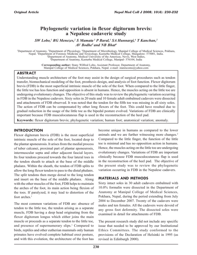
Original Article Nepal Med Coll J 2008; 10(4): 230-232 Phylogenetic variation in flexor digitorum brevis: a Nepalese cadaveric study SW Lobo,1 RG Menezes,2 S Mamata 3 P Baral,1 SA Hunnargi,4 T Kanchan,2 AV Bodhe5 and NB Bhat6 1 Department of Anatomy, 5Department of Physiology, 6Department of Microbiology, Manipal College of Medical Sciences, Pokhara, Nepal. 2Department of Forensic Medicine and Toxicology, Kasturba Medical College, Mangalore- 575001, India. 3 Department of Anatomy, Medical University of the Americas, Nevis, West Indies, 4 Department of Anatomy, Kasturba Medical College, Manipal- 576104, India Corresponding author: Stany Wilfred Lobo, Assistant Professor, Department of Anatomy, Manipal College of Medical Sciences, Pokhara, Nepal. e-mail: stanmcoms@yahoo.com ABSTRACT Understanding muscle architecture of the foot may assist in the design of surgical procedures such as tendon transfer, biomechanical modeling of the foot, prosthesis design, and analysis of foot function. Flexor digitorum brevis (FDB) is the most superficial intrinsic muscle of the sole of the foot. When compared to the little finger, the little toe has less function and opposition is absent in humans. Hence, the muscles acting on the little toe are undergoing evolutionary changes. The objective of this study was to review the phylogenetic variation occurring in FDB in the Nepalese cadavers. Sixty soles in 20 male and 10 female adult embalmed cadavers were dissected and attachments of FDB observed. It was noted that the tendon for the fifth toe was missing in all sixty soles. The action of FDB can be compensated by other long flexors of the foot. This could have resulted due to gradual reduction in the usage of the little toe as the bipedal posture evolved. Variations of FDB are clinically important because FDB musculocutanous flap is used in the reconstruction of the heel pad. Keywords: flexor digitorum brevis; phylogenetic variation; human foot; anatomical variation; anomaly. INTRODUCTION Flexor digitorum brevis (FDB) is the most superficial intrinsic muscle of the sole of the foot, located deep to the plantar aponeurosis. It arises from the medial process of tuber calcanei, proximal part of plantar aponeurosis, intermuscular septa and other adjacent fascial layers. Its four tendons proceed towards the four lateral toes in the tendon sheath to attach at the base of the middle phalanx. Within the sheath, the tendon of FDB splits to allow the long flexor tendon to pass to the distal phalanx. The split tendons then merge dorsal to the long tendon and insert on the base of the middle phalanx. Along with the other muscles of the foot, FDB helps to maintain the arches of the foot, its main action being flexion of the toes. If paralyzed, it may lead to distortion of the foot arches.1 The most common variations of FDB are: absence of tendon to the little toe, the tendon arising as a separate muscle, FDB having a deep head originating from the flexor digitorum longus which either joins the main muscle or proceeds as a separate tendon to the little toe, and presence of supernumerary slips.1 Compared to birds, reptiles and other eutherian mammals only human primates have evolved complete habitual erect posture, and with this evolution, the architecture of the foot has become unique in humans as compared to the lower animals and we are further witnessing more changes.2 Compared to the little finger, the function of the little toe is minimal and has no opposition action in humans. Hence, the muscles acting on the little toe are undergoing evolutionary changes. Variations of FDB are important clinically because FDB musculocutanous flap is used in the reconstruction of the heel pad. The objective of the present study was to review the phylogenetic variation occurring in FDB in the Nepalese cadavers. MATERIALS AND METHODS Sixty intact soles in 30 adult cadavers embalmed with 10.0% formalin were dissected in the Department of Anatomy at Manipal College of Medical Sciences, Pokhara, Nepal, during the period extending from July 2004 to December 2007. Twenty of the cadavers were males and ten females. All the cadavers were devoid of any gross foot deformity. The dissected soles were examined in detail for attachments of FDB. The present research study did not include any specific issue that needed to be approved by our Institutional Ethics Committees. The study conformed to the provisions of the Declaration of Helsinki in 1995 (as revised in Edinburgh 2000). 230 SW Lobo et al Fig. 1. The muscle belly of flexor digitorum brevis (FDB) and its three tendons to the second (I I), third (I I I), and fourth (I V) toes. RESULTS In all the 30 cadavers (sixty soles), FDB arose from the medial process of tuber calcanei, plantar aponeurosis and intermuscular septa of adjacent muscles. The muscle divided into three parts which ended in slender tendons to the second, third and forth toes (Fig. 1). The fifth toe did not receive any tendon from FDB in all 60 soles. The tendons passed through the fibrous flexor sheath of the toes and split into two parts which curved along the sides of the long flexor tendon, rejoined and were attached to the plantar surface of base of middle phalanx. DISCUSSION FDB along with the other intrinsic muscles of the foot helps to maintain the longitudinal arches of the foot. Many authors in various classic Anatomy text books have mentioned that FDB gives four tendons to the lateral four toes. Variations in FDB have been reported to occur in 63.0% of all limbs.3 Though absence of FDB tendon to the fifth toe was reported by Chaney et al.4 and Sarrafian5, in the present study we observed that FDB tendon to the fifth toe was entirely missing bilaterally in all the cadavers studied. The usage of the fifth toe in humans is minimal. According to Darwin’s disuse theory, therefore, FDB tendon to the fifth toe may be undergoing phylogenetic variation. This is supported by Reeser et al.6 in their electromyographic study of human foot which showed that FDB is not preferentially recruited over flexor digitorum longus for any normal posture or locomotion. Yalcin and Ozan7 reported anatomical variations in FDB. Of the 33 feet they7 examined, 18 differed from the classical description given in Anatomy textbooks. The muscle belly for the fifth toe was much smaller than the others in 12 feet and was absent in six feet. Present findings of our study are also supported by Claassen and Wree.8 They observed variations in FDB of both feet in a 75-year-old male cadaver. In the left foot, the fourth tendon of FDB to the little toe was atrophied, and the respective tendon in the right foot was missing. The importance of these anatomical variants, during surgery and diagnostic imaging such as computed tomography and magnetic resonance imaging, should be known to the podiatric surgeon. Awareness of these variants will decrease the chances of confusion while considering treatment options.4 In contrast with the findings in the present study, Asomugha et al.9 have reported accessory flexor of the fifth toe. This muscle originated from the tendon of tibialis posterior and inserted into the middle phalanx of the fifth toe. 9 Variations of FDB are clinically important because myocutaneous flap of FDB is used for reconstruction of the heel pad and soft tissue defects of the heel.10,11 Awareness of the variations of FDB is important to the anatomists, surgeons, and radiologists especially while dealing with computed tomography scans for soft tissue injuries of the foot. Its significance also lies in the studies related to hominid evolution. In conclusion, in our study FDB in all the 60 soles had 231 Nepal Medical College Journal only three tendons - to the second, third and fourth toes, but fifth toe did not receive any tendon. The action of FDB being flexion of the toes can be compensated by other long flexors of the foot. Due to the lesser function of the fifth toe in humans, has the tendon of FDB to the fifth toe become extinct? This could be the fact for more number of variations observed in FDB. In the distant future, some more variations in FDB can be anticipated and total disappearance of FDB may be a possibility. REFERENCES 1. Rosse C, Gaddum-Rosse P. Hollinshead’s Textbook of Anatomy. 5th ed. Philadelphia: Lippincott- Raven 1997. 2. Williams PL, Warwick R, Dyson M, Bannister LH. Gray’s Anatomy. 37th ed. New York: ELBS- Churchill Livingstone 1989. 3. Bergman RA, Thompson SA, Afifi AK, Saadeh FA. Compendium of Human Anatomic Variations. Baltimore: Urban and Schwarzenberg 1988. 4. Chaney DM, Lee MS, Khan MA, Krueger WA, Mandracchia VJ, Yoho RM. 1996. Study of ten anatomical variants of the foot and ankle. J Amer Podiatr Med Assoc 1996; 86: 532-7. 5. Sarrafian SK. Anatomy of the Foot and Ankle. Philadelphia: Lippincott Williams and Wilkins 1983. 6. Reeser LA, Susman RL, Stern JT Jr. Electromyographic studies of the human foot: experimental approaches to hominid evolution. Foot Ankle 1983; 3: 391-407. 7. Yalcin B, Ozan H. Some variations of the musculus flexor digitorum brevis. Anat Sci Int 2005; 80: 189-92. 8. Claassen H, Wree A. Isolated flexor muscles of the little toe in the feet of an individual with atrophied or lacking 4th head of the M. extensor digitorum brevis and lacking the 4th tendon of the M. extensor digitorum longus. Ann Anat 2003; 185: 81-4. 9. Asomugha AL, Chukwuanukwu TO, Nwajagu GI, Ukoha U. An accessory flexor of the fifth toe. Niger J Clin Pract 2005; 8: 130-2. 10. Hartrampf CR Jr, Scheflan M, Bostwick J 3rd. The flexor digitorum brevis muscle island pedicle flap: a new dimension in heel reconstruction. Plast Reconstr Surg 1980; 66: 264-70. 11. Ikuta Y, Murakami T, Yoshioka K, Tsuge K. Reconstruction of the heel pad by flexor digitorum brevis musculocutaneous flap transfer. Plast Reconstr Surg 1984; 74: 86-96. 232


