C-Spine Radiology A-B-C-D-E-F Adequacy Swimmer`s View
advertisement
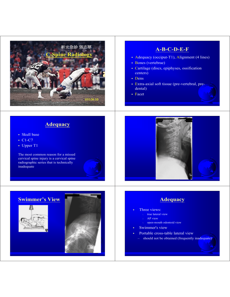
新光急診 張志華 C-Spine Radiology A-B-C-D-E-F Adequacy (occiput-T1), Alignment (4 lines) Bones (vertebrae) Cartilage (discs, epiphyses, ossification centers) Dens Extra-axial soft tissue (pre-vertebral, predental) Facet 103.06.10 Adequacy Skull base C1-C7 Upper T1 The most common reason for a missed cervical spine injury is a cervical spine radiographic series that is technically inadequate Adequacy Swimmer’s View Three views: 1. 2. 3. true lateral view AP view open-mouth odontoid view Swimmer's view Portable cross-table lateral view – should not be obtained (frequently inadequate) Alignment Anterior spinal line Posterior spinal line Spinolaminal line Spinous process tips Alignment Anterior spinal line Posterior spinal line Spinolaminal line Spinous process tips Bones Anterior vertebral line Posterior vertebral line Spinolaminal line Spinous process tips Bones Bones Anterior components – Vertebral body (cortices, endplates) – Transverse process Posterior components – – – – Articular masses and pedicles Facet joints Lamina Spinous process Cartilage Intervertebral discs Epiphyses (childhood) – Subdental synchondrosis Ossification centers (childhood) – Tapered anterior vertebrae – Absent C1 anterior ring Extra-axial Soft Tissue Parameter Predental space Adults < 3 mm Prevertebral space - C2 < 7 mm Prevertebral space - C6 < 21 mm Children < 5 mm < 1/2 vertebral body < 1 vertebral body Angulation < 11 degrees < 11 degrees Cord dimension 10 to 13 mm Adult size by 6 yr Soft tissue Subluxation C2/3 Subluxation Angulation between two adjoining vertebrae > 11 degrees Overriding of vertebra by > 3 mm Case: 6-year-old boy C2 posterior spinal line v.s. Swischuk line C2/3 Pseudosubluxation Swischuck line < 2 mm off Pseudosubluxation (< 8-16Y) : – Displacement < 1.5-2 mm Hangman fracture : – Displacement > 1.5-2 mm Odontoid View Jefferson fracture (C1) Blowout of the ring Axial loading Open-mouth (odontoid) view 1/3 associated with C-2 fracture Unstable Usually not associated with cord injury Lateral offset of C1 lateral masses > 1mm from C2 vertebral body Normal C-1 Rotary Subluxation Odontoid not equidistant from lateral masses Children Torticolis (chin toward uninvolved side) Immobilize in place Consult NS Extension view Flexion view Odontoid Fractures Odontoid Subluxation / dislocation Ruptured transverse ligament Predental space : – Ad > 3mm – Pd > 5mm (symptomatic if > 7-10mm) Odontoid fractures C1 spinal canal (Steel rule of 3) : – Odontoid – Free space – Cord Unstable type I - involves only the upper part of the dens type II (most common) occurs where the dens and the vertebral body join type III - through the upper body C2 vertebra Some odontoid fractures can be treated with external support (such as C collar or halo traction) alone while others (especially type II) require surgery Type I : Avulsion of tip – Stable Type II : At the base – Unstable – D/D : Synchondrosis if < 6Y Type III : Through vertebral body – Unstable Hangman Fracture Traumatic spondylolisthesis of C2 Mechanism : – Extension + Distraction – Extension + Axial compression X-ray : C2/3 subluxation Unstable Traction contraindicated Hangman fracture - a hyperextension injury involving bilateral pars interarticularis fractures of the axis 使用鏟子工作的人 Clay shoveler fracture C7>C6>T1 Unilateral Facet Dislocation (Bowtie Sign) Unilateral Facet Dislocation (AP) Oblique view Facet Dislocation Bilateral Facet Dislocation Unilateral (UFD) : – Stable – < 25% translation Bilateral (BFD) : – Unstable – > 50% translation Teardrop Fractures Extension teardrop : – Stable in flexion, unstable in extension – Cortices : Same length Flexion teardrop : – Extremely unstable – Cortices : Unequal length Flexion teardrop Extension teardrop Wedge fracture of C5 Interspinous widening* * C6 Narrowed C5-C6 intervertebral disc space Burst Vertebral Body Mechanically stable Spinal cord injury can occur (even total transection) * Atlanto-occipital dislocation (AOD)* longitudinal distraction with separation of the occiput from the atlas Gap between occipital condyles and atlas > 5 mm Fracture lines: # Odontoid type II # Mandibular ramus Oblique C-spine Pedicles Articular mass Intervertebral foramen Transverse process Laminae - aligned in the fashion of shingles Oblique views show the pedicle in profile, and also allows assesment of the intervertebral foramina (and osteophytes encroaching along their margins) SCIWORA Spinal Cord Injury WithOut Radiographic Abnormality 67%-80% of pediatric SCI Mainly < 8 Y Plain films / tomograms / CT (-) May have transient neurologic symptoms and apparently recover then return 1d later with significant neurologic abnormalities Poor prognosis SCIWORA Etiology : – Vascular injuries (occlusion, spasm, infarction) – Ligamentous injury – Disc impingement – Incomplete neuronal destruction Spinal EDH Venous bleeds Minor traumas Ascending neurologic symptoms Hours or days MRI Clear neck collar SPINAL Severe pain Point of tenderness Injury mechanism Neurologic deficit Altered level of consciousness Limitation of motion Thank You
