950
Value of Cardiac Monitoring and Echocardiography
in TIA and Stroke Patients
J . A . R E M , M.D.,
V.C.
HACHINSKI, M . D . ,
D.R.
AND H.J.M. BARNETT,
BOUGHNER, M . D . ,
PH.D.,
M.D.
Downloaded from http://stroke.ahajournals.org/ by guest on September 30, 2016
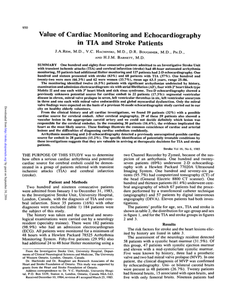
SUMMARY One hundred and eighty-four consecutive patients admitted to an Investigative Stroke Unit
with transient ischemic attacks (TIA) and cerebral infarction (stroke) had 48-hour automated arrhythmia
monitoring, 55 patients had additional Holter monitoring and 127 patients had 2-D echocardiography. One
hundred and sixteen presented with stroke (63%) and 68 patients with TIA (37%). One hundred and
twenty-two were men (66.3%) and 62 were women (33.7%), mean age 63.5 years, range 25-86.
The monitoring identified twelve (6.5%) patients with significant arrhythmias undetected by history,
examination and admission electrocardiogram: six with atrial fibrillation (AF), four with 2° heart block type
Mobitz II and one each with 3° heart block and sick sinus syndrome. Two-D echocardiography showed a
previously unknown potential source for cardiac emboli in 22 patients (17.3%): segmental ventricular
disease in eleven, mitral valve prolapse in seven, left ventricular thrombus in six, left ventricular aneurysm
in three and one each with mitral valve endocarditis and global myocardial dysfunction. Only the mitral
valve findings were expected on the basis of a previous M-mode echocardiographic study carried out in our
city on healthy elderly volunteers.
From the clinical history and all cardiac investigations, we found 59 patients (32%) with a possible
cardiac source for cerebral emboli. After cerebral angiography, 29 of these 59 patients also showed a
vascular lesion in the appropriate carotid artery and we could not decide definitely which lesion was
responsible for the cerebral embolus. In the remaining 30 patients (16.4%), the evidence implicated the
heart as the most likely source. These findings illustrate the common coincidence of cardiac and arterial
lesions and the difficulties of diagnosing cardiac embolism confidently.
Arrhythmia monitoring and 2-D echocardiography detected a previously unrecognized possible cardiac
source for emboli in 28 patients (15.2%). The specific identification of potentially treatable conditions by
these investigations suggests that they are valuable in arriving at therapeutic decisions for TIA and stroke
patients.
Stroke Vol 16, No 6, 1985
THE PURPOSE OF THIS STUDY was to determine
how often a serious cardiac arrhythmia and potential
cardiac source for cerebral emboli could be demonstrated in a group of patients referred with transient
ischemic attacks (TIA) and cerebral infarction
(stroke).
Patient and Methods
Two hundred and nineteen consecutive patients
were admitted from January 1 to December 31, 1983,
to the Investigative Stroke Unit, University Hospital,
London, Canada, with the diagnosis of TIA and cerebral infarction. Since 35 patients (16%) with other
diagnoses were excluded (table 1) 184 patients were
the subject of this study.
The history was taken and the general and neurological examinations were carried out by a neurology
resident (specialty trainee). There were 182 patients
(98.9%) who had an admission electrocardiogram
(ECG). All patients were monitored for a minimum of
48 hours with a Hewlett Packard 78525 Arrhythmia
Monitoring System. Fifty-five patients (29.9%) also
had additional 24 to 48 hour Holter monitoring using a
From the Investigative Stroke Unit, University Hospital, Departments of Clinical Neurological Sciences and Medicine, The University
of Western Ontario, London, Ontario, Canada.
Dr. Hachinski and Dr. Boughner are Research Associates of the
Heart and Stroke Foundation of Ontario. This study was supported by
grants from the Heart and Stroke Foundation of Ontario.
Address correspondence to: Dr. V.C. Hachinski, University Hospital, P.O. Box 5339, Station A, London, Ontario, Canada N6A-5A5.
Received December 10, 1984; revision #1 accepted March 25, 1985.
two Channel Recorder by Zymed, because of the suspicion of an arrhythmia. One hundred and twentyseven patients (69%) underwent 2-D echocardiography with a Hewlett Packard 77020A Ultrasound
Imaging System. One hundred and seventy-six patients (95.7%) had computerized tomography (CT) of
the head (General Electric 8800 CT scanner). One
hundred and thirteen patients (61.4%) underwent cerebral angiography of which 67 patients had the procedure performed by a transfemoral catheter technique
(angiography) and 57 patients by digital intravenous
angiography (DIVA). Eleven patients had both investigations.
The patients' profile for age, sex, TIA and stroke is
shown in table 2, the distribution for age-group and sex
in figure 1, and for the TIA and stroke groups in figures
2 and 3.
Results
The risk factors for stroke and the heart lesions elicited by history are listed in table 3.
The assessment of the neurology resident detected
58 patients with a systolic heart murmur (31.5%). Of
this group, 47 patients with systolic ejection murmur
and eleven with a mid-systolic/late systolic murmur.
Five were known by history, three had a prosthetic
valve and two had mitral valve prolapse (MVP). In one
patient, the clinical diagnosis of MVP was confirmed
by echocardiography. Uni- or bilateral carotid bruits
were present in 48 patients (26.7%). Twenty patients
had femoral bruits, 15 associated with open bruits, and
five with only femoral bruits. Nineteen patients had
CARDIAC MONITORING AND ECHOCARDIOGRAPHY IN TIA AND STROKEIRem et al
DISTRIBUTION AGE AND SEX
TABLE 1 Exclusions
Downloaded from http://stroke.ahajournals.org/ by guest on September 30, 2016
Intracerebral hemorrhage
Tumor
Syncope
Subarachnoid hemorrhage
Seizure
Transient global amnesia (atypical)
Acute labyrinthitis
Complicated migraine
Meniere syndrome
Syringomyelia
Cause undetermined:
Vertigo and visual disturbances
Vertigo
Visual disturbance
Unsteadiness
Total
6
5
5
4
4
2
1
1
1
1
N
TIA
Stroke
Mean age
Range
W MALE
35
H FEMALE
30
25
20
2
1
1
1
35
carotid bruits and a heart murmur. An irregular pulse
(extrasystoles, atrial fibrillation) was noted in 21 patients (11 .4%). Fifteen were known to have an arrhythmia and six had a normal history. One of these six
patients had atrial fibrillation (AF) on ECG, the remaining patients had normal cardiac monitoring.
One hundred and eighty-two patients (98.9%) had
ECG on admission. In 108 patients, the ECG was
abnormal (59.3%) and in 74 patients (40.7%) normal.
The detected abnormalities are listed in table 4. Thirteen patients had AF, ten of them known previously.
Forty-two patients had signs of myocardial infarct
(MI) on ECG. Nineteen patients had a history of a
recent or remote MI. Twenty-three had a silent MI.
Thirty-one of 42 patients with MI (73.8%) were nondiabetics (NDM) and eleven patients (26.2%) suffered
from diabetes mellitus (DM) (table 5). Patients with
DM had significantly more Mi's than NDM (Chisquare test, p < 0.05). By comparing only the silent
MI, the significance for having an MI in the DM-group
becomes even higher (Chi-square test, p < 0.01). The
mean age in NDM-group with MI on the ECG was
67.0 years (±SD 9.6), range 40-81 and 63.3 years
(±SD 8.2), range 48-77 in the DM-group. The patients in the DM-group with MI on ECG were significantly younger (t-test,/j <0.05). Fourteen non-diabetics with a silent MI (9.1%) had a mean age of 68.1
years (±SD 7.8), range 48-76 (10 patients 65 years)
and nine diabetics with a silent MI (30%) had a mean
age of 61.1 years (± SD 7.9), range 52-77 (2 patients
TABLE 2
951
15
10
1
<40 41-50 51-60 61-70 71-80 2 81
AGE
FIGURE 1. Age and sex distribution of study group.
65 years). Diabetics with a silent MI were significantly
younger than non-diabetics (t.test, p < 0.05).
The 48-hour cardiac monitoring was abnormal in 94
patients (51.1%) and normal in 90 patients (48.9%).
T I A GROUP:
AGE AND SEX
^
40
MALE
E l FEMALE
35
30
J
25
20
15
10
Age and Sex of the Patients
Men
Women
Total
122 (66.3%)
47
75
62.8
25-86
62 (33.7%)
21
41
64.8
30-83
184
68 (37%)
116(63%)
63.5
25-86
-•-AGE
<40
4 1 - 5 0 5 1 - 6 0 6 1 - 7 0 7 1 - 8 0 >8I
FIGURE 2. Age and sex distribution of the transient ischemic
attack (TIA) patients.
STROKE
952
40
W MALE
H FEMALE
30
25
20
15
Downloaded from http://stroke.ahajournals.org/ by guest on September 30, 2016
10
£40 41-50 51 60 61 70 71 80 2 81
FIGURE 3. Age and sex distribution of the cerebral infarct
(stroke) patients.
The abnormalities are listed in table 6. Of the seventeen patients with AF, eleven were persistent and six
paroxysmal. Eleven of these (eight persistent, three
paroxysmal) were known by history, two patients with
persistent AF were detected by the ECG and confirmed
by the cardiac monitoring, and in four patients the
abnormalities were detected by the monitoring. Two
patients had a 2° heart block type Mobitz II. One of
them had a normal ECG and the other had a 1° AVblock. One patient with a transient 3° heart block had a
normal resting ECG.
TABLE 3
Risk Factors
Hypertension
Smoking
Coronary artery disease
Previous TIA/stroke
Diabetes mellitus
Atrial fibrillation — paroxysmal
— persistent
Rheumatic fever/rheumatic heart disease
Congestive heart failure
Prosthetic valve — aortic
— mitral
Tachyarrhythmia
Mitral valve prolapse
Pacemaker
Hemiplegic migraine
No known risk factors
1985
TABLE 4 Admission Electrocardiogram (ECG) Abnormalities in
108 Patients
STRODE GROUP: AGE AND SEX
35
VOL 16, No 6, NOVEMBER-DECEMBER
104
72
58
58
30
12
10
6
5
1
2
3
2
2
1
18
(56.5%)
(39.1%)
(31.5%)
(31.5%)
(16.3%)
(6.5%)
(5.4%)
(3.3%)
(2.7%)
(0.5%)
(1.1%)
(1.6%)
(1.1%)
(1.1%)
(0.5%)
(9.8%)
Arrhythmias:
Sinus arrhythmia
Sinus bradycardia 55 beats/min
Sinus tachycardia 100 beats/min
Atrial extrasystoles
Atrial fibrillation
Ventricular extrasystoles
Conduction defect:
Right bundle branch block
Left anterior fascicular block
Left posterior fascicular block
Left bundle branch block
1° AV-block
Artificial pacemaker rhythm
Accelerated AV-conduction
Myocardial abnormalities
Myocardial ischemia
Myocardial infarction
Left atrial hypertension
Left ventricular hypertrophy
Total
2
16
7
5
13 (10)
7
5
12
1
2
20
2 (2)
1
4 (1)
42 (19)
13
12
164 (32)
() known by history.
The additional Holter monitoring was abnormal in
48 patients (87.3%) and normal in seven patients
(12.7%). Results are shown in table 7. Six patients had
atrial fibrillation, four paroxysmal and two persistent.
Four of these had arrhythmias suspected but not proven
by the 48 hour cardiac monitoring. Two patients with
persistent atrial fibrillation also had a positive ECG
and one of them was known to have atrial fibrillation
by history and by 48 hour cardiac monitoring. One
patient with paroxysmal atrial fibrillation had a positive history and one had positive cardiac monitoring.
One patient with sick sinus syndrome (normal history,
ECG: myocardial infarct, time indeterminate), and
two patients with 2° heart block type Mobitz II were
detected. One of them had a 1° AV-block on the ECG.
The other patient had a normal ECG. After these investigations three patients had a permanent pacemaker
inserted.
The 2-D echocardiography was abnormal in 61 patients (48%), normal in 56 patients (44.1%) and ten
investigations (7.9%) were difficult to evaluate beTABLE 5 Diabetes Mellitus — Myocardial Infarction on Admission ECG
Known
Unknown
Total
MI
NonDM
154 (83.7%)
19
23
42 (25%)
17
14
31 (20.1%)
DM
30 (16.3%)
2
9
11 (36.7%)
MI = myocardial infarction; DM = diabetes mellitus.
Chi-square test: p < 0.05 for all MI; p < 0.01 for silent MI.
CARDIAC MONITORING AND ECHOCARDIOGRAPHY IN TIA AND STROKEJRem et al
TABLE 6 48-Hour Cardiac Monitoring Abnormalities In 94 Patients
Downloaded from http://stroke.ahajournals.org/ by guest on September 30, 2016
Arrhythmias:
Sinus arrhythmia
4
Sinus pause (2-4 sec)
2
Sinus bradycardia 55 beats/min
35
Sinus tachycardia 100 beats/min
7
Atrial extrasystoles
3
Supraventricular rhythm
4
Supra ventricular tachycardia
2
Atrial fibrillation — paroxysmal
6 (3)
— persistent
11 (8)
Ventricular extrasystoles
51
Ventricular tachycardia
7
Conduction defect:
Heart block — 1° AV-block
3
— 2° Mobitz type II block
2
— 3° AV-block
1
Pacemaker rhythm (demand)
2
(2)
Total
140 (13)
((13))
((2))
((2))
((2))
((19))
() known by history; (()) known by admission ECG.
cause of technical problems. The abnormalities are
listed in table 8.
The mean age of the 61 patients was 65.3 years
(± SD 11.2), range 30-83. There are only two patients
younger than 45 years, both in the MVP group. The
following eight patients were known to have abnormalities by history or previous 2-D echocardiography:
MVP (2), prosthetic mitral (2) and aortic (1) valves,
aortic valve sclerosis (2) and segmental ventricular
disease (hypokinetic segment) (1). A previously unknown possible cardiac source for emboli was detected
in 22 patients (17.3%), table 9, mean age 64.7 years
(±SD 9.8), range 43-81, only one of these patients
was less than 45 years old (in the MVP group).
One hundred and seventy-six patients had a CT of
the head (95.7%). In 98 patients (55.7%), the CT was
TABLE 7 Hotter Monitoring Abnormalities in 48 Patients
Sinus pause (2-4 sec)
Sinus bradycardia 55 beats/min
Sick sinus syndrome
Atriai extrasystoies
Supraventricular tachycardia
100 beats/min
Atrial fibrillation — paroxysmal
— persistent
Ventricular extrasystoles
Ventricular tachycardia
Heart block — 2° mobitz type
II block
ST — depression
Total
3
1
1
i9
TABLE 8 Echocardiographic Abnormalities In 61 Patients
Valvular abnormalities:
mitral valve
piolapse
9 (2)*
sclerosis
10
stenosis
7
thickening
1
endocarditis
1
mitral annulus calcification
6
aortic valve
19 (2)*
sclerosis
stenosis
2
prolapse
1
aortic annulus calcification
1
tricuspid valve prolapse
1
Prosthetic valve
aortic
1 (1)*
mitral
2 (2)*
Myocardial abnormalities:
1
global myocardial dysfunction
segmental ventricular disease
12 (1)*
left ventricular aneurysm
3
thrombus
6
left ventricular hypertrophy
7
dilation
5
left atrial dilation
5(1)*
Miscellaneous:
calcified papillary muscle
1
dilated aortic root
1
Total
102 (9)*
*Known by history or previous echocardiography.
abnormal and in 78 patients (44.3%) normal. In 80
patients (81.6%) of whom 71 had stroke and 9 had
TIA, the CTfindingswere related to the clinical presentation and in 18 patients (18.4%) of whom eleven
had TIA and seven had stroke, they were not related.
One hundred and thirteen patients (61.4%) had cerebral angiography. The investigations were abnormal in
TABLE 9
Echocardiography and Possible Cardiac Source for
Emboli
11 Segmental ventricular
disease
4
2
37
2
(1) 1*
(1)
«1*,1))
7 Mitral valve prolapse
2 Aneurysm
1 Thrombus
2
2
82
953
(2) 2*
() known by history; (()) known by admission ECG.
*48-hour cardiac monitoring.
((2))
1 Global myocardial
dysfunction
22 patients (17.3%)
—
—
—
—
—
—
—
2 with thrombus
1 with aneurysm and thrombus
1 mitral annulus calcification
1 with vegetation (endocarditis)
1 mitral annulus calcification
2 with thrombus
with CAD known because of
previous echocardiogram
954
STROKE
90 patients (78.6%) and normal in 23 patients
(21.4%). The angiographic lesions were related to the
clinical presentation in 73 patients (81.1%) and unrelated in seventeen patients (18.9%). Sixty-seven patients had cerebral angiography done by the transfemoral catheter technique (59.3%) and 57 patients
(50.4%) DIVA. Eleven patients had both investigations, two of them with normal results. Stenosis from
mild (0-30% narrowing) to very severe (90 narrowing)
was present in 48 patients (42.5%), occlusion of an
artery in fourteen patients (12.4%) and atherosclerotic
changes without stenosis in eleven patients (9.7%). All
these lesions were in vessels appropriate to the symptoms and signs. There were 29 patients who had a
lesion in the appropriate carotid and they also had a
possible cardiac source for embolus.
Downloaded from http://stroke.ahajournals.org/ by guest on September 30, 2016
Discussion
The use of cardiac investigations in TIA and stroke
patients remains controversial.1"" Significantly more
cardiac arrhythmias were found in patients with acute
stroke as compared to nonstroke patients admitted to
an investigative stroke care unit.12 These arrhythmias
are rarely (2%) responsible for hemodynamic ischemic
cerebrovascular lesions, but may have been associated
with cerebral embolism in up to 17% of all cases.12
With 48 hour cardiac monitoring and additional Holter
monitoring we detected 12 patients (6.5%) with significant arrhythmias. There were six patients with
atrial fibrillation and six patients with conduction defects. Persistent or paroxysmal atrial fibrillation unaccompanied by any recognizable underlying heart disease is associated with a substantial increase in the
incidence of emboli.8 Wolf et al13 reported that the risk
of stroke from idiopathic atrialfibrillationis increased
5-fold and 17-fold with atrial fibrillation associated
with rheumatic heart disease.13 According to de Bono9
routine ambulatory ECG monitoring is not indicated in
patients with focal neurological symptoms unless the
occurrence of paroxysmal arrhythmia is suggested by
history. No patients were detected with atrial fibrillation and negative history and ECG with Holter monitoring was detected in one study.10 We detected with
the ECG three patients with atrial fibrillation and a
negative history. In an ECG study, atrial fibrillation
was found in 115 patients (33%) with ischemic cerebrovascular disease and in 35 patients (10%) of a control group.14 The opinion of some writers is that patients presenting with "CNS insufficiency of any
degree" should receive a complete medical work-up
including continuous Holter monitoring.15 Lavy et al16
monitored acute stroke patients over 24 hours and
demonstrated that electrocardiographic disturbances
are frequent within 24 hours of a stroke. They also
found that the prognosis of patients with co-existing
stroke and cardiac abnormalities is grave. They suggested that stroke victims should be watched closely
and treated promptly when complications arise. Therefore, their opinion was that the introduction of the
investigative stroke care unit may lead to a better outcome for patients with stroke.
VOL 16, No 6, NOVEMBER-DECEMBER
1985
Of interest is the high incidence of silent MI, 23 of
42 patients (54.8%). We found that diabetic patients
have significantly (p < 0.05) more silent MI and are
significantly younger (p < 0.05) than non-diabetics.
In the non-DM group, ten of 31 patients were aged 65
or more and in the DM group only two of eleven
patients. Fisch3 studied individuals without cardiovascular disease under 25 years of age and individuals 65
and older. In the group aged 65 and older, he found
that 30 patients (4.4%) with unequivocal ECG evidence of MI showed no correlation with clinical heart
disease. He stated that: "This reflects the inherent difficulty of obtaining a reliable history of this group of
individuals". Rothbaum17 states: "Painless subendocardial infarction is commonly encountered in the elderly patients, due to profound hypotension, anemia or
hypoxia." It is known that the incidence of MI is increased in diabetic patients.18 In one series 93 diabetic
patients (32.6%) with acute MI presented without
chest pain.19 These patients presented with heart failure, uncontrolled diabetes, vomiting, collapse, confusion and cerebrovascular events.19 Faerman et al20
studied the sympathetic and parasympathetic nerve fibers of the heart in five diabetics who had died of
painless MI. They assumed that the nerve lesions
found could be blamed for the absence of pain during
the attack. Thus, afferent impulses could have been
interrupted by diabetic visceral neuropathy.
Ourfindingsthat ECG and 48 hour cardiac monitoring detected significant arrhythmias in 6.5% of patients who had a negative history for arrhythmia and
unhelpful admission ECG's, suggests that routine
monitoring is warranted in patients who are candidates
for preventive treatment.
Physical examination identified only 58 patients
(31.5%) with a heart murmur by the admitting neurology resident. Half of the patients were also seen by
cardiologists but usually after cardiac investigations
were available so that theirfindingsare not listed in our
study.
We identified with 2-D echocardiography 22 patients (17.3%) with a previously unknown potential
source of cardiac emboli. Of interest is the fact that six
of seven patients with MVP were older than 45 years.
Mitral valve prolapse,21 segmental ventricular disease,
rheumatic heart disease, global myocardial dysfunction are known to be potential sources of cardiac emboli. In contrast, Greenland et al examined 100 consecutive hospitalized TIA and stroke patients and
found no evidence of atrial thrombi, mitral stenosis,
cardiac tumor, or vegetations suggesting endocarditis.5
Therefore, routine echocardiography was not recommended. It was suggested that the test may be of more
value in patients with clinical, electrocardiographic or
x-ray evidence of heart disease.5 Come et al10 also
examined the value of ECG's and echocardiography
and found that electrocardiography demonstrated cardiac abnormalities that might predispose to emboli in
47% of patients with and 14% without evident cardiovascular disease. Lesions that might be directly responsible for emboli, including thrombi, myxomas
CARDIAC MONITORING AND ECHOCARDIOGRAPHY IN TIA AND STROKEIRem et al
Downloaded from http://stroke.ahajournals.org/ by guest on September 30, 2016
and vegetations, were identified in only eleven patients
(4%), all of whom had clinically apparent disease.
Patients less than 45 years of age with clinically evident cardiovascular disease were especially likely to
have an identifiable potential source of embolism. According to their results, they suggested that echocardiography should be reserved for those patients with
clinically apparent cardiovascular disease, because patients without clinically evident heart disease are particularly unlikely to have thrombi or vegetation demonstrated by echocardiography.
de Bono states that echocardiography should be reserved for those patients who have heart murmurs and
probably any stroke patient under the age of 45. 9 While
some other authors share these opinions, 4 ' 6 - 7 we do not
agree. Only one (8.3%) of our 22 patients was younger
than 45 years. Moreover, Barnett and colleagues found
that in their series of MVP cases, none had known
heart disease and 75% of their patients had a normal
cardiac examination. Robbins et al" says that echocardiography has a low yield, high cost and the findings
do not influence early therapy with systemic embolism. With 2-D echocardiography they identified the
heart as a high probable source for emboli in thirteen of
116 patients (11.2%) studied. The sensitivity of 2-D
echocardiography in detecting left ventricular clots
was found to be 72% and the specificity 90%. 2 Franco
et al22 demonstrated with their study that patients with
cerebrovascular accident and TIA frequently have
echocardiographic abnormalities (58%), many of
which were clinically unsuspected. They suggested
that echocardiography should be performed in these
patients since the cardiac abnormalities identified may
be contributory to the cerebrovascular event. Barnett8
commented that the treatment and long-term management of stroke patients must be based on an accurate
diagnosis in a given patient and a generous approach to
cardiac studies, rather than their occasional use, would
appear justifiable. Our results support this approach.
The incidence of unsuspected echocardiographic abnormalities in an older adult population remains unclear. Most echocardiographic studies exclude cardiovascular disease when studying an aging population,
focusing on alterations in ventricular function and
chamber size. A study carried out in our city with
participation by one of the authors (D.R. B.) did list
the incidence of abnormalities on M-mode echocardiography in the elderly.23 Among 146 asymptomatic
volunteers with a mean age of 72 years (range 60 to 94
years), 38 had findings on history and physical examination that excluded them from the echocardiographic
study. Of this excluded group, 10 (6.8%) were hypertensive, two (1.4%) were diabetic, six (4.1%) had left
ventricular hypertrophy on their electrocardiogram,
nine (6.1%) had previous myocardial infarcts and two
had evidence of aortic stenosis. An additional nine
were excluded because of obstructive lung disease,
hyperthyroidism and intermittent claudication. Of the
remaining 108 patients who underwent M-mode echocardiography, 14 (13%) produced unsatisfactory studies compared with the 7.9% failure rate in our two-
955
dimensional echocardiographic study. The M-mode
studies showed nine patients (9.5%) to have unsuspected mitral valve prolapse compared with 7.6% in
our series. No examples of mitral stenosis were found
and three patients showed mitral annular calcification.
One structurally abnormal aortic valve was noted.
Also in that study, there were no instances of unsuspected septal or posterior wall motion abnormalities
compatible with previous infarction. However, the
ability of M-mode echocardiography to detect coronary artery disease is limited since it only images the
left ventricular minor axis and does not examine either
the apex or the antero-lateral wall. Conceivably, silent
myocardial infarction in those two areas may have
been missed. Our present study showed a much higher
incidence of unsuspected wall motion abnormalities in
the TIA patients as well as various valvular lesions.
Only the mitral valve prolapse cases were expected on
the basis of the results from the healthy elderly volunteers.
From the clinical history and all cardiac investigations we found 59 patients (32%) with a possible cardiac source for cerebral emboli. After cerebral angiography, 29 of those 59 patients were found also to have a
vascular lesion in the appropriate carotid and we could
not decide definitely which lesion really was responsible for the cerebral embolus. In the remaining 30 patients (16.4%), we believe that the heart was the most
likely source.
In summary, our results show that cardiac investigations are worthwhile in TIA and stroke patients, who
are considered candidates for preventive treatment. It
must be emphasized, however, that we are a referral
center and that most patients are admitted for further
investigations and may be younger and have less severe neurological deficits than those seen in a general
hospital.
Editors Note: In accordance with Stroke policy, this
article was guest edited by J.P. Mohr.
References
1. Wolf PA, Dawber TR, Kannel WB: Heart disease as a precursor of
stroke. Advances in Neurology 19: 567-577, 1978
2. Al-Nouri MB, Patel K, Johnson DW, et al: The sensitivity and
specificity of two-dimensional echocardiography in detecting left
ventricular thrombi. (Abstract). Circulation 62 (Suppl. Ill): 21,
1980
3. Fisch C: The electrocardiogram in the aged. Geriatric cardiology,
Cardiovascular clinics 12/1: 65-71, 1981
4. Donaldson RM, Emanuel RW, Earl CJ: The role of two-dimensional echocardiography in the detection of potentially embolic intracardiac masses in patients with cerebral ischemia. J Neurol Neurosurg and Psych 44: 803-809, 1981
5. Greenland P, Knopman DS, Mikell FL, et al: Echocardiography in
diagnostic assessment of stroke. Ann Intern Med 95:51-53, 1981
6. Lovett JL, Sandok BA, Giuliani ER, Nasser FN: Two-dimensional
echocardiography in patients with focal cerebral ischemia. Ann
Intern Med 95: 1-4, 1981
7. Knopman DS, Anderson DC, Asinger RW, et al: Indication for
echocardiography in patients with ischemic stroke. Neurology 32:
1005-1011, 1982
8. Barnett HJM: Heart is ischemic stroke — a changing emphasis. IN:
Barnett HJM (ed) Neurologic Clinics, volume 1, number 1, Philadelphia, W.B. Saunders, 291-336, 1983
STROKE
956
Downloaded from http://stroke.ahajournals.org/ by guest on September 30, 2016
9. de Bono DP: Cardiac causes of stroke. In: Ross Russell RW (ed):
Vascular Disease of the Central Nervous System, 2nd edition,
London, Churchill Livingstone, 324-336, 1983
10. Come PC, Riley MF, Bivas NK: Roles of echocardiography and
arrhythmia monitoring in the evaluation of patienls with suspected
systemic embolism. Ann Neural 13: 527-531, 1983
11. RobbinsJA, SagarKB, French BS, Smith PJ: Influence of echocardiography on management of patients with systemic emboli. Stroke
14: 564-549, 1983
12. Norris JW, Froggatt GM, Hachinski VC: Cardiac arrhythmias in
acute stroke. Stroke 9: 392-396, 1978
13. Wolf PA, Dawber TR, Thomas EH, Kannel WB: Epidemiologic
assessment of chronic atrial fibrillation and risk of stroke. The
Framingham Study. Neurolgy 28: 973-977, 1978
14. Nishide M, Irino T, Gotoh M, et al: Cardiac abnormalities in
ischemic cerebrovascular disease studied by two-dimensional
echocardiography. Stroke 14: 541-545, 1983
15. Levin EB: Use of Holter echocardiographic monitor in the diagnosis of transient ischemic attacks. J Amer Geriat Soc 24: 516-512,
1976
16. Lavy S, Yaar I, Melamed E, Stern S: The effect of acute stroke on
17.
18.
19.
20.
21.
22.
23.
VOL
16,
No
6,
NOVEMBER-DECEMBER
1985
cardiac function as observed in an intensive stroke care unit. Stroke
5: 775-780, 1974
Rothbaum DA: Coronary artery disease. Geriatric cardiology, Cardiovascular clinics 12/1: 105-118, 1981
Fein FS, Scheuer J: Heart disease in diabetes. In: Ellenberg M,
Rifkin H (eds), Diabetes Mellitus, 3rd edition, New York, Medical
Examination Publishing Co., 851-861, 1983
Soler NG, Bennett MA, Pentecost BL, et al: Myocardial infarction
in diabetics. Quart J Med 19: 125-132, 1975
Faerman I, Faccio E, Millei J, et al: Autonomic neuropathy and
painless myocardial infarction in diabetic patients. Diabetes 26:
1147-1158, 1977
Barnett HJM, Boughner DR, Taylor DW, et al: Further evidence
relating mitral-valve prolapse to cerebral ichemic events. N Engl J
Med 302: 139-144, 1980
Franco R, Alam M, Ausman J et al: Echocardiography in cerebrovascular accidents and cerebral transient ischemic attacks. (Abstract). Circulation 62 (Suppl III): 22, 1980
Manyari D, Patterson C, Johnson D, et al: An echocardiographic
study on resting left ventricular function in healthy elderly subjects.
J Clin Exp Gerontology 4(4): 504-420, 1982
Internal Carotid Artery Dissection After Childbirth
DAVID O.
WIEBERS, M . D . ,
AND BAHRAM MOKRI,
M.D.
SUMMARY A 44-year-old woman developed a left cerebral infarction secondary to internal carotid
artery dissection 6 days after childbirth. A cesarean section had been carried out after 14 hours of strenuous
unsuccessful labor. Although in the past some authors have implicated oral contraceptives as a cause for
carotid dissection, carotid dissection associated with childbirth has not been previously described.
Stroke Vol 16, No 6, 1985
DISSECTIONS OF INTERNAL CAROTID ARTERIES (ICAs) occur most frequently in patients less than
50 years of age. Ipsilateral head and face pain, with or
without neck pain, is the most common sign. Other
common manifestations include oculosympathetic paresis, focal cerebral ischemic symptoms, and bruits.MZ
Although many of the dissections are thought to occur
spontaneously, the role of trivial trauma such as
coughing, straining, and abrupt or exaggerated neck
movements or neck postures cannot be entirely excluded. In some cases there is evidence of an arterial
disease such as fibromuscular dysplasia,3' l3~19 or cystic
medial necrosis.1'2-7> 12 Spontaneous carotid dissection
has also been described in association with Marfan's
syndrome.20 Traumatic dissections of the ICAs have
been well recognized as the result of penetrating trauma such as that caused by an angiographic needle, or
as the result of blunt injuries associated with such
factors as motor vehicle accidents and whiplash injuries, chiropractic manipulations, falls, strangulation,
and sports activities.5' 21~28
We report the occurrence of ICA dissection after
childbirth. In this patient none of the previously recognized predisposing factors were present.
From the Department of Neurology, Mayo Clinic and Mayo Foundation, Rochester, Minnesota.
Address correspondence to: David O. Wiebers, M.D., Mayo Clinic,
200 First Street SW, Rochester, Minnesota 55905.
Received March 26, 1985; accepted September 9, 1985.
Report of Case
The patient was a 44-year-old right-handed white
woman, gravida II, para II. The first pregnancy was
uneventful and ended in a normal vaginal delivery of a
healthy baby. The second pregnancy, which took place
22 years later, was uncomplicated except that this otherwise healthy woman, into the 8th month of her pregnancy, developed a flu-like illness for 1 week from
which she completely recovered. The labor began
spontaneously at 37 weeks, but vaginal delivery was
not successful; after 14 hours, a cesarean section had to
be carried out. This was accomplished without complications under general anesthesia on January 14, 1983,
and the patient and her normal baby did well.
In the morning of the 6th day after the childbirth, the
patient awoke with a moderately severe left-sided
headache. She went back to sleep but when she awoke
again about 3 hours later, she noted inability to speak
and profound right-sided weakness involving the face
and arm more than the leg. There was moderately
severe right hemiparesis, left oculosympathetic palsy,
and severe aphasia. A computed tomographic (CT)
scan of the head later that day showed an area of
decreased attenuation in the left posterior frontal and
anterior temporal regions, suggestive of cerebral infarction. The patient was treated with intravenous heparin and over the following 2 weeks the neurologic
deficits improved to the point of a slight to moderate
right hemiparesis and slight to moderate aphasia.
Value of cardiac monitoring and echocardiography in TIA and stroke patients.
J A Rem, V C Hachinski, D R Boughner and H J Barnett
Stroke. 1985;16:950-956
doi: 10.1161/01.STR.16.6.950
Downloaded from http://stroke.ahajournals.org/ by guest on September 30, 2016
Stroke is published by the American Heart Association, 7272 Greenville Avenue, Dallas, TX 75231
Copyright © 1985 American Heart Association, Inc. All rights reserved.
Print ISSN: 0039-2499. Online ISSN: 1524-4628
The online version of this article, along with updated information and services, is located on the
World Wide Web at:
http://stroke.ahajournals.org/content/16/6/950
Permissions: Requests for permissions to reproduce figures, tables, or portions of articles originally published in
Stroke can be obtained via RightsLink, a service of the Copyright Clearance Center, not the Editorial Office.
Once the online version of the published article for which permission is being requested is located, click Request
Permissions in the middle column of the Web page under Services. Further information about this process is
available in the Permissions and Rights Question and Answer document.
Reprints: Information about reprints can be found online at:
http://www.lww.com/reprints
Subscriptions: Information about subscribing to Stroke is online at:
http://stroke.ahajournals.org//subscriptions/


