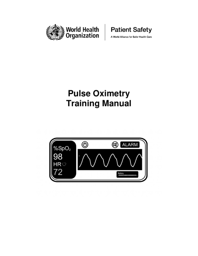
Pulse Oximetry
Training Manual
WHO Library Cataloguing-in-Publication Data
Pulse oximetry training manual.
1.Oximetry - instrumentation. 2.Oximetry - methods. 3.Anesthesia. 4.Monitoring, Physiologic methods. 5.Anoxia - prevention and control. 6.Safety management. 7.Surgical procedures,
Operative - standards. 8.Teaching materials. I.World Health Organization. II.WHO Patient Safety.
ISBN 978 92 4 150113 2
(NLM classification: WO 178)
© World Health Organization 2011
All rights reserved. Publications of the World Health Organization can be obtained from WHO
Press, World Health Organization, 20 Avenue Appia, 1211 Geneva 27, Switzerland (tel.: +41
22 791 3264; fax: +41 22 791 4857; e-mail: bookorders@who.int). Requests for permission to
reproduce or translate WHO publications – whether for sale or for noncommercial distribution
– should be addressed to WHO Press, at the above address (fax: +41 22 791 4806; e-mail:
permissions@who.int).
The designations employed and the presentation of the material in this publication do not
imply the expression of any opinion whatsoever on the part of the World Health Organization
concerning the legal status of any country, territory, city or area or of its authorities, or
concerning the delimitation of its frontiers or boundaries. Dotted lines on maps represent
approximate border lines for which there may not yet be full agreement.
The mention of specific companies or of certain manufacturers’ products does not imply that
they are endorsed or recommended by the World Health Organization in preference to others
of a similar nature that are not mentioned. Errors and omissions excepted, the names of
proprietary products are distinguished by initial capital letters.
All reasonable precautions have been taken by the World Health Organization to verify the
information contained in this publication. However, the published material is being distributed
without warranty of any kind, either expressed or implied. The responsibility for the
interpretation and use of the material lies with the reader. In no event shall the World Health
Organization be liable for damages arising from its use.
2
TABLE OF CONTENTS
Page
The WHO Pulse Oximetry Training Manual
4
Glossary of terms
6
Understanding the Physiology of Oxygen Transport
o Pulse Oximeter Quiz 1
o Oxygen
o Oxygen transport to the tissues
o How much oxygen does the blood carry?
o What is Oxygen Saturation?
7
Knowing the Pulse Oximeter
o Pulse Oximeter Quiz 2
o What does the pulse oximeter measure?
o The Pulse Oximeter monitor
o The Pulse Oximeter probe
o Practical use of the Pulse Oximeter
o What do the alarms tell you?
o What factors can interfere with the oximeter readings?
o What is not measured by the pulse oximeter?
9
Understanding How Oxygen Desaturation Occurs
o Pulse Oximeter Quiz 3
o Causes of hypoxia during anaesthesia
o What should be done when the saturation falls
o Management plan for Sp02 below 94%
o Actions to be taken when SpO2 is 94% and below
o Pulse Oximeter Quiz 4
o Pulse Oximeter Quiz 5
13
Appendix
Further readings and the haemoglobin oxygen dissociation curve
22
3
THE WHO PULSE OXIMETRY TRAINING MANUAL
Welcome to the World Health Organization pulse oximeter training manual. WHO has recently
introduced the WHO Surgical Safety Checklist as part of the Safe Surgery Saves Lives initiative.
Measures to improve anaesthesia safety are integral to the programme. In many countries pulse
oximetry is mandatory for monitoring patients during anaesthesia.
Although pulse oximetry is a simple and reliable technology that can detect low levels of oxygen in the
blood, it is only effective if the anaesthesia provider understands how an oximeter works and what to
do when hypoxia is detected. This manual describes a simple plan to respond to this situation, and
explains how oximeters work and how to use them.
The manual contains essential information for all anaesthesia providers who are not experienced in
using pulse oximetry and would be useful reading for all members of the theatre team.
The content of this manual can be studied on its own or can be taught in a classroom. Additional
learning materials about pulse oximetry and information on the WHO Surgical Safety Checklist can be
freely obtained at http://www.who.int/patientsafety/safesurgery/pulse_oximetry/en/index.html and at
http://www.who.int/patientsafety/safesurgery/en/index.html respectively. The material may be freely
distributed for educational purposes.
4
ACKNOWLEDGEMENTS
William Berry, MD, MPH
Harvard School of Public Health, Boston, USA
Gonzalo Barreiro MD
Sanatorio Americano, Montevideo, Uruguay
Gerald Dziekan MD, MSc
World Health Organization, Geneva, Switzerland
Angela Enright MB, FRCPC (Editor)
Royal Jubilee Hospital, Victoria, Canada
Peter Evans
Special Advisor
Luke Funk MD, MPH
Harvard School of Public Health, Boston, USA
Atul Gawande MD MPH
Harvard School of Public Health, Boston, USA
Iain H Wilson MB ChB, FRCA
Royal Devon and Exeter NHS Foundation Trust, Exeter, UK
Robert J McDougall MBBS, FANZCA, GradCetHlthProfEd
Royal Children's Hospital, Parkville, Victoria, Australia
Alan Merry ONZM, FANZCA, FFPMANZCA, FRCA, Hon FFFLM
University of Auckland, New Zealand
Florian Nuevo MD, DPBA, FPBCA
University of Santo Tomas, Manila, Philippines
Rafael Ortega MD
Boston University Medical Center, Boston, USA
Michael Scott MB ChB, FRCP, FRCA
Royal Surrey County Hospital, Guildford, UK
Stephen Ttendo MB ChB, MMed (Anaesth)
Mbarara University of Science and Technology, Mbarara, Uganda
Isabeau Walker BSc MB BChir FRCA (Editor)
Great Ormond Street Hospital NHS Trust, London, UK
David J Wilkinson MBBS, PGDip(AP), FFARCS
St Bartholomew's Hospital, London, UK
The editors would also like to acknowledge the assistance of Drs Regina Aleeva (Russia), Yoo Kuen
Chan (Malaysia), Sarah Hodges (Uganda), Vjacheslav Istomin (Russia), Fawaz Kateb (Syria), Mikhail
Kirov (Russia), Sergey Komarov (Russia), Konstantin Lebedinskiy (Russia), Jin Liu (China), Christine
Manning (Canada), Isabelle Murat (France), Haydn Perndt (Australia), Dmitry Shestakov (Russia),
Alexey Smetkin (Russia), Olaitan Soyannwo (Nigeria), Marina Venchikova (Russia), Lize Xiong
(China) and Buwei Yu (China).
5
GLOSSARY OF TERMS
Anaphylaxis
A severe life threatening allergic reaction to a drug or
other substance such as latex in surgical gloves
Arrhythmia
An abnormal heart rhythm
Atelectasis
Partial or total collapse of a lung or segment of a lung
which has been previously expanded
Bradycardia
A heart rate that is too slow for the patient. Adults less
than 60 beats / min; children according to age – see
page 9 of this manual
Capnograph
A monitor that detects the amount of carbon dioxide in
each breath
Cyanosis
Dusky blue appearance of the skin, tongue or mucous
membranes due to a low level of oxygenated
haemoglobin in blood vessels near the skin surface
Desaturated haemoglobin
Haemoglobin without attached oxygen
Hypotension
Low blood pressure
Hypothermia
Low body temperature (less than 36°C)
Hypoventilation
Breathing at a rate and/or depth that is less than
required
Hypovolaemia
Reduced blood volume
Hypoxia
Abnormally low levels of oxygen in the body
Microprocessor
A mini-computer that can calculate readings of pulse
rate and peripheral haemoglobin saturation from signals
detected by the probe
Oesophageal intubation
A tracheal tube that is incorrectly inserted into the
oesophagus
Oximeter / Oximetry
Pulse oximeter /Pulse oximetry
A device that can detect a pulsatile signal in an extremity
such as the finger or toe and can calculate the amount
of oxygenated haemoglobin and the pulse rate
Pneumothorax
Lung collapse caused by air leaking from the lung,
usually following trauma. Air enters the space outside
the lung (pleural space) and stops the lung from
expanding (also see Tension pneumothorax)
Pyrexia
Raised body temperature (greater than 37°C)
Tension pneumothorax
In a tension pneumothorax, the pressure in the pleural
space is very high, the patient has severe breathing
difficulties and distortion of the heart may cause cardiac
arrest
Vasopressors
Drugs such as adrenaline, ephedrine or phenylephrine
that raise the blood pressure by causing constriction of
blood vessels or increased cardiac output
6
UNDERSTANDING THE PHYSIOLOGY OF OXYGEN TRANSPORT
PULSE OXIMETER QUIZ 1
Before reading the manual, we would like you to assess your knowledge about pulse oximetry.
The correct answers are in the next section.
1. How is oxygen transported from the atmosphere to the tissues?
2. What is the normal oxygen saturation in arterial blood?
3. What is preoxygenation?
4. A patient undergoing general anaesthesia for hernia repair has an oxygen saturation of 82%
during surgery. Is this reading high or low? Is any action required?
__________________________________________________________________________
OXYGEN
Human beings depend on oxygen for life. All organs require oxygen for metabolism but the brain and
heart are particularly sensitive to a lack of oxygen. Shortage of oxygen in the body is called hypoxia.
A serious shortage of oxygen for a few minutes is fatal.
During anaesthesia, patients’ airways may become obstructed, their breathing may become
depressed, their circulation may be affected by blood loss or an abnormal heart rhythm or the
anaesthetic equipment may develop a problem such as an accidental disconnection or obstruction of
the breathing circuit. These factors can result in a reduction of oxygen delivery to the tissues which, if
not managed correctly, could lead to injury or death. The earlier the anaesthesia provider detects a
problem, the sooner it can be treated so that no harm comes to the patient.
OXYGEN TRANSPORT TO THE TISSUES
Oxygen is carried around the body attached to an iron-containing protein called haemoglobin, (Hb)
contained in red blood cells. After oxygen is breathed into the lungs, it combines with the
haemoglobin in red blood cells as they pass through the pulmonary capillaries. The heart pumps
blood continuously around the body to deliver oxygen to the tissues.
There are five important things that must happen in order to deliver enough oxygen to the tissues:
•
Oxygen must be breathed in (or inspired) from the air or anaesthesia circuit into the lungs.
•
Oxygen must pass from the air spaces in the lung (called the alveoli) to the blood. This is called
alveolar gas exchange.
•
The blood must contain enough haemoglobin to carry sufficient oxygen to the tissues.
•
The heart must be able to pump enough blood to the tissues to meet the patient’s oxygen
requirements.
•
The volume of blood in the circulation must be adequate to ensure oxygenated blood is
distributed to all the tissues.
HOW MUCH OXYGEN DOES THE BLOOD CARRY?
In a patient who is in good health:
•
Each gram of haemoglobin combines with 1.34 ml of oxygen. Therefore, in blood with a normal
haemoglobin concentration of 15g/dl, 100 ml of blood carries approximately 20 ml of oxygen
combined with haemoglobin. In addition, a small quantity of oxygen is dissolved in the blood.
•
The heart normally pumps approximately 5000 ml of blood per minute to the tissues in an
average sized adult. This delivers about 1000 ml of oxygen to the tissues per minute.
•
The cells in the tissues extract oxygen from the blood for metabolism, normally around 250ml of
oxygen per minute. This means that if there is no oxygen being exchanged in the lung, there is
only enough oxygen stored in the blood for around 3 minutes (only 75% of the oxygen carried by
7
the haemoglobin is available to the tissues).
•
Breathing 100% oxygen prior to induction of anaesthesia (preoxygenation) increases the oxygen
stores in the lungs. If a patient stops breathing and is not ventilated, the amount of oxygen in the
lungs will rapidly diminish. If the patient has been given 100% oxygen to breathe for several
minutes prior to induction of anaesthesia, the increased oxygen reservoir will supply much
needed oxygen, adding potentially life-saving minutes. There are many situations where this may
be important. One example is in the pregnant mother where the enlarged uterus reduces lung
volume and the metabolic demands are increased by the foetus. Another example is in young
children who have small lung volumes and high metabolic demands. They can use up oxygen
very quickly and can sometimes be resistant to efforts to preoxygenate them.
•
Anaemic patients have lower levels of haemoglobin and are therefore unable to carry as much
oxygen in the blood. At a haemoglobin concentration of less than 6g/dl, delivery of oxygen to the
tissues may become too low to meet the metabolic demands. Patients who suffer major blood
loss during surgery and become acutely anaemic should be given 100% oxygen to breathe. This
will increase the amount of dissolved oxygen in the blood and will improve tissue oxygen delivery
by a small amount. Blood transfusion may be life saving.
WHAT IS OXYGEN SATURATION?
Red blood cells contain haemoglobin. One molecule of haemoglobin can carry up to four molecules
of oxygen after which it is described as “saturated” with oxygen. If all the binding sites on the
haemoglobin molecule are carrying oxygen, the haemoglobin is said to have a saturation of 100%.
Most of the haemoglobin in blood combines with oxygen as it passes through the lungs. A healthy
individual with normal lungs, breathing air at sea level, will have an arterial oxygen saturation of 95%
– 100%. Extremes of altitude will affect these numbers. Venous blood that is collected from the
tissues contains less oxygen and normally has a saturation of around 75%. (See appendix 1 for more
details about this).
Arterial blood looks bright red whilst venous blood looks dark red. The difference in colour is due to
the difference in haemoglobin saturation. When patients are well saturated, their tongues and lips
appear pink in colour; when they are desaturated, they appear blue. This is called cyanosis. It can be
difficult to see cyanosis clinically, particularly in a dark skinned patient. You may not notice this sign
until the oxygen saturation is less than 90%. Detecting cyanosis is even more difficult in a poorly lit
operating theatre.
Cyanosis is only visible when the deoxygenated haemoglobin concentration is greater than 5 g/dl. A
severely anaemic patient may not appear cyanosed even when extremely hypoxic as there is very
little haemoglobin circulating through the tissues.
During anaesthesia the oxygen saturation should always be 95 - 100%. If the oxygen saturation is
94% or lower, the patient is hypoxic and needs to be treated quickly. A saturation of less than 90%
is a clinical emergency.
Learning point: It is difficult to detect cyanosis clinically until the saturation is <90%.
A patient who is severely anaemic may not appear cyanosed, even if the oxygen
saturation is very low.
8
KNOWING THE PULSE OXIMETER
PULSE OXIMETER QUIZ 2:
The correct answers are in the next section.
1. What two things does a pulse oximeter measure?
2. What is displayed on a pulse oximeter screen?
3. An oximeter probe has two parts. What are they?
__________________________________________________________________________
WHAT DOES A PULSE OXIMETER MEASURE?
There are TWO numerical values obtained from the pulse oximeter monitor:
•
The oxygen saturation of haemoglobin in arterial blood. The value of the oxygen saturation
is given together with an audible signal that varies in pitch depending on the oxygen saturation. A
falling pitch indicates falling oxygen saturation. Since the oximeter detects the saturation
peripherally on a finger, toe or ear, the result is recorded as the peripheral oxygen saturation,
described as SpO2.
•
The pulse rate in beats per minute, averaged over 5 to 20 seconds. Some oximeters display a
pulse waveform or indicator that illustrates the strength of the pulse being detected. This display
indicates how well the tissues are perfused. The signal strength falls if the circulation becomes
inadequate.
Learning point: A pulse oximeter is an early-warning device.
A pulse oximeter continuously measures the level of oxygen saturation of
haemoglobin in the arterial blood. It can detect hypoxia much sooner than the
anaesthesia provider can see clinical signs of hypoxia such as cyanosis. This ability
to provide an early warning has made the pulse oximeter essential for safe
anaesthesia.
THE PULSE OXIMETER:
A pulse oximeter consists of the monitor containing the batteries and display, and the probe that
senses the pulse.
This picture shows a pulse oximeter. The screen
shows that the Sp02 is 98% and the pulse rate is
72 beats per minute.
THE PULSE OXIMETER MONITOR
The monitor contains the microprocessor and
display. The display shows the oxygen saturation, the pulse rate and the waveform detected by the
sensor. The monitor is connected to the patient via the probe.
During use, the monitor updates its calculations regularly to give an immediate reading of oxygen
saturation and pulse rate. The pulse indicator is continuously displayed to give information about the
9
circulation. The audible beep changes pitch with the value of oxygen saturation and is an important
safety feature. The pitch drops as the saturation falls and rises as it recovers. This allows you to
hear changes in the oxygen saturation immediately, without having to look at the monitor all the time.
The monitor is delicate. It is sensitive to rough handling and excessive heat and can be damaged by
spilling fluids on it. The monitor can be cleaned by gently wiping with a damp cloth. When not in use,
it should be connected to an electrical supply to ensure that the battery is fully charged.
THE PULSE OXIMETER PROBE
The oximeter probe consists of two parts, the light emitting diodes (LEDs) and a light detector (called
a photo-detector). Beams of light are shone through the tissues from one side of the probe to the
other. The blood and tissues absorb some of the light emitted by the probe. The light absorbed by
the blood varies with the oxygen saturation of haemoglobin. The photo-detector detects the light
transmitted as the blood pulses through the tissues and the microprocessor calculates a value for the
oxygen saturation (SpO2).
In order for the pulse oximeter to function, the probe must be placed where a pulse can be detected.
The LEDs must face the light detector in order to detect the light as it passes through the tissues.
The probe emits a red light when the machine is switched on; check that you can see this light to
make sure the probe is working properly.
Probes are designed for use on the finger, toe or ear lobe. They are of different types as shown in the
diagram. Hinged probes are the most popular, but are easily damaged. Rubber probes are the most
robust. The wrap around design may constrict the blood flow through the finger if put on too tightly.
Ear probes are lightweight and are useful in children or if the patient is very vasoconstricted. Small
probes have been designed for children but an adult hinged probe may be used on the thumb or big
toe of a child. For finger or toe probes, the manufacturer marks the correct orientation of the nail bed
on the probe.
The oximeter probe is the most delicate part of a pulse oximeter and is easily damaged. Handle the
probe carefully and never leave it in a place where it could be dropped on the floor. The probe
connects to the oximeter using a connector with a series of very fine pins that can be easily damaged
– see diagram. Always align the connector correctly before attempting to insert it into the monitor.
Never pull the probe from the machine by pulling on the cable; always grasp the connector firmly
between finger and thumb.
Hinged finger probe showing connector
Rubber finger probes and ear sensor
that can only be connected to the oximeter
one way by aligning the gap on the probe connector
with the corresponding notch on the machine.
10
When not in use, the oximeter probe cable may be loosely coiled for storage or carrying, but should
not be coiled too tightly as this will damage the wires inside the cable. The lens and detector should
be kept clean. Use soapy water or an alcohol soaked swab to gently clean dust, dirt or blood from
the probe.
Learning point: In order to get a satisfactory reading the probe must be emitting a red
light and must be correctly positioned to detect pulsatile blood flow.
PRACTICAL USE OF THE PULSE OXIMETER
•
•
•
•
•
•
•
•
•
•
•
•
Turn the pulse oximeter on: it will go through internal calibration and checks.
Select the appropriate probe with particular attention to correct sizing and where it will go (usually
finger, toe or ear). If used on a finger or toe, make sure the area is clean. Remove any nail
varnish.
Connect the probe to the pulse oximeter.
Position the probe carefully; make sure it fits easily without being too loose or too tight.
If possible, avoid the arm being used for blood pressure monitoring as cuff inflation will interrupt
the pulse oximeter signal.
Allow several seconds for the pulse oximeter to detect the pulse and calculate the oxygen
saturation.
Look for the displayed pulse indicator that shows that the machine has detected a pulse. Without
a pulse signal, any readings are meaningless.
Once the unit has detected a good pulse, the oxygen saturation and pulse rate will be displayed.
Like all machines, oximeters may occasionally give a false reading - if in doubt, rely on your
clinical judgement, rather than the machine.
The function of the oximeter probe can be checked by placing it on your own finger.
Adjust the volume of the audible pulse beep to a comfortable level for your theatre – never use on
silent.
Always make sure the alarms are on.
If no signal is obtained on the oximeter after the probe has been placed on a finger, check the
following:
•
•
Is the probe working and correctly positioned? Try another location.
Does the patient have poor perfusion?
o Check for low cardiac output especially due to hypovolemia, cardiac problems or septic
shock. If hypotension is present, resuscitation of the patient is required immediately. The
signal will improve when the clinical condition of the patient improves.
o Check the temperature of the patient. If the patient or the limb is cold, gentle rubbing of
the digit or ear lobe may restore a signal.
Tip: If you are uncertain that the probe is working properly, check it by testing it on
your own finger.
11
WHAT DO THE ALARMS ON A PULSE OXIMETER TELL YOU?
Alarms alert the anaesthetist to clinical problems. The alarms are as follows:
•
•
•
•
Low saturation emergency (hypoxia) i.e. SpO2 <90%
No pulse detected
Low pulse rate
High pulse rate
Low saturation alarm. The oxygen saturation in healthy patients of any age should be 95% or
above.
Learning point: During anaesthesia the SpO2 should be 95% or above. If SpO2 is 94%
or below, the patient must be assessed quickly to identify and treat the cause.
SpO2 OF < 90% IS A CLINICAL EMERGENCY AND SHOULD BE TREATED URGENTLY.
‘No pulse detected’ alarm is commonly caused by the probe coming off the finger, but it may also
be triggered if the patient is hypotensive, hypovolaemic, or has suffered a cardiac arrest. Check the
probe site quickly and then assess the patient - ABC.
Pulse rate alarms are useful to let the anaesthetist know that the heart is beating too fast or too slow.
However, alert anaesthetists will have already noticed the abnormal heart rate before the alarms
sound. Children normally have higher heart rates than adults, but the same oxygen saturation – see
table below.
Age
Normal Heart Rate
Normal
(SpO2)
oxygen
saturation
Newborn – 2 years
2-10 years
10 years -adult
100 - 180
60 - 140
50 - 100
All patients should have an
SpO2 of 95% or above during
anaesthesia or during recovery
from anaesthesia*
* Exception: premature babies receiving oxygen therapy in the neonatal ICU should have an SpO2 between 89-94% to avoid
toxicity to the retina. During surgery the oxygen saturation of premature babies should be maintained at >95%, as with all other
patients.
Light anaesthesia, inadequate pain relief, atropine, ketamine, hypovolaemia, fever, or arrhythmia may
trigger the high pulse alarm. The low pulse alarm may be triggered by bradycardia secondary to
vagal stimulation due to e.g. peritoneal retraction, the oculocardiac reflex or intubation (particularly in
babies) or from deep anaesthesia (particularly halothane) or severe hypoxia. A highly trained athlete
or a patient who is taking ß-blockers may have a slow pulse rate.
12
WHAT FACTORS CAN INTERFERE WITH THE PULSE OXIMETER READING?
Several factors can interfere with the correct function of a pulse oximeter including:
•
•
•
•
•
Light – bright light (such as the operating theatre light or sunlight) directly on the probe may affect
the reading. Shield the probe from direct light.
Shivering – movement may make it difficult for the probe to pick up a signal.
Pulse volume – the oximeter only detects pulsatile flow. When the blood pressure is low due to
hypovolaemic shock or the cardiac output is low or the patient has an arrhythmia, the pulse may
be very weak and the oximeter may not be able to detect a signal
Vasoconstriction reduces blood flow to the peripheries. The oximeter may fail to detect a signal
if the patient is very cold and peripherally vasoconstricted.
Carbon monoxide poisoning may give a falsely high saturation reading. Carbon monoxide
binds very well to haemoglobin and displaces oxygen to form a bright red compound called
carboxyhaemoglobin. This is only an issue in patients following smoke inhalation from a fire.
Learning point: Hypovolaemia is the most common cause of a weak pulse oximeter
signal during anaesthesia. Hypothermia should also be considered.
WHAT IS NOT MEASURED BY A PULSE OXIMETER?
A pulse oximeter does not give direct information about respiratory rate, tidal volume, cardiac output
or blood pressure. However, it does so indirectly and if these factors lead to desaturation, this will be
detected by the pulse oximeter.
Learning point: Supplemental oxygen is often essential during anaesthesia. However,
be aware that it can mask the effects of hypoventilation on oxygen saturation. Clinical
vigilance will be necessary to ensure that ventilation is adequate especially if a
capnograph is not available.
Pulse oximeters function normally in anaemic patients. In an extremely anaemic patient, the oxygen
saturation will still be normal (95%-100%), but there may not be enough haemoglobin to carry
sufficient oxygen to the tissues. In cases of severe anaemia, the patient should be given 100%
oxygen to breathe during anaesthesia to try to improve tissue oxygen delivery by increasing the
amount of dissolved oxygen in the blood.
UNDERSTANDING HOW OXYGEN DESATURATION OCCURS
PULSE OXIMETER QUIZ 3
The causes of hypoxia during anaesthesia can be attributed to problems with the Airway, Breathing,
Circulation, Drugs or Equipment. By remembering to check the patient in this order, most of the
causes of hypoxia can be identified and treated.
Using the headings below, consider what could go wrong during anaesthesia to cause hypoxia.
Compare your answers to the table on the next page.
Airway
Breathing
Circulation
Drugs
Equipment
What do you think is the most common cause of hypoxia in theatre or recovery?
13
CAUSES OF HYPOXIA DURING ANAESTHESIA:
The causes of hypoxia during anaesthesia are summarised in the table below. Airway obstruction
is the most common cause of hypoxia.
Causes of hypoxia in theatre – ‘ABCDE’
Source of problem
Common problem
A. AIRWAY
•
•
•
An obstructed airway prevents oxygen from reaching the lungs
The tracheal tube can be misplaced e.g. in the oesophagus
Aspirated vomit can block the airway
B. BREATHING
•
Inadequate breathing prevents enough oxygen from reaching the
alveoli.
Severe bronchospasm may not allow enough oxygen to reach the
lungs nor carbon dioxide to be removed from the lungs.
A pneumothorax may cause the affected lung to collapse
High spinal anaesthesia may cause inadequate breathing
•
•
•
C. CIRCULATION
•
•
Circulatory failure prevents oxygen from being transported to the
tissues
Common causes include hypovolemia, abnormal heart rhythm or
cardiac failure
D. DRUGS
•
•
•
•
Deep anaesthesia may depress breathing and circulation
Many anaesthetic drugs cause a drop in blood pressure
Muscle relaxants paralyse the muscles of respiration
Anaphylaxis can cause bronchospasm and low cardiac output
E. EQUIPMENT
•
Problems with the anaesthetic equipment include disconnection or
obstruction of the breathing circuit
Problems with oxygen supply include an empty cylinder or an
inadequately functioning oxygen concentrator
Problems with the monitoring equipment include battery failure in the
oximeter or a faulty probe
•
•
Learning point: When hypoxia occurs, it is essential to decide whether the problem is
with the patient or the equipment. After a quick check of the common patient
problems, make sure the equipment is working properly. Always have a self-inflating
bag available in case of problems with the breathing circuit.
WHAT SHOULD BE DONE WHEN THE SATURATION FALLS?
During anaesthesia, low oxygen saturations must be treated immediately and appropriately. The
patient may become hypoxic at any time during induction, maintenance or emergence from
anaesthesia. The appropriate response is to administer 100% oxygen, make sure that ventilation is
adequate by using hand ventilation and then correct the factor that is causing the patient to become
hypoxic. For example, if the patient has an obstructed airway and is unable to breathe oxygen into
the lungs, the problem will only be cured when the airway is cleared.
14
Whenever the patient has low oxygen saturations, administer high flow oxygen and consider
‘ABCDE’:
•
•
•
•
•
A - airway clear?
B - breathing adequately?
C - circulation working normally?
D - drugs causing a problem?
E - equipment working properly?
You must respond to hypoxia immediately by giving more oxygen, ensuring adequate ventilation by
hand, calling for help, and proceeding through the ‘ABCDE’ sequence. Treat each element of the
sequence as you check it. After you have been through all the checks for the first time, go back and
recheck them until you are satisfied that the patient’s condition has improved. WHO has put this into
a chart (below) to help you remember what to look for in a logical sequence. In an emergency, there
may not be time to read through the protocol. You should ask a colleague to read it aloud for you to
make sure that you have not forgotten anything.
Learning point: If SpO2 is <94%, give 100% oxygen, hand ventilate, consider ABCDE
15
MANAGEMENT OF SPO2 < 94%
16
ACTIONS TO TAKE IF THE OXYGEN SATURATION IS 94% OR BELOW
If the oxygen saturation is 94% or below, you should administer 100% oxygen, ventilate by hand,
consider whether the problem is with the patient or the equipment, then move through the plan
‘ABCDE’, assessing each factor and correcting it immediately as you go.
Oxygen
Administer high flow oxygen if SpO2 is <94%
A – Is the airway clear?
•
•
•
•
Is the patient breathing quietly without signs of obstruction?
Are there signs of laryngospasm? (mild laryngospasm – high pitched inspiratory noise; severe
laryngospasm – silent, no gas passes between the vocal cords)
Is there any vomit or blood in the airway?
Is the tracheal tube in the right place?
Action
•
•
•
•
•
Ensure that there is no obstruction.
o If breathing via a facemask - chin lift, jaw thrust
Consider an oropharyngeal or nasopharyngeal airway
Check for laryngospasm and treat if necessary
o Check the tracheal tube/LMA - if any doubt about the position, remove and use a
facemask.
Suction the airway to clear secretions.
Consider waking the patient up if you have difficulty maintaining the airway immediately after
induction of anaesthesia.
Consider intubation.
If you ‘can’t intubate, can’t ventilate’, an emergency surgical airway is required.
Airway obstruction is the most common cause of hypoxia in theatre. Airway obstruction is a clinical
diagnosis and must be acted upon swiftly. Unrecognised oesophageal intubation is a major cause of
anaesthesia morbidity and mortality. An intubated patient who has been previously well saturated
may become hypoxic if the tracheal tube becomes displaced, kinked or obstructed by secretions.
Check the tracheal tube and - ‘If in doubt, take it out’
B - Is the patient breathing adequately?
Look, listen and feel:
• Are the chest movement and tidal volume adequate?
• Listen to both lungs – is there normal bilateral air entry? Are the breath sounds normal? Any
wheeze or added sounds?
• Is the chest movement symmetrical?
• Is anaesthesia causing respiratory depression?
• Is a high spinal causing respiratory distress?
Bronchospasm, lung consolidation/collapse, lung trauma, pulmonary oedema or pneumothorax may
prevent oxygen from getting into the alveoli to combine with haemoglobin. Drugs such as opioids,
poorly reversed neuromuscular blocking agents or deep volatile anaesthesia may depress breathing.
A high spinal anaesthetic may paralyse the muscles of respiration. In an infant, stomach distension
from facemask ventilation may splint the diaphragm and interfere with breathing. The treatment
should address the specific problem.
17
Action
• Assist ventilation with adequate tidal volumes to expand both lungs until the problem is diagnosed
and treated appropriately.
• If there is sufficient time, consider a chest X-ray to aid diagnosis.
The patient should be ventilated via a facemask, LMA or tracheal tube if respiration is inadequate.
This will rapidly reverse hypoventilation due to drugs or a high spinal and cause a collapsed lung to
re-expand. The lower airway should be suctioned with suction catheters to remove any secretions. A
nasogastric tube should be passed to relieve stomach distension.
A pneumothorax may occur following trauma, central line insertion or a supraclavicular brachial
plexus block. It should be suspected if there is reduced air entry on the affected side. In thin patients
a hollow note on percussion may also be detected. A chest X-ray is diagnostic. A chest drain should
be inserted to prevent the pneumothorax from worsening. When there is associated hypotension
(tension pneumothorax), the pneumothorax should be treated by emergency needle decompression
nd
through the 2 intercostal space in the mid-clavicular line without waiting for an X-ray. A definitive
chest drain should be inserted later. Always maintain a high index of suspicion in trauma cases.
C - Is the circulation normal?
•
•
•
•
•
•
•
Feel for a pulse and look for signs of life, including active bleeding from the surgical wound
Check the blood pressure
Check the peripheral perfusion and capillary refill time.
Observe for signs of excessive blood loss in the suction bottles or wound swabs
Is anaesthesia too deep? Is there a high spinal block?
Is venous return impaired by compression of the vena cava (gravid uterus, surgical compression)
Is the patient in septic or cardiogenic shock?
An inadequate circulation may be revealed by the pulse oximeter as a loss or reduction of pulsatile
waveform or difficulty obtaining a pulse signal.
Action
•
•
•
•
•
•
If the blood pressure is low, correct it
Check for hypovolaemia
Give IV fluids as appropriate (normal saline or blood as indicated)
Consider head down or leg up position, or in the pregnant mother, left lateral positioning.
Consider a vasoconstrictor such as ephedrine or phenylephrine
If the patient has suffered a cardiac arrest, commence cardiopulmonary resuscitation (CPR) and
consider reversible causes (4 H’s, 4T’s: Hypotension, Hypovolaemia, Hypoxia, Hypothermia;
Tension pneumothorax, Tamponade (cardiac), Toxic effects (deep anaesthesia, sepsis, drugs),
Thromboemboli (pulmonary embolism).
D – Drug effects
Check that all anaesthesia drugs are being given correctly.
• Excessive halothane (or other volatile agent) causes cardiac depression.
• Muscle relaxants will depress the ability to breathe if not reversed adequately at the end of
surgery.
• Opioids and other sedatives may depress breathing.
• Anaphylaxis causes cardiovascular collapse, often with bronchospasm and skin flushing (rash).
This may occur if the patient is given a drug, blood or artificial colloid solution that he/she is
allergic to. Some patients are allergic to latex rubber.
18
Action
•
•
Look for an adverse drug effect and treat as appropriate.
In anaphylaxis, stop administering the causative agent, ventilate with 100% oxygen, give
intravenous saline starting with a bolus of 10ml/kg, administer adrenaline and consider giving
steroids, bronchodilators and an antihistamine.
E - Is the equipment working properly?
•
•
Is there a problem with the oxygen delivery system to the patient?
Does the oximeter show an adequate pulse signal?
Action
•
•
•
•
•
Check for obstruction or disconnection of the breathing circuit or tracheal tube.
Check that the oxygen cylinder is not empty
Check that the oxygen concentrator is working properly
Check that the central hospital oxygen supply is working properly
Change the probe to another site; check that it is working properly by trying it on your own finger.
If it is felt that the anaesthesia equipment is faulty, use a self-inflating bag to ventilate the patient
with air while new equipment or oxygen supplies are obtained. If equipment is missing, mouth to
tracheal tube, or mouth-to-mouth ventilation may be lifesaving.
19
Quiz 4. Pulse Oximeter - demonstration
Demonstrate to the instructor or to a colleague:
1. How to charge the oximeter battery and store the accessories ready for clinical use.
2. How to select the most appropriate sensor for the patient.
3. How to apply the sensor to the patient correctly.
4. The battery condition indicator – what does the reading show?
5. How to switch on the monitor and describe the self-check routine.
6. The features of the main display.
7. The features of the waveform or pulse indicator.
8. How to adjust the alarm limits.
9. How to adjust the pulse sound volume.
10. How to turn the backlight on or off.
Answer the following two questions.
11. What conditions could cause inaccurate readings?
12. How should the sensor site be selected?
20
Quiz 5. Knowledge about pulse oximetry
Answer these questions about pulse oximetry – the answers are at the bottom of the page. More than
one answer may be correct.
1. The pulse oximeter measures:
a. Haemoglobin level in blood
b. The amount of oxygen contained in the blood
c. Percentage of haemoglobin saturated with oxygen
d. Pulse rate
e. Cardiac output
2. Which of the following (if any) statements is true about oximeter probes?
a. Ear probes tend to read higher than finger probes
b. Probes are expensive
c. The probe can be cleaned gently with soapy water
d. If a signal is not present, the probe is always faulty
e. Nail varnish does not affect probe function
3. Which of the following can cause false readings on a pulse oximeter?
a. Dark skinned patients
b. Fast pulse rates with normal blood pressures
c. Overhead lights shining on probes
d. Carbon monoxide poisoning
e. Oxygen treatment
4. Oxygen saturation:
a. Should always be 100% during anaesthesia
b. Is normally above 95% in a healthy 2-year-old
c. Is normally less than 93% in a 70-year-old
d. Only becomes seriously low when under 75%
e. Is not worth measuring during spinal anaesthesia for Caesarean section
5. The following may reduce the chance of a successful oximeter reading:
a.
b.
c.
d.
e.
Fever
Hypertension
Sickle cell disease
Arrhythmia
Hypovolaemia
The correct answers are
1.
2.
3.
4.
5.
c, d
b, c
c, d
b
d, e
21
APPENDIX: FURTHER READING AND THE HAEMOGLOBIN OXYGEN DISSOCIATION CURVE
This section contains extra information about the way haemoglobin functions and how SpO2 relates to
arterial blood gases. There are also a number of references for further reading which may be
accessed via the Internet.
Arterial blood gases and SpO2
As previously explained, the pulse oximeter measures the oxygen saturation of haemoglobin in
arterial blood. A blood gas analyser may be used to measure the oxygen content in a blood sample
(‘arterial blood gases’). The blood gas analyser describes the gas content as a partial pressure. It
measures the partial pressure of oxygen (PaO2) and carbon dioxide (PaCO2), the pH of the blood and
the bicarbonate concentration.
What is partial pressure? – The atmosphere is made up of a mixture of gases at a
pressure of one atmosphere, 101kPa or 760mmHg. Oxygen is 21% of the atmosphere and
the partial pressure of oxygen in air is 21kPa or 150mmHg. When blood is exposed to
gases, the gas crosses into the blood down the pressure gradient. The partial pressure of
oxygen and carbon dioxide in blood can be measured by analysing a sample of blood in a
‘blood-gas machine’ to assess the efficiency of oxygenation and ventilation. Oxygen
saturation measured with a pulse oximeter gives a more useful minute-to-minute
measurement of oxygenation, but gives no information about CO2 or pH.
22
THE HAEMOGLOBIN OXYGEN DISSOCIATION CURVE
The relationship between the partial pressure of oxygen and the oxygen saturation is shown by the
oxygen dissociation curve. As the partial pressure of oxygen in blood increases, so does the oxygen
saturation. The sigmoid shape of the oxygen dissociation curve reflects the cooperative interaction
between haemoglobin and oxygen molecules.
Some arterial gas analysers use the partial pressure of oxygen to estimate the haemoglobin
saturation from a computer in the analyser, but this measurement is not as accurate as measured by
a co-oximeter.
Gas exchange occurs in the lungs. The lungs are reloaded with fresh oxygen with each breath.
Oxygen at a high partial pressure (PaO2 13kPa or 100 mmHg) drives oxygen on to the haemoglobin
until 95 – 100% is saturated. Haemoglobin releases oxygen as the blood passes through the tissues.
The partial pressure of oxygen in blood returning from the tissues (mixed venous blood) is much lower
than in arterial blood (PaO2 5.3 kPa or 40mmHg).
The oxygen dissociation curve is initially steep, and then flattens out (sigmoid shape). The most
important aspect of the oxygen dissociation curve is that, as the oximeter reading falls below 90%, the
partial pressure of oxygen in the blood drops very rapidly and oxygen delivery to the tissues is
reduced and may lead to cardiac arrest. You must intervene swiftly if the oxygen saturation drops
below 90%.
Oxygen delivery to the
tissues falls rapidly if arterial
SaO2 is less than 90%
Arterial point. Blood fully
oxygenated: SaO2 100%, PaO2
13.3.kPa (100 mmHg)
Mixed venous point.
Deoxygenated blood returning
to the heart: SvO2 75%, PvO2
5.3 kPa (40 mmHg)
FURTHER READING ABOUT PULSE OXIMETRY
1. Fearnley SJ. Pulse Oximetry. Update in Anaesthesia
http://www.nda.ox.ac.uk/wfsa/html/u05/u05_003.htm
2. Hill E, Stoneham MD. Practical applications of pulse oximetry.
http://www.nda.ox.ac.uk/wfsa/html/u11/u1104_01.htm
3. Principles of pulse oximetry. http://www.oximeter.org/pulseox/principles.htm
23
24

