NEURONAL MODELING
Single-trial spike trains in parietal
cortex reveal discrete steps during
decision-making
Kenneth W. Latimer,1,2 Jacob L. Yates,1,2 Miriam L. R. Meister,2,3
Alexander C. Huk,1,2,4,5 Jonathan W. Pillow1,2,5,6*
Neurons in the macaque lateral intraparietal (LIP) area exhibit firing rates that appear to
ramp upward or downward during decision-making. These ramps are commonly assumed
to reflect the gradual accumulation of evidence toward a decision threshold. However,
the ramping in trial-averaged responses could instead arise from instantaneous jumps at
different times on different trials. We examined single-trial responses in LIP using
statistical methods for fitting and comparing latent dynamical spike-train models. We
compared models with latent spike rates governed by either continuous diffusion-to-bound
dynamics or discrete “stepping” dynamics. Roughly three-quarters of the choice-selective
neurons we recorded were better described by the stepping model. Moreover, the inferred
steps carried more information about the animal’s choice than spike counts.
R
(10, 11). Although decision-making at the behavioral level is well described as an accumulation
process (12, 13), whether the brain computes decisions through a direct neural correlate (ramping)
or a discrete implementation (stepping) remains a central, unresolved question in systems neuroscience.
We used advanced statistical methods to identify the single-trial dynamics governing spike
trains in the lateral intraparietal (LIP) area of
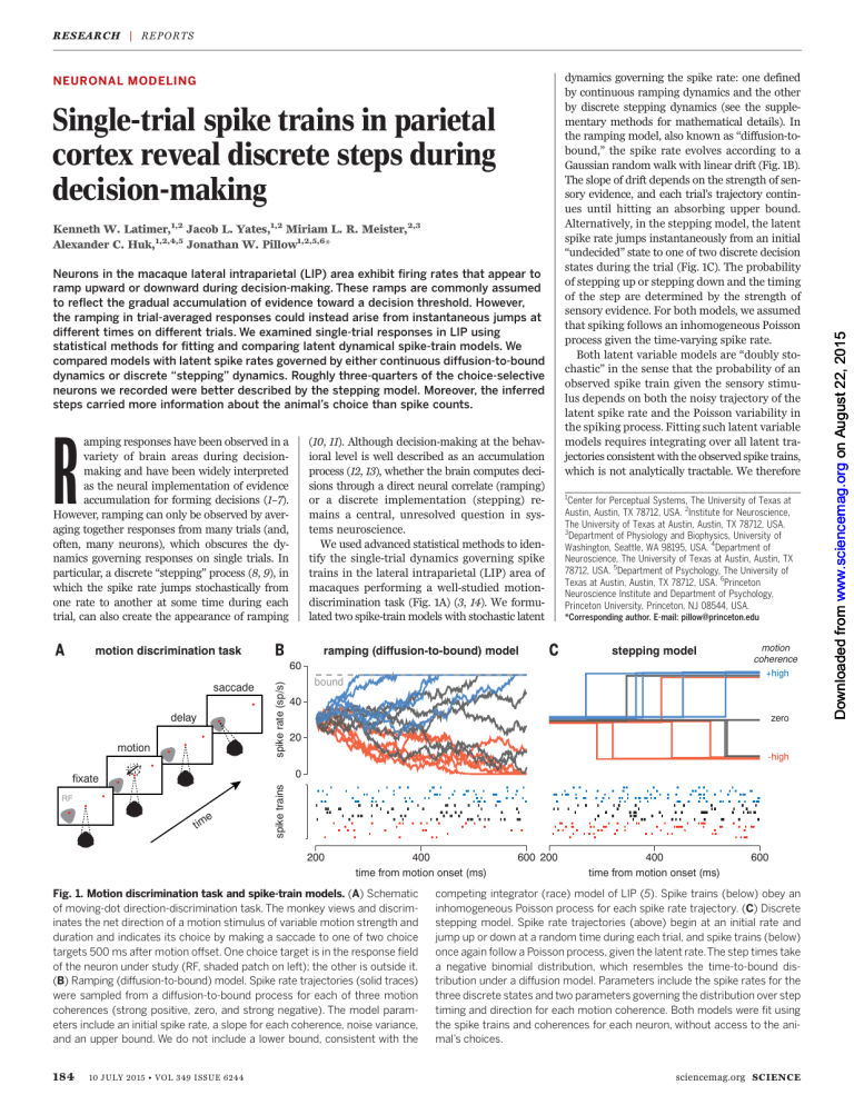
macaques performing a well-studied motiondiscrimination task (Fig. 1A) (3, 14). We formulated two spike-train models with stochastic latent
amping responses have been observed in a
variety of brain areas during decisionmaking and have been widely interpreted
as the neural implementation of evidence
accumulation for forming decisions (1–7).
However, ramping can only be observed by averaging together responses from many trials (and,
often, many neurons), which obscures the dynamics governing responses on single trials. In
particular, a discrete “stepping” process (8, 9), in
which the spike rate jumps stochastically from
one rate to another at some time during each
trial, can also create the appearance of ramping
ramping (diffusion-to-bound) model
motion discrimination task
dynamics governing the spike rate: one defined
by continuous ramping dynamics and the other
by discrete stepping dynamics (see the supplementary methods for mathematical details). In
the ramping model, also known as “diffusion-tobound,” the spike rate evolves according to a
Gaussian random walk with linear drift (Fig. 1B).
The slope of drift depends on the strength of sensory evidence, and each trial’s trajectory continues until hitting an absorbing upper bound.
Alternatively, in the stepping model, the latent
spike rate jumps instantaneously from an initial
“undecided” state to one of two discrete decision
states during the trial (Fig. 1C). The probability
of stepping up or stepping down and the timing
of the step are determined by the strength of
sensory evidence. For both models, we assumed
that spiking follows an inhomogeneous Poisson
process given the time-varying spike rate.
Both latent variable models are “doubly stochastic” in the sense that the probability of an
observed spike train given the sensory stimulus depends on both the noisy trajectory of the
latent spike rate and the Poisson variability in
the spiking process. Fitting such latent variable
models requires integrating over all latent trajectories consistent with the observed spike trains,
which is not analytically tractable. We therefore
1
Center for Perceptual Systems, The University of Texas at
Austin, Austin, TX 78712, USA. 2Institute for Neuroscience,
The University of Texas at Austin, Austin, TX 78712, USA.
3
Department of Physiology and Biophysics, University of
Washington, Seattle, WA 98195, USA. 4Department of
Neuroscience, The University of Texas at Austin, Austin, TX
78712, USA. 5Department of Psychology, The University of
Texas at Austin, Austin, TX 78712, USA. 6Princeton
Neuroscience Institute and Department of Psychology,
Princeton University, Princeton, NJ 08544, USA.
*Corresponding author. E-mail: pillow@princeton.edu
stepping model
saccade
delay
motion
spike rate (sp/s)
60
bound
40
zero
20
-high
RF
e
tim
spike trains
0
fixate
200
400
time from motion onset (ms)
Fig. 1. Motion discrimination task and spike-train models. (A) Schematic
of moving-dot direction-discrimination task. The monkey views and discriminates the net direction of a motion stimulus of variable motion strength and
duration and indicates its choice by making a saccade to one of two choice
targets 500 ms after motion offset. One choice target is in the response field
of the neuron under study (RF, shaded patch on left); the other is outside it.
(B) Ramping (diffusion-to-bound) model. Spike rate trajectories (solid traces)
were sampled from a diffusion-to-bound process for each of three motion
coherences (strong positive, zero, and strong negative). The model parameters include an initial spike rate, a slope for each coherence, noise variance,
and an upper bound. We do not include a lower bound, consistent with the
184
motion
coherence
+high
10 JULY 2015 • VOL 349 ISSUE 6244
600 200
400
time from motion onset (ms)
600
competing integrator (race) model of LIP (5). Spike trains (below) obey an
inhomogeneous Poisson process for each spike rate trajectory. (C) Discrete
stepping model. Spike rate trajectories (above) begin at an initial rate and
jump up or down at a random time during each trial, and spike trains (below)
once again follow a Poisson process, given the latent rate.The step times take
a negative binomial distribution, which resembles the time-to-bound distribution under a diffusion model. Parameters include the spike rates for the
three discrete states and two parameters governing the distribution over step
timing and direction for each motion coherence. Both models were fit using
the spike trains and coherences for each neuron, without access to the animal’s choices.
sciencemag.org SCIENCE
Downloaded from www.sciencemag.org on August 22, 2015
R ES E A RC H | R E PO R TS
RE S EAR CH | R E P O R T S
developed sampling-based Markov chain Monte
Carlo methods, which provide samples from
the posterior distribution over model parameters and allow us to perform Bayesian model
comparison.
We focused on a population of 40 neurons
with highly choice-selective responses that exhibited ramping in their average responses (14),
typically increasing during trials in which the
monkey eventually chose the target inside the
response field (RF) of the neuron and decreasing when the monkey chose the target outside
the RF. We fit each neuron with both ramping
and stepping models, using the spike-train data
from 200 ms after motion onset (15) until 200 ms
after motion offset (300 ms before the monkey
received the “go” signal). Figure 2A shows the
raster of spike trains from an example LIP neuron plotted in two different ways: first, aligned
to the time of motion stimulus onset (left); and
second, aligned to the step time inferred under
the stepping model (right). The traditional raster and peristimulus time histogram (PSTH) at
left show that the average response ramps upward or downward depending on choice, as expected. The step-aligned raster at right, however,
shows that these data are also consistent with
discrete steplike transitions with variable timing
across trials. Additional panels show the distribution of step times inferred under the model
(Fig. 2B), and the dependence of step direction
(up or down) on the motion signal (Fig. 2C). Discrete steps in the instantaneous spike rate could
thus plausibly underlie the gradual ramping ac-
fraction of trials
step-aligned
out choice in choice
trials
stimulus-aligned
tivity seen in stimulus-aligned and averaged LIP
spike responses.
We applied the same analysis to the full set of
LIP neurons and observed similar structure in
step-aligned rasters (figs. S13 to S15). Figure 3A
shows population-averaged PSTHs computed
from stimulus-aligned (left) and step-aligned
responses, sorted by motion strength (middle),
or motion strength and step direction (right).
The middle and right plots show that spike rate
is effectively constant when spike trains are
aligned to the inferred step time on each trial.
The multiple step heights observed in the middle plot result from the fact that the proportion
of up and down steps varies with motion strength.
The right plot confirms that the firing rate,
once conditioned on stepping up or down, is
step up
step down
no step
15
out-RF choice
0
200
600
1000 -300
time from motion onset (ms)
0
300
time from step (ms)
Fig. 2. Model-based analysis of spike responses from an example LIP
neuron. (A) Spike rasters sorted by the monkey’s choice in or out of the RF
of the neuron under study (black, in-RF; gray, out-RF) and their associated
averages (PSTHs, below). (Left) Conventional stimulus-aligned rasters with
each trial aligned to the time of motion onset exhibit commonly observed
ramping in the PSTH. Blue and red triangles indicate the inferred time of an up
or down step on each trial under the fitted stepping model. Yellow triangles
600
probability of up step
spike rate (sp/s)
in-RF choice
model
0.2
0.1
0
45
30
inferred step times
0.3
data
200 450 700 950 1200
time after motion onset (ms)
1
0.75
0.5
0.25
0
-high -low zero +low +high
coherence
indicate that no step was found during the trial and are placed at the end of the
trial segment we analyzed (200 ms after motion offset). (Right) The same
spike trains aligned to the inferred step time for each trial. The estimated step
direction of the neuron does not always match the animal’s decision on each
trial. (B) The distribution of inferred step times shown in (A) (histogram) and
the distribution over step times under the fitted parameters (black trace).
(C) The probability of an up step for each coherence level. Error bars, 95% CIs.
population average
25
+low
20
zero
15
-low
10
-high
“up” step
“down” step
5
200
600
1000 -300
time from motion onset (ms)
0
300
600 -300
time from step (ms)
Fig. 3. Stepping model captures LIP responses. (A) Population average
PSTH sorted by motion coherence computed from spike trains: (Left) Aligned
to motion onset and sorted by motion strength. (Middle) Aligned to step
times inferred under the stepping model and sorted by motion strength.
(Right) Aligned to step times and sorted by both motion strength and inferred
step direction. Simulated results from the stepping model (dashed lines)
provide a close match to the real data under all types of alignment and
SCIENCE sciencemag.org
0
300
time from step (ms)
ramping
6
number of cells
30
spike rate (sp/s)
model comparison
motion
coherence
+high
600
stepping
4
2
0
-250
-50 -25
0
25
50
250
DIC difference
conditioning. (B) Quantitative model comparison using DIC reveals a superior
fit of the stepping model over the ramping model for the majority of cells
(31 out of 40). A DIC difference greater than T10 (gray region) is commonly
regarded as providing strong support for one model over the other (22).
We found substantially more cells with strong evidence for stepping over
ramping (25 cells versus 6 cells; median DIC difference = 22.1; sign test
P < 0.001).
10 JULY 2015 • VOL 349 ISSUE 6244
185
R ES E A RC H | R E PO R TS
independent of motion strength. Furthermore,
simulated spike responses, based on the fitted
stepping models, resemble the real data under
both kinds of alignment (dashed traces).
Although these analyses provide a visually compelling illustration of the plausibility of stepping
dynamics in LIP, they do not by themselves definitively rule out the ramping model (see fig. S16).
Using our latent variable models, we can formally
address this issue using statistical model comparison. Both models give a probability distribution over spike trains, and the model that better
represents the data should place more probability
mass over the observed spike trains. We compared the model fits using the deviance information criterion (DIC) (16), which integrates
over the entire posterior distribution of model
mean rate (sp/s)
simulation: stepping
data
30
parameters given the data, thereby taking into
account the uncertainty in the model fit as well
as the number of parameters in each model.
The stepping model provided a superior account of LIP responses for 78% (31 out of 40) of
the cells compared to the ramping model (Fig.
3B). The stepping model therefore not only accounts for the ramplike activity observed in averaged LIP responses, but its qualitative ability to
reveal step times is bolstered by quantitative superiority in accounting for the statistical structure of spike trains for a majority of LIP neurons.
The superiority was supported not just by DIC
but also by other model comparison metrics, such
as Bayes factors (fig. S1).
We subsequently examined how well the two
models account for the time-varying mean and
simulation: ramping
20
motion
coherence
+high
+low
zero
-low
-high
10
variance
30
20
10
200
450
700 200
450
700 200
time from motion onset (ms)
700
1
model-based CP
0.6
choice probability
450
0.55
0.5
200
450
700 -250
0
250
time from motion onset (ms)
time from step (ms)
0.75
0.5
0.5
0.75
1
conventional CP
Fig. 4. Stepping model better explains variance of responses and can be used to decode choices.
(A) Comparison of model fits to average population activity, sorted by stimulus strength. Motion coherence and direction are indicated by color (blue, in-RF; red, out-RF). Average spike rate (top) and spike
count variance (bottom) for the population aligned to motion onset. The data (left) and simulations from
the stepping model (center) and the diffusion-to-bound model (right) fits to all 40 cells are shown. Spike
rates and variances were calculated with a 25-ms sliding window. (B) Population average CP aligned to
stimulus onset (left) and average CP aligned to estimated step times (right). Gray region indicates mean T
1 SEM. CPs were calculated with a sliding 25-ms window. Conventional alignment suggests a ramp in
choice selectivity, and the model-based alignment indicates a rapid transition. (C) Conventional CP based
on spike counts using responses 200 to 700 ms after motion onset versus model-based CP using the
probability of stepping to the up state by the end of the same period. Model-based CP is greater than
conventional CP in the population (Wilcoxon signed rank test; P < 0.05). Stepping models were fit using
10-fold cross-validation. Error bars show mean T 1 SE of CPs, as computed on each training data set.
Black points indicate cells with significant differences between model-based and conventional CP
(Student’s t test; P < 0.05), and gray indicates that differences were not significant.
186
10 JULY 2015 • VOL 349 ISSUE 6244
the variance of neural responses. Figure 4A shows
the comparison for the mean responses (top row)
and variance (bottom row) for the data (left column), stepping model (middle column), and ramping model (right column). Although the models
were fit to predict the spike responses on each
trial, as opposed to these summary statistics, both
models did an acceptable job of accounting for
the mean response [fraction of variance in the
PSTHs explained: stepping R2 = 0.94, 95% credible interval (CI) (0.90, 0.94); ramping R2 = 0.78,
95% CI (0.71, 0.79)]. This is consistent with the
long-standing difficulty in distinguishing between
these two mechanisms. However, the stepping
model provided a more accurate fit to the variance of neural responses [stepping R2 = 0.40,
95% CI (0.09, 0.45); ramping R2 = −0.49, 95% CI
(−0.86, −0.27)]. In particular, the stepping model
captured the decreasing variance observed in trials
with strong negative motion much better than
the ramping model. (A similar result held for estimates of variance of the underlying spike rate;
see fig. S21).
Finally, the stepping model provides a platform for neural decoding, because the posterior
distribution over the step can be used for reading out decisions from the spikes on a single
trial. We first quantified decoding performance
using choice probability (CP), a popular metric for
quantifying the relationship between choice and
spike counts. Aligned to motion onset, CP grows
roughly linearly with time (Fig. 4B, left). However, the CP relative to the inferred step times
(Fig. 4B, right) was consistent with an abrupt
emergence of choice-related activity. We then compared classical CP with a model-based CP measure, which assumed that the direction of the
neuron’s step predicted the animal’s choice. We
reiterate that the model was fit to the spike trains
without access to the animal’s choices. The modelbased CP was on average greater than classical CP,
indicating that the states estimated under the
stepping model were more informative about the
animal’s choice than raw spike counts (Fig. 4C).
In conclusion, we have developed tractable,
principled methods for fitting and comparing
statistical models of single-neuron spike trains in
which spike rates are governed by a latent stochastic process. We have applied these methods
to determine the dynamics underlying neural activity in area LIP. Although neurons in this area
have been largely assumed to exhibit ramping
dynamics, reflecting the temporal accumulation
of evidence posited by models of decision-making,
statistical model comparison supports an alternative hypothesis: LIP responses were better described by randomly timed, discrete steps between
underlying states. [In a supplementary analysis,
we examined data from a response-time version
of the dots task and found results consistent with
the fixed duration version; this initial comparison will be strengthened by extending the models to account for overlapping decision and
motor events and application to larger data sets
(figs. S23 to S25) (17)]. In addition to accounting
better for the dynamics of the mean firing rates,
only the stepping model accounts accurately for
sciencemag.org SCIENCE
RE S EAR CH | R E P O R T S
the variance of neural responses. Finally, the
estimation of single-trial step times provides a
novel view of choice-related activity, revealing
that choice-correlated fluctuations in response
are also dominated by discrete steplike dynamics.
Although these results challenge the canonical
perspective of LIP dynamics during decisionmaking, the approach facilitates new avenues of
investigation. Our analyses suggest that accumulation may be implemented by stochastic steps,
but simultaneous recordings of multiple neurons
will be required to investigate whether population activity ramps or discretely transitions between states on single trials (8); population-level
ramping could still be implemented via step
times that vary across neurons, even on the same
trial. Fortunately, the statistical techniques reported here are scalable to simultaneously recorded samples of multiple neurons, and newer
recording techniques are starting to yield these
multineuron data sets (18–21). It is also possible
that single neurons with ramping dynamics implement evidence integration elsewhere in the
brain and that LIP neurons are postdecisional or
premotor indicators of the binary result of this
computation. More generally, we believe that
these techniques will have broad applicability for
identifying and interpreting the latent factors
governing multineuron spike responses, allowing
for principled tests of the dynamics governing
cognitive computations in many brain areas.
RE FE RENCES AND N OT ES
1. M. E. Mazurek, J. D. Roitman, J. Ditterich, M. N. Shadlen,
Cereb. Cortex 13, 1257–1269 (2003).
2. J. I. Gold, M. N. Shadlen, Annu. Rev. Neurosci. 30, 535–574
(2007).
3. R. Kiani, T. D. Hanks, M. N. Shadlen, J. Neurosci. 28,
3017–3029 (2008).
4. R. Kiani, M. N. Shadlen, Science 324, 759–764 (2009).
5. M. N. Shadlen, R. Kiani, Neuron 80, 791–806 (2013).
6. M. N. Shadlen, W. T. Newsome, Proc. Natl. Acad. of Sci. 93,
628–633 (1996).
7. T. D. Hanks et al., Nature 520, 220–223 (2015).
8. P. Miller, D. B. Katz, J. Neurosci. 30, 2559–2570 (2010).
9. D. Durstewitz, G. Deco, Eur. J. Neurosci. 27, 217–227 (2008).
10. M. S. Goldman, Encyclopedia of Computational Neuroscience,
D. Jaeger, R. Jung, Eds. (Springer, New York, 2015),
pp. 1177–1182.
11. A. K. Churchland et al., Neuron 69, 818–831 (2011).
12. R. Ratcliff, J. N. Rouder, Psychol. Sci. 9, 347–356 (1998).
13. B. W. Brunton, M. M. Botvinick, C. D. Brody, Science 340,
95–98 (2013).
14. M. L. R. Meister, J. A. Hennig, A. C. Huk, J. Neurosci. 33,
2254–2267 (2013).
15. A. K. Churchland, R. Kiani, M. N. Shadlen, Nat. Neurosci. 11,
693–702 (2008).
16. D. J. Spiegelhalter, N. G. Best, B. P. Carlin, A. van der Linde,
J. R. Stat. Soc. Series B Stat. Methodol. 64, 583–639 (2002).
17. J. D. Roitman, M. N. Shadlen, J. Neurosci. 22, 9475–9489
(2002).
18. I. H. Stevenson, K. P. Kording, Nat. Neurosci. 14, 139–142 (2011).
19. A. Bollimunta, D. Totten, J. Ditterich, J. Neurosci. 32,
12684–12701 (2012).
20. R. Kiani, C. J. Cueva, J. B. Reppas, W. T. Newsome, Curr. Biol.
24, 1542–1547 (2014).
21. M. T. Kaufman, M. M. Churchland, S. I. Ryu, K. V. Shenoy, eLife
4, e04677 (2015).
22. K. P. Burnham, D. R. Anderson, Model Selection and Multimodel
Inference: A Practical Information-Theoretic Approach (Springer
Science & Business Media, New York, 2002).
of data. This research was supported by grants from the National
Eye Institute (EY017366 to A.C.H.) and the National Institute of
Mental Health (MH099611 to J.W.P. and A.C.H.), by the Sloan
Foundation (J.W.P.), McKnight Foundation (J.W.P.), a National
Science Foundation CAREER award (IIS-1150186 to J.W.P.), and
the National Institutes of Health under Ruth L. Kirschstein National
Research Service Awards T32DA018926 from the National
Institute on Drug Abuse and T32EY021462 from the National
Eye Institute. All behavioral and electrophysiological data are
presented in (14) and are archived at the Center for Perceptual
Systems, The University of Texas at Austin.
www.sciencemag.org/content/349/6244/184/suppl/DC1
Materials and Methods
Supplementary Text
Figs. S1 to S25
Tables S1 and S2
References (23–33)
1 December 2014; accepted 10 June 2015
10.1126/science.aaa4056
PROTEIN STRUCTURE
Crystal structure of a mycobacterial
Insig homolog provides insight into
how these sensors monitor sterol levels
Ruobing Ren,1,2,3* Xinhui Zhou,1,2,3* Yuan He,1,2,3 Meng Ke,1,2,3 Jianping Wu,1,2,3
Xiaohui Liu,4 Chuangye Yan,1,2,3 Yixuan Wu,1,2,3 Xin Gong,1,2,3 Xiaoguang Lei,4
S. Frank Yan,5 Arun Radhakrishnan,6 Nieng Yan1,2,3†
Insulin-induced gene 1 (Insig-1) and Insig-2 are endoplasmic reticulum membrane–embedded
sterol sensors that regulate the cellular accumulation of sterols. Despite their physiological
importance, the structural information on Insigs remains limited. Here we report the
high-resolution structures of MvINS, an Insig homolog from Mycobacterium vanbaalenii.
MvINS exists as a homotrimer. Each protomer comprises six transmembrane segments
(TMs), with TM3 and TM4 contributing to homotrimerization. The six TMs enclose a
V-shaped cavity that can accommodate a diacylglycerol molecule. A homology-based
structural model of human Insig-2, together with biochemical characterizations, suggest
that the central cavity of Insig-2 accommodates 25-hydroxycholesterol, whereas TM3
and TM4 engage in Scap binding. These analyses provide an important framework for
further functional and mechanistic understanding of Insig proteins and the sterol
regulatory element–binding protein pathway.
C
holesterol homeostasis is essential for human physiology. Aberrant accumulation
of sterols contributes to the initiation and
progression of atherosclerosis that can
lead to heart attack and stroke (1). Cellular
sterol levels are monitored by several membraneembedded proteins, including insulin-induced
gene 1 (Insig-1) and Insig-2, which are essential
components of the sterol regulatory element–
binding protein (SREBP) pathway that controls
cellular lipid homeostasis through a feedback
inhibition mechanism (2–5).
SREBPs are a family of membrane-anchored
transcription factors that activate genes encod1
ACKN OW LEDG MEN TS
State Key Laboratory of Membrane Biology, Tsinghua
University, Beijing 100084, China. 2Center for Structural
Biology, School of Life Sciences, School of Medicine,
Tsinghua University, Beijing 100084, China. 3Tsinghua-Peking
Center for Life Sciences, Tsinghua University, Beijing
100084, China. 4National Institute of Biological Sciences,
Beijing 102206, China. 5Molecular Design and Chemical
Biology, Therapeutic Modalities, Roche Pharma Research and
Early Development, Roche Innovation Center Shanghai,
Shanghai 201203, China. 6Department of Molecular Genetics,
University of Texas Southwestern Medical Center, Dallas, TX
75390-9046, USA.
We thank I. Memming Park and C. Carvalho for constructive
discussions and J. Roitman and M. Shadlen for public sharing
*These authors contributed equally to this work. †Corresponding
author. E-mail: nyan@tsinghua.edu.cn
SCIENCE sciencemag.org
SUPPLEMENTARY MATERIALS
ing low-density lipoprotein receptor and enzymes for sterol synthesis (6–8). SREBP forms a
stable complex with SREBP cleavage-activating
protein (Scap) through their respective C domains (9–13). The complex is anchored on the
endoplasmic reticulum (ER) through interactions between the membranous domain of Scap
and Insig-1/-2 in a sterol-dependent manner
(14, 15). Upon cholesterol deprivation, Scap dissociates from Insig-1/-2 and associates with
COPII, which translocates the SREBP-Scap complex from the ER to the Golgi (16, 17). In the
lumen of the Golgi, SREBP is cleaved by the
membrane-anchored site-1 protease (S1P) and
then by the intramembrane site-2 protease (S2P)
(18, 19), allowing its soluble N-terminal transcription factor domain to enter the nucleus for gene
activation (20–23).
Insig-1/-2 negatively regulate the cellular
accumulation of sterols, mainly through two distinct mechanisms. First, upon binding to 25hydroxycholesterol (25HC), Insig-1/-2 inhibit the
exit of the SREBP-Scap complex from the ER,
hence preventing transcriptional activation of
genes for cholesterol synthesis and uptake (24).
Second, during sterol repletion, Insig-1 recruits
the protein degradation machinery to quickly
10 JULY 2015 • VOL 349 ISSUE 6244
187
Single-trial spike trains in parietal cortex reveal discrete steps during
decision-making
Kenneth W. Latimer et al.
Science 349, 184 (2015);
DOI: 10.1126/science.aaa4056
This copy is for your personal, non-commercial use only.
If you wish to distribute this article to others, you can order high-quality copies for your
colleagues, clients, or customers by clicking here.
The following resources related to this article are available online at
www.sciencemag.org (this information is current as of August 22, 2015 ):
Updated information and services, including high-resolution figures, can be found in the online
version of this article at:
http://www.sciencemag.org/content/349/6244/184.full.html
Supporting Online Material can be found at:
http://www.sciencemag.org/content/suppl/2015/07/08/349.6244.184.DC1.html
This article cites 31 articles, 13 of which can be accessed free:
http://www.sciencemag.org/content/349/6244/184.full.html#ref-list-1
This article appears in the following subject collections:
Neuroscience
http://www.sciencemag.org/cgi/collection/neuroscience
Science (print ISSN 0036-8075; online ISSN 1095-9203) is published weekly, except the last week in December, by the
American Association for the Advancement of Science, 1200 New York Avenue NW, Washington, DC 20005. Copyright
2015 by the American Association for the Advancement of Science; all rights reserved. The title Science is a
registered trademark of AAAS.
Downloaded from www.sciencemag.org on August 22, 2015
Permission to republish or repurpose articles or portions of articles can be obtained by
following the guidelines here.


