3D Petri Dish of Microtissues - Sigma
advertisement
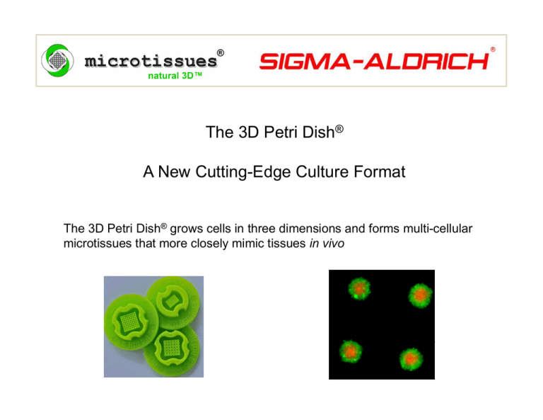
The 3D Petri Dish® A New Cutting-Edge Culture Format The 3D Petri Dish® grows cells in three dimensions and forms multi-cellular microtissues that more closely mimic tissues in vivo The 3D Petri Dish® is now available from Sigma-Aldrich® The 3D Petri Dish® is an easy-to-use y 3D cell culture system y that creates a natural 3D environment by maximizing cell-cell interactions. Its scaffold-free. Autoclavable precision micro-molds for the creation of spheroids, neurospheres, hepatospheres, cardiospheres, embryoid bodies, and much more. A growing list of over 40 different cell types (primary cells & cell lines) form 3D microtissues in the 3D Petri Dish® Z764000-6EA Z764019 6EA Z764019-6EA Z764094-6EA Z764035-6EA Z764027-6EA 12 well format 12-256 256 small spheroids 12 81 12-81 81 llarge spheroids h id 12-1 spheroids, mixed pack 12-36TO 36 toroids 12-60TR 60 rods Z764043-6EA Z764051-6EA Z764108-6EA Z764078-6EA Z764086-6EA 24 well format 24-96 96 small spheroids 24-35 35 large spheroids 24-1 spheroids, mixed pack 24-24TR 24 rods 24-H 1 honeycomb Z764116-8EA Tech pack 8-1 1 of each micro-mold, pk of 8 precision micro-molds spheroids of human mesenchymal stem cells Micro-mold casting system A. Autoclave sterilize micro-mold B. Fill micro-mold with sterile molten agarose (2%) C. Separate gelled agarose from flexible micro-mold D. Place gel in multi-well plate and equilibrate with cell culture medium E. Seed with mono-dispersed cells F. Cells settle into micro-wells of agarose, aggregate and self-assemble spheroids Remove gelled agarose from micro-mold Fill large chamber with mono-dispersed cells (190l) Cells settle by gravity to bottoms of many micro-wells C ll aggregate Cells t and d self-assemble lf bl many spheroids h id Remove agarose from micro-mold Fill large chamber with mono-dispersed cells (190l) Cells settle by gravity to bottoms of many micro-wells C ll aggregate Cells t and d self-assemble lf bl many spheroids h id Remove agarose from micro-mold Fill large chamber with mono-dispersed cells (190l) Cells settle by gravity to bottoms of many micro-wells C ll aggregate Cells t and d self-assemble lf bl many spheroids h id Remove agarose from micro-mold Fill large chamber with mono-dispersed cells (190l) Cells settle by gravity to bottoms of many micro-wells C ll aggregate Cells t and d self-assemble lf bl many spheroids h id The 3D Petri Dish® is scaffold-free Multi-cellular spheroids form at the bottom of nonadhesive agarose micro-wells Cells aggregate and self-assemble 3D spheroids No scaffold, no added ECM, no attachment factors Cell-cell interactions and cell-cell communication are maximized Cell density is akin to normal tissue No lot-lot to variation N No-synthetic h i scaffold ff ld No artificially stiff substrate spheroids of human mesenchymal stem cells Culture spheroids in a stable uniform environment won’t dry out won’t spill out won’t fall from a drop surrounded by culture medium on all sides culture long term replenish media, test drugs, add growth factors, sequential differentiation cells on plastic cells on substrate cells in gel or scaffold cells in 3D Petri Dish® Make 100s of spheroids in a single pipeting step 12 well format Z764000-6EA 12-256 256 small spheroids Z764019-6EA 12-81 81 large spheroids 24 well format Z764043-6EA 24-96 96 small spheroids Z764051-6EA 24-35 35 large spheroids Control spheroid size by micro-mold choice Z764019-6EA 12-81 81 large spheroids micro-well diameter ~800m large spheroids of human mesenchymal stem cells micro-well depth ~800m Z764000-6EA 12 256 12-256 256 small spheroids micro-well diameter ~400m small spheroids of human mesenchymal stem cells micro-well depth ~800m Control spheroid size by number of cells seeded Cell seeding numbers for different sized spheroids. # 12-256 12 well plate Small spheroids: 256 # 24-96 24 well plate Small spheroids: 96 Spheroid S i diameter (m) ~Cells/spheroid Total cells seeded (cells/190 l) Total cells seeded (cells/75 l) 50 100 150 15 125 421 3,840/190 l 32 000/190 l 32,000/190 107,000/190 l 1,440/75 l 12 000/75 l 12,000/75 40,000/75 l 200 1,000 256,000/190 l 96,000/75 l 250 1,953 500,000/190 l 187,000/75 l Form spheroids in an array on the same optical plane arrayy of spheroids p of mesenchymal stem cells same optical plane ordered, addressable array image replicates automation time lapse, phase contrast, fluorescent microscopy Form mixed spheroids with different cell types 3 cell types DIC microscopy; hepatocytes (blue) fibroblasts (green); HUVEC)(red); and their composite image 1:1 mix, hepatocytes (green) fibroblasts (red) control ratio of cells maximize heterotypic cell interactions tumor cell–stromal cell interactions Culture spheroids long term in a stable environment Grow clonal spheroids from single cells n = 10 Seed ~1 cell/micro-well, quantify spheroid growth over 2 weeks won’t dry out, won’t spill out, won’t fall from a drop surrounded by culture medium on all sides replenish media, test drugs, add growth factors f sequential differentiation cancer stem cells Harvest by flushing spheroids out of micro-wells with a pipette embryoid bodies Image courtesy of J Bruder Max Planck no gel to dissolve, no enzymatic treatment spheroids h id remain i intact i t t and d unaltered lt d harvest spheroids for RT-PCR, Western blots, histology, immuno-staining harvest spheroids for transplantation, for tumor implantation, for cell therapy Form complex shapes for novel applications 12 well format Z764035-6EA 12-36TO 36 toroids Z764027-6EA 12-60TR 60 rods 24 well format Z764078-6EA 24-24TR 24 rods Z764086-6EA 24-H 1 honeycomb y Y27632 drug washout human fibroblasts seeded in troughs form multi-cellular rods that self assemble and contract over 12 hours n = 10 Troughs: 2,200 m long 400 m wide 800 m deep round bottom trough ~ 25,000 cells/trough quantify cell-cell adhesion drug testing, genetic manipulations cell-cell adhesion of cell mixtures toroid outer diameter 1 1.4 4 mm diameter of inner peg 600 m circular trough width 400 m circular trough depth 800 m honeycomb size 3.4 mm diameter of pegs 600 m trough width 400 m trough depth 800 m developmental biology tissue engineering cell migration A growing list of cell types have been used in the 3D Petri Dish® Primary cells Cell lines Human dermal fibroblasts Rat cardiac myocytes Rat cardiac fibroblasts Human umbilical vein endothelial cells (HUVEC) Calf pulmonary artery endothelial cells (CPAE) Murine fibroblasts (3T3) Murine endothelial cells (bEnd3) Rat endothelial cells (RBE4) Human breast cancer cells (MCF-7) Human breast cancer cells (T47D) Human granulosa cells (KGN) Human trophoblast cells (TCL) Rat neuroblastoma cells (RG2) Rat glioma cells (9L) Rat glioma cells (C6) Human theca cells Human breast cancer cells (MDA-MB-231) Rat astrocytes (A7) Murine neurons from the hippocampus Human breast cancer cells (Hs-578T) Murine neuroblastoma cells (B104) Human mesenchymal y stem cells Human cervical cancer cells ((HeLa)) Retinal p pigment g epithelial p cells ((ARPE-19)) Zebrafish ectoderm progenitor cells Human epithelial carcinoma cells (A431) Human embryonic kidney cells (HEK) Zebrafish mesoderm progenitor cells Human mesenchymal stem cells Embryoid bodies Human hepatocytes (HepG2) Rat hepatocytes (H35) Human mesothelioma (M28) Rat endothelial cells ( RBE4) Murine endothelial cells (bEnd3) Human non-small lung cancer cells (A549) Murine neural stem cells Human mesothelioma ((REN)) Human non-small lung g cancer cell s ((H1299)) Human chondrocytes Bovine chondrocytes Rat aortic smooth muscle cells If your cells ll are adherent dh t adhesive dh i cells, ll they will self-assemble in the 3D Petri Dish® The 3D Petri Dish® is now available from Sigma-Aldrich® PRODUCT SUPPORT Visit our website (www.microtissues.com) Detailed protocols FAQs Published papers List of cells Tech support Email your questions
