Use of Pit and Fissure Sealants: Evidence-Based Clinical Recommendations1
Levels of evidence and strength of recommendations:
Each recommendation is based on the best available evidence. The level of evidence available to support each recommendation
may differ. Lower levels of evidence do not mean the recommendation should not be applied for patient treatment.
Correlate these colors with the text below.
A
B
C
D
Recommendation based on higher
levels of evidence
Recommendation based on lower
levels of evidence
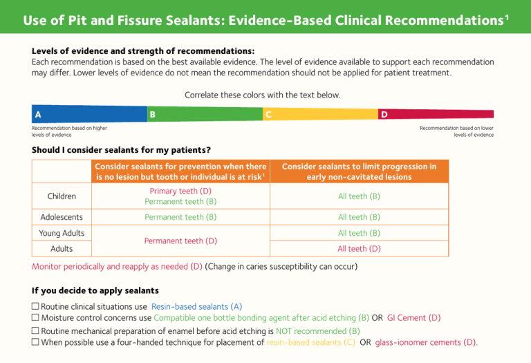
Should I consider sealants for my patients?
Consider sealants for prevention when there
is no lesion but tooth or individual is at risk1
Consider sealants to limit progression in
early non-cavitated lesions
Children
Primary teeth (D)
Permanent teeth (B)
All teeth (B)
Adolescents
Permanent teeth (B)
All teeth (B)
Young Adults
Adults
Permanent teeth (D)
All teeth (B)
All teeth (D)
Monitor periodically and reapply as needed (D) (Change in caries susceptibility can occur)
If you decide to apply sealants
Routine clinical situations use Resin-based sealants (A)
Moisture control concerns use Compatible one bottle bonding agent after acid etching (B) OR GI Cement (D)
Routine mechanical preparation of enamel before acid etching is NOT recommended (B)
When possible use a four-handed technique for placement of resin-based sealants (C) OR glass-ionomer cements (D).
Use of Pit and Fissure Sealants: Evidence-Based Clinical Recommendations1
These images2 are examples of non-cavitated lesions that may be considered for sealants. Non-cavitated lesion refers
to pits and fissures in fully erupted teeth that may display discoloration not due to extrinsic staining, developmental opacities or
fluorosis. The discoloration may be confined to the size of a pit or fissure or may extend to the cusp inclines surrounding a pit or
fissure. The tooth surface should have no evidence of a shadow indicating dentinal caries, and, if radiographs are available, they
should be evaluated to determine that neither the occlusal nor the proximal surfaces have signs of dentinal caries.
Tooth surface with an
early (noncavitated)
carious lesion that
exhibits a white
demineralization line
around the margin
of the pit and fissure
and /or a light brown
discoloration within
the confines of the
pit-and-fissure area.
A small, distinct,
dark brown early
(noncavitated)
carious lesion within
the confines of the
fissure.
A deep fissure
area (arrow 1)
and another area
exhibiting a small
light brown pit and
fissure (arrow 2).
Note that the lesion
does not extend
beyond the confines
of the pit and fissure.
A more distinct
early (noncavitated)
carious lesion (arrow)
that is larger than the
normal anatomical
size of the fissure
area.
A more distinct
early (noncavitated)
carious lesion (arrow)
that is larger than the
normal anatomical
size of the fissure.
ADA Council on Scientific Affairs. Use of Pit and Fissure Sealants: Evidence-based clinical recommendations. JADA 2008;139(3):257-68. Copyright © 2008 American Dental Association.
All rights reserved. Adapted with permission. To see the full text of this article, please go to http://jada.ada.org/cgi/content/abstract/139/3/257.
2
Images provided courtesy of Dr. Amid I. Ismail, the Detroit Dental Health Project (National Institute of Dental and Craniofacial Research grant U-54 DE 14261-01).
1
This page may be used, copied, and distributed for non-commercial purposes without obtaining prior approval from the ADA. Any other use, copying, or distribution, whether in printed or electronic format, is strictly
prohibited without the prior written consent of the ADA.


