Genetic Disruption of Kir6.2, the Pore-Forming Subunit of
advertisement

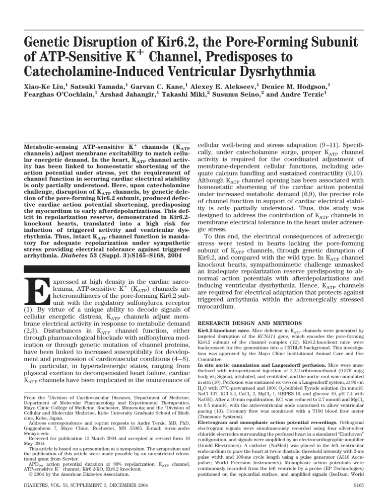
Genetic Disruption of Kir6.2, the Pore-Forming Subunit of ATP-Sensitive Kⴙ Channel, Predisposes to Catecholamine-Induced Ventricular Dysrhythmia Xiao-Ke Liu,1 Satsuki Yamada,1 Garvan C. Kane,1 Alexey E. Alekseev,1 Denice M. Hodgson,1 Fearghas O’Cochlain,1 Arshad Jahangir,1 Takashi Miki,2 Susumu Seino,2 and Andre Terzic1 Metabolic-sensing ATP-sensitive Kⴙ channels (KATP channels) adjust membrane excitability to match cellular energetic demand. In the heart, KATP channel activity has been linked to homeostatic shortening of the action potential under stress, yet the requirement of channel function in securing cardiac electrical stability is only partially understood. Here, upon catecholamine challenge, disruption of KATP channels, by genetic deletion of the pore-forming Kir6.2 subunit, produced defective cardiac action potential shortening, predisposing the myocardium to early afterdepolarizations. This deficit in repolarization reserve, demonstrated in Kir6.2knockout hearts, translated into a high risk for induction of triggered activity and ventricular dysrhythmia. Thus, intact KATP channel function is mandatory for adequate repolarization under sympathetic stress providing electrical tolerance against triggered arrhythmia. Diabetes 53 (Suppl. 3):S165–S168, 2004 E xpressed at high density in the cardiac sarcolemma, ATP-sensitive K⫹ (KATP) channels are heteromultimers of the pore-forming Kir6.2 subunit with the regulatory sulfonylurea receptor (1). By virtue of a unique ability to decode signals of cellular energetic distress, KATP channels adjust membrane electrical activity in response to metabolic demand (2,3). Disturbances in KATP channel function, either through pharmacological blockade with sulfonylurea medication or through genetic mutation of channel proteins, have been linked to increased susceptibility for development and progression of cardiovascular conditions (4 – 8). In particular, in hyperadrenergic states, ranging from physical exertion to decompensated heart failure, cardiac KATP channels have been implicated in the maintenance of From the 1Division of Cardiovascular Diseases, Department of Medicine, Department of Molecular Pharmacology and Experimental Therapeutics, Mayo Clinic College of Medicine, Rochester, Minnesota; and the 2Division of Cellular and Molecular Medicine, Kobe University Graduate School of Medicine, Kobe, Japan. Address correspondence and reprint requests to Andre Terzic, MD, PhD, Guggenheim 7, Mayo Clinic, Rochester, MN 55905. E-mail: terzic.andre @mayo.edu. Received for publication 12 March 2004 and accepted in revised form 18 May 2004. This article is based on a presentation at a symposium. The symposium and the publication of this article were made possible by an unrestricted educational grant from Servier. APD90, action potential duration at 90% repolarization; KATP channel, ATP-sensitive K⫹ channel; Kir6.2-KO, Kir6.2 knockout. © 2004 by the American Diabetes Association. DIABETES, VOL. 53, SUPPLEMENT 3, DECEMBER 2004 cellular well-being and stress adaptation (9 –11). Specifically, under catecholamine surge, proper KATP channel activity is required for the coordinated adjustment of membrane-dependent cellular functions, including adequate calcium handling and sustained contractility (9,10). Although KATP channel opening has been associated with homeostatic shortening of the cardiac action potential under increased metabolic demand (6,9), the precise role of channel function in support of cardiac electrical stability is only partially understood. Thus, this study was designed to address the contribution of KATP channels in membrane electrical tolerance in the heart under adrenergic stress. To this end, the electrical consequences of adrenergic stress were tested in hearts lacking the pore-forming subunit of KATP channels, through genetic disruption of Kir6.2, and compared with the wild type. In KATP channel knockout hearts, sympathomimetic challenge unmasked an inadequate repolarization reserve predisposing to abnormal action potentials with afterdepolarizations and inducing ventricular dysrhythmia. Hence, KATP channels are required for electrical adaptation that protects against triggered arrhythmia within the adrenergically stressed myocardium. RESEARCH DESIGN AND METHODS Kir6.2-knockout mice. Mice deficient in KATP channels were generated by targeted disruption of the KCNJ11 gene, which encodes the pore-forming Kir6.2 subunit of the channel complex (12). Kir6.2-knockout mice were backcrossed for five generations into a C57BL/6 background. This investigation was approved by the Mayo Clinic Institutional Animal Care and Use Committee. In situ aortic cannulation and Langendorff perfusion. Mice were anesthetized with intraperitoneal injection of 2,2,2-tribromoethanol (0.375 mg/g body wt; Sigma), intubated, and ventilated, and the aortic root was cannulated in situ (10). Perfusion was sustained ex vivo on a Langendorff system, at 90 cm H2O with 37°C-prewarmed and 100% O2-bubbled Tyrode solution (in mmol/l: NaCl 137, KCl 5.4, CaCl2 2, MgCl2 1, HEPES 10, and glucose 10, pH 7.4 with NaOH). After a 10-min equilibration, KCl was reduced to 2.7 mmol/l and MgCl2 to 0.5 mmol/l, with the atrioventricular node cauterized to allow ventricular pacing (13). Coronary flow was monitored with a T106 blood flow meter (Transonic Systems). Electrogram and monophasic action potential recordings. Orthogonal electrogram signals were simultaneously recorded using four silver-silver chloride electrodes surrounding the perfused heart in a simulated “Einthoven” configuration, and signals were amplified by an electrocardiographic amplifier (Gould Electronics). A catheter (NuMed) was placed in the left ventricular endocardium to pace the heart at twice diastolic threshold intensity with 2-ms pulse width and 100-ms cycle length using a pulse generator (A310 Accupulser; World Precision Instruments). Monophasic action potentials were continuously recorded from the left ventricle by a probe (EP Technologies) positioned on the epicardial surface, and amplified signals (IsoDam; World S165 KATP CHANNELS AND TRIGGERED ARRHYTHMIA FIG. 1. Isoproterenol challenge induced action potential shortening (APD90) in wild-type (WT) (A) but not Kir6.2-KO (B) hearts, which developed early afterdepolarizations (EAD) (C). D: Incidence of EAD in the initial 50 action potentials after 5 min of isoproterenol infusion. Precision Instruments) were acquired at 11.8 kHz and stored for off-line digital analysis (9). Whole-cell patch clamp recording from isolated cardiomyocytes. Cardiomyocytes were enzymatically dissociated from the ventricular myocardium (10). Action potentials were recorded at 30 ⫾ 1°C from current-clamped isolated cells paced at 1 Hz, and were superfused with Tyrode solution (pH 7.2 adjusted with KOH) using the whole-cell patch clamp technique with 5–10 mol/l⍀ pipettes containing (in mmol/l) KCl 120, MgCl2 1, Na2ATP 5, HEPES 10, EGTA 0.5, and CaCl2 0.01 (14). Statistics. Comparisons were made using the Student’s t test. A significance level of 0.05 was preselected. Data are reported as means ⫾ SE. RESULTS AND DISCUSSION Whereas at baseline the action potentials were similar, the metabolic challenge of adrenergic stimulation induced distinct outcomes depending on the presence of functional KATP channels, with significant shortening of the action potential duration observed in wild-type hearts but not in age- and sex-matched counterparts lacking the Kir6.2 pore-forming channel subunit (Kir6.2-KO) (Fig. 1A and B). S166 After a 10-min perfusion with the sympathomimetic isoproterenol (1 mol/l), monophasic action potential duration at 90% repolarization (APD90) shortened from 82 ⫾ 2 to 74 ⫾ 2 ms in wild-type hearts (P ⬍ 0.01, n ⫽ 6; Fig. 1A). In contrast, APD90 remained at 79 ⫾ 3 and 80 ⫾ 3 ms before and following isoproterenol treatment, respectively, in Kir6.2-KO hearts (n ⫽ 6) (Fig. 1B). This deficit in repolarization led to distorted action potential profiles with characteristic phase 3 early afterdepolarizations manifested as distinct humps in hearts lacking functional KATP channels (Fig. 1B and C). In all Kir6.2-KO hearts (n ⫽ 8), adrenergic challenge induced early afterdepolarizations, which occurred in 97 ⫾ 2% of the action potentials examined (Fig. 1D). This is in contrast to the action potential profile of the wild type (n ⫽ 6) that maintained a smooth repolarization contour following isoproterenol challenge (Fig. 1A) without significant afterdepolarizations (1 ⫾ 1%; P ⬍ 0.01 vs. Kir6.2-KO) (Fig. 1D). DIABETES, VOL. 53, SUPPLEMENT 3, DECEMBER 2004 X.-K. LIU AND ASSOCIATES FIG. 2. A: Similar coronary flow in the absence and presence of isoproterenol (1 mol/l) in wild-type (WT) and Kir6.2-KO hearts. B: Isoproterenol-induced abnormal repolarization with early afterdepolarization in an isolated current-clamped Kir6.2-KO cardiomyocyte. Abnormal electrical response of Kir6.2-KO hearts under adrenergic challenge was not associated with an isoproterenol-induced deficit in coronary perfusion (Fig. 2A). In fact, abnormal electrical activity during repolarization observed at the whole-heart level was reproduced at the single-cell level using action potential recording in isoproterenol-stressed current-clamped Kir6.2-KO cardiomyocytes (Fig. 2B). Afterdepolarizations in isoproterenol-challenged Kir6.2KO hearts translated into increased electrical vulnerability (Fig. 3). In the absence of functional KATP channels, afterdepolarizations induced triggered activity, disrupting regular rhythm and manifesting as premature ventricular complexes on the electrogram (Fig. 3A). On average, isoproterenol-induced afterdepolarizations complicated by triggered activity were observed in six of eight Kir6.2-KO (75%) compared with one of six wild-type (16%) hearts, translating into a 16-fold higher risk (P ⬍ 0.05) of developing premature ventricular complexes (Fig. 3B). Hence, absence of KATP channels produces a deficit in the repolarization reserve leading to a pronounced susceptibility of Kir6.2-KO hearts to isoproterenol-induced ventricular dysrhythmia. Thus, sarcolemmal KATP channels provide for membrane electrical stability that reduces the risk for arrhythmia under hyperadrenergic conditions. Imposed catecholamine stress is a well-established precipitator of triggered activity and arrhythmia (15,16). Anal- ogous to hearts with genetic and/or environmental compromise of repolarizing currents, as observed in congenital or acquired long QT syndrome (17), here isoproterenol challenge was proarrhythmic in the KATP channel– deficient myocardium provoking afterdepolarizations and triggered activity. This is in line with pharmacological studies that demonstrate that KATP channel activation with potassium channel openers prevents—whereas channel blockade with sulfonylurea drugs enhances—afterdepolarizations and triggered activity (18 –20). In fact, in a recent randomized clinical trial in patients with type 2 diabetes, the sulfonylurea glyburide, but not the alternative oral hypoglycemic agent metformin, caused an increase in QT prolongation with QT dispersion on the electrocardiogram (21). Moreover, mutations in the cardiac sulfonylurea receptor inducing deficits in KATP channel function have been identified in patients with cardiomyopathy and ventricular arrhythmia (8). Suppression of KATP channel activity, whether by genetic deletion of channel subunits or through use of channel antagonists, predisposes to inadequate calcium handling and intracellular calcium overload (5,9). In turn, excessive cytosolic calcium acts as a trigger for early depolarizations (22,23). Conversely, in the intact heart, where KATP channel opening is promoted by -adrenergic– mediated phosphorylation of channel proteins (24,25) or subsarcolemmal ATP depletion (26), shortening of cardiac FIG. 3. A: Kir6.2-KO hearts with early afterdepolarizations (EAD) demonstrated triggered activity on monophasic action potentials (MAP) and premature ventricular complexes (PVC) on electrograms (EG). For comparison, an EG is shown from wild-type hearts that are not prone to EAD-triggered activity/PVC. B: Incidence of triggered activity with accompanying PVC in the initial 50 action potentials following 5 min of isoproterenol infusion. DIABETES, VOL. 53, SUPPLEMENT 3, DECEMBER 2004 S167 KATP CHANNELS AND TRIGGERED ARRHYTHMIA action potentials would balance catecholamine-induced increase in calcium influx protecting against triggered arrhythmia. In summary, the present study underscores the homeostatic requirement for functional KATP channels in the adaptation of cardiac repolarization under adrenergic stress. In this regard, a deficit in KATP channel function is here identified as a previously unrecognized risk factor for the development of catecholamine-induced afterdepolarizations and triggered arrhythmia. ACKNOWLEDGMENTS Supported by the National Institutes of Health (HL64822, GM08685, HL07111, and AG21201), American Heart Association, Miami Heart Research Institute, Marriott Foundation, Mayo Foundation Clinician-Investigator Program, Japan Heart Foundation, Mayo-Dubai Healthcare City Research Project, and Japanese Ministry of Education, Science, Sports, Culture and Technology. A.T. is an Established Investigator of the American Heart Association. REFERENCES 1. Inagaki N, Gonoi T, Clement JP, Wang CZ, Aguilar-Bryan L, Bryan J, Seino S: A family of sulfonylurea receptors determines the pharmacological properties of ATP-sensitive K⫹ channels. Neuron 16:1011–1017, 1996 2. Carrasco AJ, Dzeja PP, Alekseev AE, Pucar D, Zingman LV, Abraham MR, Hodgson D, Bienengraeber M, Puceat M, Janssen E, Wieringa B, Terzic A: Adenylate kinase phosphotransfer communicates cellular energetic signals to ATP-sensitive potassium channels. Proc Natl Acad Sci U S A 98:7623– 7628, 2001 3. Abraham MR, Selivanov VA, Hodgson DM, Pucar D, Zingman LV, Wieringa B, Dzeja PP, Alekseev AE, Terzic A: Coupling of cell energetics with membrane metabolic sensing: integrative signaling through creatine kinase phosphotransfer disrupted by M-CK gene knockout. J Biol Chem 277: 24427–24434, 2002 4. Meinert CL, Knatterud GL, Prout TE, Klimt C: A study of the effects of hypoglycemic agents on vascular complications in patients with adultonset diabetes: mortality results. Diabetes 19:789 – 830, 1970 5. Brady PA, Terzic A: The sulfonylurea controversy: more questions from the heart. J Am Coll Cardiol 31:950 –956, 1998 6. Suzuki M, Sasaki N, Miki T, Sakamoto N, Ohmoto-Sekine Y, Tamagawa M, Seino S, Marban E, Nakaya H: Role of sarcolemmal KATP channels in cardioprotection against ischemia/reperfusion injury in mice. J Clin Invest 109:509 –516, 2002 7. Gumina RJ, Pucar D, Bast P, Hodgson DM, Kurtz CE, Dzeja PP, Miki T, Seino S, Terzic A: Knockout of Kir6.2 negates ischemic preconditioninginduced protection of myocardial energetics. Am J Physiol 284:H2106 – H2113, 2003 8. Bienengraeber M, Olson TM, Selivanov VA, Kathmann EC, O’Coclain F, Gao F, Karger AB, Ballew JD, Hodgson DM, Zingman LV, Pang Y-P, Alekseev AE, Terzic A: ABCC9 mutations identified in human dilated cardiomyopathy disrupt catalytic KATP channel gating. Nat Genet 36:382– 387, 2004 S168 9. Zingman LV, Hodgson DM, Bast PH, Kane GC, Perez-Terzic C, Gumina RJ, Pucar D, Bienengraeber M, Dzeja PP, Miki T, Seino S, Alekseev AE, Terzic A: Kir6.2 is required for adaptation to stress. Proc Natl Acad Sci U S A 99:13278 –13283, 2002 10. Hodgson DM, Zingman LV, Kane GC, Perez-Terzic C, Bienengraeber M, Ozcan C, Gumina RJ, Pucar D, O’Coclain F, Mann DL, Alekseev AE, Terzic A: Cellular remodeling in heart failure disrupts KATP channel-dependent stress tolerance. EMBO J 22:1732–1742, 2003 11. Kane GC, Behfar A, Yamada S, Perez-Terzic C, O’Cochlain F, Reyes S, Dzeja PP, Miki T, Seino S, Terzic A: ATP-sensitive K⫹ channel knockout compromises the metabolic benefit of exercise training, resulting in cardiac deficits. Diabetes 53 (Suppl. 3):S169 –S175, 2004 12. Miki T, Nagashima K, Tashiro F, Kotake K, Yoshitomi H, Tamamoto A, Gonoi T, Iwanaga T, Miyazaki J, Seino S: Defective insulin secretion and enhanced insulin action in KATP channel-deficient mice. Proc Natl Acad Sci U S A 95:10402–10406, 1998 13. Liu XK, Wang W, Ebert SN, Franz MR, Katchman A, Woosley RL: Female gender is a risk factor for torsades de pointes in an in vitro animal model. J Cardiovasc Pharmacol 34:287–294, 1999 14. Chen YJ, Chen SA, Chen YC, Yeh HI, Chang MS, Lin CI: Electrophysiology of single cardiomyocytes isolated from rabbit pulmonary veins: implication in initiation of focal atrial fibrillation. Basic Res Cardiol 97:26 –34, 2002 15. Zeng J, Rudy Y: Early afterdepolarizations in cardiac myocytes. Biophys J 68:949 –964, 1995 16. Volders PG, KulcSar A, Vos MA, Sipido KR, Wellens HJ, Lazzara R, Szabo B: Similarities between early and delayed afterdepolarizations induced by isoproterenol in canine ventricular myocytes. Cardiovasc Res 34:348 –359, 1997 17. Anderson ME, Al-Khatib SM, Roden DM, Califf RM: Cardiac repolarization. Am Heart J 144:769 –781, 2002 18. Antzelevitch C, Di Diego JM: Role of K⫹ channel activators in cardiac electrophysiology and arrhythmias. Circulation 85:1627–1629, 1992 19. Shimizu W, Antzelevitch C: Effects of a K⫹ channel opener to reduce transmural dispersion of repolarization and prevent torsade de pointes in LQT1, LQT2, and LQT3 models of the long-QT syndrome. Circulation 102:706 –712, 2000 20. Pasnani JS, Ferrier GR: Differential effects of glyburide on premature beats and ventricular tachycardia in an isolated tissue model of ischemia and reperfusion. J Pharmacol Exp Ther 262:1076 –1084, 1992 21. Najeed SA, Khan IA, Molnar J, Somberg JC: Differential effect of glyburide (glibenclamide) and metformin on QT dispersion: a potential adenosine triphosphate sensitive K⫹ channel effect. Am J Cardiol 90:1103–1106, 2002 22. Choi BR, Burton F, Salama G: Cytosolic Ca2⫹ triggers early afterdepolarizations and torsade de pointes in rabbit hearts with type 2 long QT syndrome. J Physiol 543:615– 631, 2002 23. Volders PG, Vos MA, Szabo B, Sipido KR, de Groot SH, Gorgels AP, Wellens HJ, Lazzara R: Progress in the understanding of cardiac early afterdepolarizations and torsades de pointes. Cardiovasc Res 46:376 –392, 2000 24. Beguin P, Nagashima K, Nishimura M, Gonoi T, Seino S: PKA-mediated phosphorylation of the human KATP channel. EMBO J 18:4722– 4732, 1999 25. Lin YF, Jan YN, Jan LY: Regulation of ATP-sensitive potassium channel function by protein kinase A-mediated phosphorylation in transfected HEK293 cells. EMBO J 19:942–955, 2000 26. Schackow TE, Ten Eick RE: Enhancement of ATP-sensitive potassium current in cat ventricular myocytes by -adrenoreceptor stimulation. J Physiol 474:131–145, 1994 DIABETES, VOL. 53, SUPPLEMENT 3, DECEMBER 2004


![Anti-ABCC9 antibody [S323A31] - C-terminal ab174631](http://s2.studylib.net/store/data/012696516_1-ac50781de55479848678303901c47ff1-300x300.png)