The Whipple Operation – Illustrations
advertisement

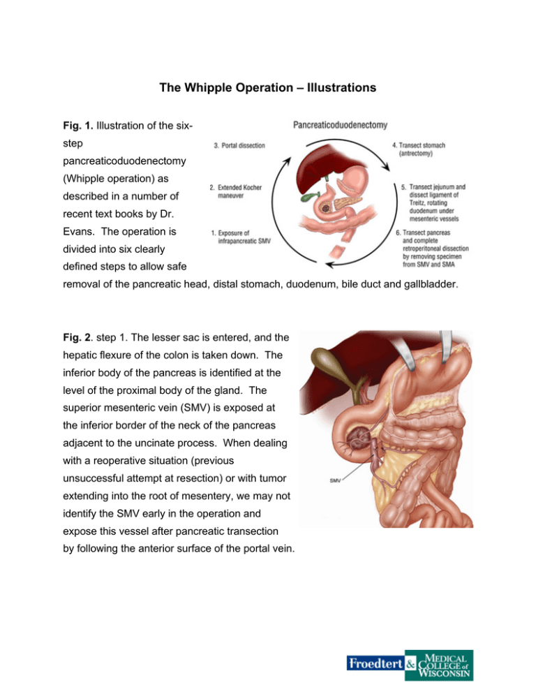
The Whipple Operation – Illustrations Fig. 1. Illustration of the sixstep pancreaticoduodenectomy (Whipple operation) as described in a number of recent text books by Dr. Evans. The operation is divided into six clearly defined steps to allow safe removal of the pancreatic head, distal stomach, duodenum, bile duct and gallbladder. Fig. 2. step 1. The lesser sac is entered, and the hepatic flexure of the colon is taken down. The inferior body of the pancreas is identified at the level of the proximal body of the gland. The superior mesenteric vein (SMV) is exposed at the inferior border of the neck of the pancreas adjacent to the uncinate process. When dealing with a reoperative situation (previous unsuccessful attempt at resection) or with tumor extending into the root of mesentery, we may not identify the SMV early in the operation and expose this vessel after pancreatic transection by following the anterior surface of the portal vein. Fig. 3. Illustration of step 2. A Kocher maneuver has been performed by first identifying the inferior vena cava (IVC) at the level of the proximal portion of the transverse segment of the duodenum (D3). One can then mobilize the duodenum and pancreatic head off of the IVC in a cephalad direction thereby removing all soft tissue anterior to the IVC. Note that the Kocher maneuver is continued to the left lateral border of the aorta (AO). Fig. 4. Illustration of step 3. Dissection of the porta hepatis begins with identification of the common hepatic artery (CHA), by removal of the large lymph node which commonly sits anterior to this vessel. The CHA is then followed distally to allow identification and division of the right gastric artery (not shown) and the gastroduodenal artery (GDA). This allows the CHA-proper hepatic artery to be mobilized off of the underlying anterior surface of the portal vein (PV). The PV is always identified prior to division of the common hepatic duct (CHD). Fig. 5. Illustration of step 4. The antrum of the stomach is resected with the main specimen by dividing the stomach at the level of the third or fourth transverse vein on the lesser curvature. CHA, common hepatic artery; CHD, common hepatic duct; SMA, superior mesenteric artery; SMV, superior mesenteric vein. Sometimes the entire stomach is preserved; when this is done, the operation is called a pylorus preserving Whipple (or pancreaticoduodenectomy). Fig. 6. Illustration of step 5. Transection of the jejunum is followed by ligation and division of its mesentery. The loose attachments of the ligament of Treitz are taken down, and the fourth and third portions of the duodenum are mobilized by dividing their short mesenteric vessels. The duodenum and jejunum are then reflected underneath the mesenteric vessels in preparation for the final and most important part of pancreaticoduodenectomy. Fig. 7. Illustration of step 6. The pancreatic head and uncinate process are separated from the superior mesentericportal vein confluence. The pancreas has been transected at the level of the portal vein and the pancreatic head is reflected laterally, allowing identification of small venous tributaries from the portal vein and superior mesenteric vein (SMV). These tributaries are ligated and divided. CHA, common hepatic artery. Fig. 8. Illustration of the continuation of step 6, and the final step in resection of the tumor. Medial retraction of the superior mesenteric-portal vein confluence facilitates dissection of the soft tissues adjacent to the lateral wall of the proximal superior mesenteric artery (SMA). This is the most important step in the operation from an oncologic perspective. Fig. 9. Illustration of the important surgical anatomy of the superior mesenteric vein (SMV). The SMV usually bifurcates into two main branches, one to the ileum and one to the jejunum. Adequate venous return from the small bowel requires that one or the other of these two main SMV-tributaries is intact. The jejunal branch of the SMV (often referred to as the first jejunal branch) drains the proximal jejunum, travels posterior to the superior mesenteric artery (SMA), and enters the SMV along its posterolateral wall. Very rarely the jejunal branch will travel anterior to the SMA. Figs. 10-11. Illustration of the final step in pancreaticoduodenectomy when segmental venous resection is required and the splenic vein is preserved. The intact splenic vein tethers the portal vein, making a primary anastomosis impossible in most cases. An interposition graft is used to repair the segment of vein which is removed; the left internal jugular vein from the neck is used for the graft in most case2. Fig. 12. Illustration of the different types of venous reconstruction used at the time of pancreaticoduodenectomy. When a patch is needed we use the saphenous vein from the leg and when an interposition graft is needed we use the left internal jugular (IJ) vein. PV, portal vein; SMV, superior mesenteric vein; Spl V, splenic vein. Fig. 13-14. Illustration of pancreaticojejunostomy. A two-layer, end-to-side, duct-tomucosa retrocolic pancreaticojejunostomy is performed with (when the pancreatic duct is not dilated) or without a small stent. When used, the stent (4-5 cm long) is sewn to the pancreatic duct with a single absorbable monofilament suture. Fig. 15. Illustration of hepaticojejunostomy. A onelayer, end-to-side hepaticojejunostomy is performed with 4-0 or 5-0 absorbable monofilament sutures distal to the pancreaticojejunostomy. A stent is rarely placed in this anastomosis. Fig. 16. Illustration of the completed reconstruction after pancreaticoduodenectomy. The falciform ligament, mobilized upon opening the abdomen, is placed over the hepatic artery to cover the stump of the gastroduodenal artery, thereby separating the hepatic artery from the afferent jejunal limb. CHA, common hepatic artery Fig. 17. Our care team of nurses who work on the specialty floor at Froedtert Hospital where patients recover from their pancreatic surgery. Such highly trained nurse specialists understand all aspects of this type of cancer surgery.


