ISPY-2 BREAST updated 1/1/2013
advertisement

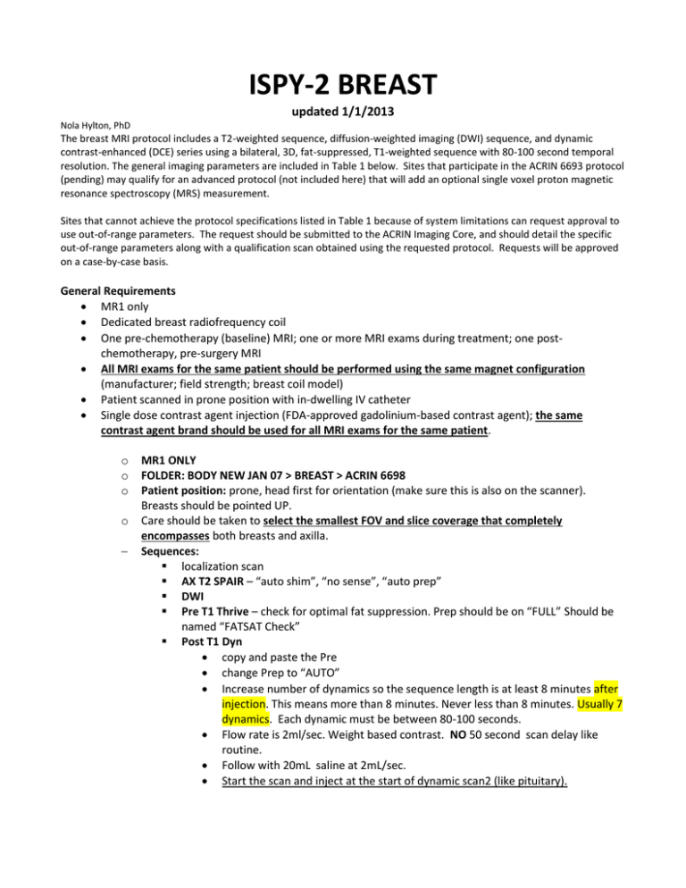
ISPY-2 BREAST updated 1/1/2013 Nola Hylton, PhD The breast MRI protocol includes a T2-weighted sequence, diffusion-weighted imaging (DWI) sequence, and dynamic contrast-enhanced (DCE) series using a bilateral, 3D, fat-suppressed, T1-weighted sequence with 80-100 second temporal resolution. The general imaging parameters are included in Table 1 below. Sites that participate in the ACRIN 6693 protocol (pending) may qualify for an advanced protocol (not included here) that will add an optional single voxel proton magnetic resonance spectroscopy (MRS) measurement. Sites that cannot achieve the protocol specifications listed in Table 1 because of system limitations can request approval to use out-of-range parameters. The request should be submitted to the ACRIN Imaging Core, and should detail the specific out-of-range parameters along with a qualification scan obtained using the requested protocol. Requests will be approved on a case-by-case basis. General Requirements MR1 only Dedicated breast radiofrequency coil One pre-chemotherapy (baseline) MRI; one or more MRI exams during treatment; one postchemotherapy, pre-surgery MRI All MRI exams for the same patient should be performed using the same magnet configuration (manufacturer; field strength; breast coil model) Patient scanned in prone position with in-dwelling IV catheter Single dose contrast agent injection (FDA-approved gadolinium-based contrast agent); the same contrast agent brand should be used for all MRI exams for the same patient. o o o o MR1 ONLY FOLDER: BODY NEW JAN 07 > BREAST > ACRIN 6698 Patient position: prone, head first for orientation (make sure this is also on the scanner). Breasts should be pointed UP. Care should be taken to select the smallest FOV and slice coverage that completely encompasses both breasts and axilla. Sequences: localization scan AX T2 SPAIR – “auto shim”, “no sense”, “auto prep” DWI Pre T1 Thrive – check for optimal fat suppression. Prep should be on “FULL” Should be named “FATSAT Check” Post T1 Dyn copy and paste the Pre change Prep to “AUTO” Increase number of dynamics so the sequence length is at least 8 minutes after injection. This means more than 8 minutes. Never less than 8 minutes. Usually 7 dynamics. Each dynamic must be between 80-100 seconds. Flow rate is 2ml/sec. Weight based contrast. NO 50 second scan delay like routine. Follow with 20mL saline at 2mL/sec. Start the scan and inject at the start of dynamic scan2 (like pituitary). Create the subtraction and send to PACS only. Send T2, DWI, Pre and Dyn to EWS MR2. Please burn images from EWS MR2 to DVD provided by Alina Send T2 and Post to Dynacad Send Only good images to PACS. Send subtraction The day prior to the scan Alina will provide the forms and DVD(the forms(TM) can also be downloaded from http://www.acrin.org/PROTOCOLSUMMARYTABLE/PROTOCOL6698/6698DataForms.a spx Contrast Agent Administration An intravenous catheter will be inserted in the arm or hand prior to the start of imaging. For the contrastenhanced study (following the T2-weighted acquisition), gadolinium contrast agent will be administered intravenously at a dose of 0.1 mmol/kg body weight and rate of 2 ml/second, followed by a 20 ml saline flush. Contrast injection will begin simultaneously with the start of data acquisition. Table 1: Pulse Sequence Parameters* Sequence type 2D or 3D sequence Slice orientation Laterality Frequency direction Phase direction FOV - frequency FOV - phase Matrix – frequency (acquired) Matrix – phase (acquired) In-plane resolution Fat-suppression TR TE Echo Train Length TI (STIR sequence) Flip Angle T2-weighted Fast spin echo (FSE) or STIR 2D Axial or sagittal Bilateral A/P R/L (axial); S/I (sagittal) 260-360 mm (axial); 180220 mm (sagittal) 260-360 mm (axial); 180220 mm (sagittal) 256-512 T1-weighted gradient echo (GE) 256 1.4 mm Active fat-sat recommended 2000-10000 ms 70-140 ms 16 150 ms (1.5T); 300 ms (3.0T) 90 degrees 3D Axial Bilateral A/P R/L 260-360 mm DWI Diffusion-weighted spin echo, echo planar imaging (DW SE-EPI) 2D Axial Bilateral A/P R/L 260-360 mm 260-360 mm 260-360 mm 384-512 192 256 1.4 mm Active fat-sat recommended 4-10 ms 1.3 or 4.2 ms (fat/water inphase) N/A N/A 192 1.9 mm Active fat-sat 10-20 degrees 90 degrees 6,000 ms Minimum TE N/A N/A B values Slice thickness (acquired, not interpolated) Number of slices Slice Gap Parallel imaging factor No. of excitations or averages k-space ordering N/A 4 mm N/A 2.5 mm 0, 800 s/mm2 5 mm Variable; complete bilateral coverage 1.0 mm 2 2 60; complete bilateral coverage No gap 2 2 Variable; complete bilateral coverage No gap 2 5 N/A -k to +k (standard, noncentric) 80 sec scan time 100 sec 8 minutes following injection N/A Sequence acquisition time 7 minutes Total post-contrast imaging duration N/A 4 minutes N/A Scan Verification Form ISPY2 PATIENT ID#________________________ ISPY visit: 1 2 3 4 MRI DATE: STUDY BREAST: ______________________________________________ Right Left T2 SERIES: series______________________ DWI SERIES: series______________________ 3D T1 SERIES: series______________________ GADOLINIUM INJECTION RATE _____________ ml/sec Flush Volume: SCAN ISSUES/COMMENTS: _____________ ml


