Abdominal Pelvic Regions Lesson w/ Anatomage Table:
advertisement

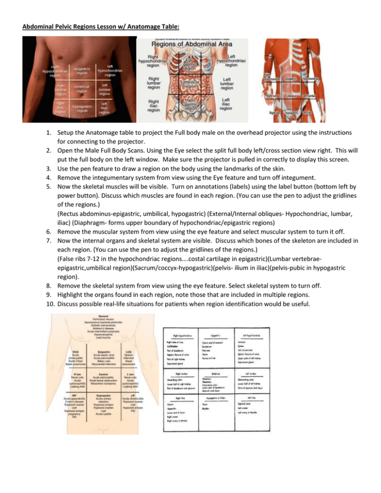
Abdominal Pelvic Regions Lesson w/ Anatomage Table: 1. Setup the Anatomage table to project the Full body male on the overhead projector using the instructions for connecting to the projector. 2. Open the Male Full Body Scans. Using the Eye select the split full body left/cross section view right. This will put the full body on the left window. Make sure the projector is pulled in correctly to display this screen. 3. Use the pen feature to draw a region on the body using the landmarks of the skin. 4. Remove the integumentary system from view using the Eye feature and turn off integument. 5. Now the skeletal muscles will be visible. Turn on annotations (labels) using the label button (bottom left by power button). Discuss which muscles are found in each region. (You can use the pen to adjust the gridlines of the regions.) (Rectus abdominus-epigastric, umbilical, hypogastric) (External/Internal obliques- Hypochondriac, lumbar, iliac) (Diaphragm- forms upper boundary of hypochondriac/epigastric regions) 6. Remove the muscular system from view using the eye feature and select muscular system to turn it off. 7. Now the internal organs and skeletal system are visible. Discuss which bones of the skeleton are included in each region. (You can use the pen to adjust the gridlines of the regions.) (False ribs 7-12 in the hypochondriac regions….costal cartilage in epigastric)(Lumbar vertebraeepigastric,umbilical region)(Sacrum/coccyx-hypogastric)(pelvis- ilium in iliac)(pelvis-pubic in hypogastric region). 8. Remove the skeletal system from view using the eye feature. Select skeletal system to turn off. 9. Highlight the organs found in each region, note those that are included in multiple regions. 10. Discuss possible real-life situations for patients when region identification would be useful. Student Worksheet Abdominal Pelvic Regions: Part I: Label the regions in the diagram. Part II: Indicate which region(s) would be important in each situation. 1. Umbilical cord _________________________ 2. Stomach __________________________ __________________________ 3. Bladder ___________________________ 4. Appendix __________________________ 5. Ovaries (female) _____________________ ___________________________ 6. Pancreatitis _________________________ 7. Peptic ulcer _________________________ 8. Myocardial Infarction (Heart attack) __________________________ 9. Ectopic pregnancy ____________________ ___________________________ 10. Liver ___________________________ ___________________________ 11. Kidney ________________________ ___________________________ ______________________________ ___________________________ 12. Spleen __________________________ Student Worksheet Abdominal Pelvic Regions: ANSWERS Part I: Label the regions in the diagram. Part II: Indicate which region(s) would be important in each situation. 1. Umbilical cord ___Umbilical______________________ 2. Stomach ___L hypochondriac_______________________ _____Epigastric_____________________ 3. Bladder ___hypogastric________________________ 4. Appendix ____R iliac ______________________ 5. Ovaries (female) ____R iliac_________________ _____L iliac______________________ 6. Pancreatitis ____Epigastric_____________________ 7. Peptic ulcer _____Epigastric____________________ 8. Myocardial Infarction (Heart attack) _____Epigastric_____________________ 9. Ectopic pregnancy ___R iliac_________________ ________L iliac___________________ 10. Liver ____R hypochondriac_______________________ _______Epigastric____________________ 11. Kidney ____R hypochondriac____________________ _____L hypochondriac______________________ __R lumbar____________________________ _____L lumbar______________________ 12. Spleen ____L hypochondriac______________________


