Development of dentition and occlusion Dr. Zuber Ahamed Naqvi
advertisement
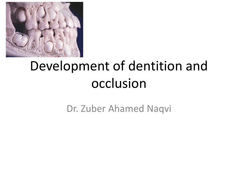
Development of dentition and occlusion Dr. Zuber Ahamed Naqvi OBJECTIVES Stages of tooth development Pre – dental period The deciduous dentition period The mixed dentition period The permanent dentition The permanent dentition period The mixed dentition period • Stages of tooth development (based on shape of enamel organ): • Bud stage • Cap stage • Bell stage Tooth formation Primary epithelial bands: Horseshoeshaped bands. Two subdivisions: vestibular lamina and dental lamina vestibule dental arches BUD STAGE 1. Tooth bud 2. Oral epithelium 3. Mesenchyme Cap Stage • Histodifferentiation (differentiation of tissues) • Morph differentiation • Dental papilla dentin and pulp tissue (mesenchymal origin) • Basement membrane separating the dental organ and the dental papilla becomes the future site for the dentinoenamel junction (DEJ) • Remaining mesenchyme surrounds the dental/enamel organ and condenses to form the dental sac or the dental follicle Bell Stage Early bell stage Advanced bell stage • Bell-like shape • differentiation produces four types of cells within the enamel/dental organ – – – – 1. inner enamel epithelium 2. outer enamel epithelium 3. stellate reticulum 4. stratum intermedium The dental papilla undergoes differentiation and produces two types of cells – 1. outer cells of the DP – forms the dentin-secreting cells (odontoblasts) – 2. central cells of the DP – forms the primordium of the pulp ROOT FORMATION • Takes place as the crown is completely shaped and the tooth begins to erupt • Root formation is through the formation of a cervical loop (CL) • Two layers consisting of Iner enamel epithelium and outer enamel epithelium • The CL begins to grow down into the dental sac • It forms a Hertwig's root sheath Periodontal ligament • The mesenchyme of the dental sac condenses to form the periodontal ligament • The cells of the disintegrating Hertwig root sheath develop into discrete islands of epithelial cells – become epithelial rests of Malassez Development of occlusion Occlusion is the relationship of the mandibular and maxillary teeth when closed or during excursive movements of the mandible; when the teeth of the mandibular arch come into contact with the teeth of the maxillary arch in any functional relationship. Periods of occlusal development • 1. Pre – dental period • 2. The deciduous dentition • period • 3. The mixed dentition period • 4. The permanent dentition • period GUM PADS • The alveolar processes at the time of birth are known as gum pads • The gum pads are pink, firm & covered by a dense layer of fibrous periosteum • They are HORSE-SHOE shaped & develop in two parts: (1) the labio-buccal portion & (2) the lingual portion • The two portions of the gum pads are separated from each other by a groove called the dental groove dental groove GUM PADS • Lateral sulcus-The transverse groove b/w canine & first deciduous molar segment . • The lateral sulcus of the mandibular arch is normally more DISTAL to that of the maxillary arch • The gum pads are divided into TEN SEGMENTS by certain grooves called TRANSVERSE GROOVES • • Each of these segments consist of one developing deciduous tooth sac • • The gingival groove separates the gum pads from the palate & floor of the mouth • There is a complete overjet all around • (1) Contact occurs b/w the upper & lower gum pads in the first molar region • (2) A space exist b/w them in the anterior region • This infantile open bite is Open bite considered normal & it helps in suckling NATAL TEETH Neonatal teeth • Teeth that are present at the time of birth are called NATAL TEETH Teeth that are erupt during the first month of age are called Neonatal teeth The natal & neonatal teeth are mostly located in the mandibular incisor region & Show a familial tendency. The deciduous dentition period • Primary teeth begin to erupt at the age of 6 months. • The eruption of all primary teeth is completed by 2.5 to 3.5 years of age when second deciduous molars come into occlusion. The mixed dentition period • The mixed dentition period begins at approximately 6 years of age with the eruption of the first permanent molars. primary + permanent teeth (6 Y – 12 Y) Classified into three phases: • 1. First transitional period • 2. Inter-transitional period • 3. Second transitional period First transitional period The first transitional period is characterized by : (1) the emergence of the first permanent molars & (2) the exchange of the deciduous incisors with the permanent incisors The mesio-distal relation b/w the Distal Surfaces of the upper & lower second deciduous molars can be of three types : • FLUSH PLANE • MESIAL STEP • DISTAL STEP • Flush to Class I molar relation This occurs by(1) utilization of the physiological spaces and primate spaces( early shift) (2) & leeway space in the lower arch ( late shift) (3) by differential forward growth of the mandible Inter – transitional period • In this period the maxillary & mandibular arches consist of Sets of deciduous & permanent teeth 6edc21 Second transitional period • Leeway space of Nance - The combined mesio-distal width of the permanent canines & premolars is usually less than that of the deciduous canines & molars Maxillary arch - 1.8mm (0.9 mm in each quadrant of the arch) Mandibular arch- 3.4mm (1.7 mm in each quadrant of the arch) The normal leeway space according to Moyers is 2.6 mm in the maxilla and 6.2 mm in the mandible. • The ugly duckling stage: • As the developing permanent canines erupt, they displace the roots of the lateral incisors mesially. • This result in transmitting of the force on to the roots of the central incisors which also get displaced mesially • A resultant distal divergence of the crowns of the two central incisors causes a midline spacing • This situation has been described by Broadbent as the ugly duckling stage. The permanent dentition period • The eruption sequence of the permanent dentition may exhibit variation • The frequently seen sequences in the maxillary arch are: – 6 – 1 – 2 – 4 – 5 – 3 – 7 or – 6–1–2––4 -3–5–7 In case of the mandibular arch the sequence is – 6 – 1 – 2 – 3 – 4 – 5 – 7 or – 6–1–2–4–3–5–7 • References: Contemporary orthodontics. 5th edition. William R Proffit Orthodontics : the art and science. 4th edition. S I Bhalaji Dentistry for the child and adolescent. 8th edition. Ralph E. McDonald, David R. Avery, Jeffrey A. Dean
