بوستر2
advertisement
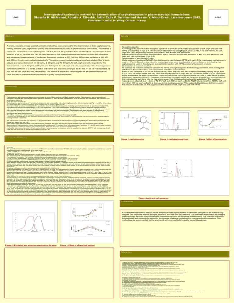
New spectrofluorimetric method for determination of cephalosporins in pharmaceutical formulations Shazalia M. Ali Ahmed, Abdalla A. Elbashir, Fakhr Eldin O. Suliman and Hassan Y. Aboul-Enein, Luminescence 2012, Published online in Wiley Online Library LOGO Abstract LOGO Results A simple, accurate, precise spectrofluorimetric method has been proposed for the determination of three cephalosporins, namely, cefixime (cefi), cephalexine (ceph), and cefotaxime sodium (cefo) in pharmaceutical formulations. This method is based on a reaction between cephalosporins with 8-hydroxy-1,3,6-pyrenetrisulfonic acid trisodium salt (HPTS) in alkaline medium, at pH 12.0 for cefi and 13.0 for ceph and cefo to give highly fluorescent derivatives extracted with chloroform and subsequent measurements of the formed fluorescent products at 520, 500 and 510nm after excitation at 480, 470 and 480 nm for cefi, ceph and cefo respectively. The optimum experimental conditions have been studied. Beer’s law is obeyed over concentrations of 10–60 ng/mL, 5–35ng/mL and 10–60ng/mL for cefi, ceph and cefo, respectively. The detection limits were 4.20ng/mL, 2.54ng/mL and 4.09 ng/mL for cefi, ceph and cefo, respectively, with a linear regression correlation coefficient of 0.99783, 0.99705 and 0.9978 and recoveries in ranges 96.96–105.77, 96.13–102.55 and 95.45– 105.39% for cefi, ceph and cefo, respectively. This method is simple and can be applied for the determination of cefi, ceph and cefo in pharmaceutical formulations in quality control laboratories. Absorption spectra According to the procedure the absorption spectrum of products produced by the reaction of cefi, ceph and cefo with HPTS are recorded in Figs 2–4. The maximum absorption wavelength peak (lmax) at 480, 470, and 480 nm, for cefi, ceph and cefo, respectively and the lmax of HPTS was 400nm. The derivatives exhibit maximum fluorescence intensities lmax at 520, 500 and 510nm after excitation at 480, 470 and 480nm for cefi, ceph and cefo respectively (Figs 5–7). Determination of stoichiometric ratio Under optimum conditions (Table 2), the stoichiometric ratio between HPTS and each of the investigated cephalosporins was1 : 1 (Fig. 8). Based on this ratio, the reaction pathways were postulated as shown in Scheme 1, indicating that cephalosporins used in this study are susceptible to reaction with HPTS producing fluorescent products. Optimization of reaction variables To optimize the reaction conditions between the HPTS and cephalosporins the following parameters were investigated: pH of the buffer, reaction time, and temperature and HPTS concentration. Effect of pH. The effect of pH on the reaction of cefi, ceph, and cefo with HPTS were examined by varying the pH from 4.0 to 13.0, the results reveal that cefi, ceph and cefo are difficult to react with HPTS in acidic media (Fig. 9). This is due to the existence of the amino group of cefi, ceph and cefo in the form of hydrochloride salt; thus, it loses its nucleophilic substitution capability. As the pH increased, the readings increased rapidly, as the amino group of cefi, ceph and cefo (in the hydrochloride salt) turns into the free amino group, thus facilitating the nucleophilic substitution. The maximum readings were attained at pH values of 12.0 for cefi and 13.0 for ceph and cefo. At pH values more than 12.0 for cefi and more than 13.0 for ceph and cefo, a decrease in the readings occurred. This was probably attributed to the increase in the amount of hydroxide ion that suppresses the reaction of cefi, ceph and cefo with HPTS. Introduction Cephalosporins are antibacterial agents commonly used to control Gram-positive and Gram-negative bacteria. Cephalosporins are the second most important b-lactams after penicillin for treating infectious diseases (1). Many of these manifestations, such as urticaria and exanthema, are cutaneous, but anaphylactic reactions have also been reported (2). Cephalosporins are derivatives of 7-aminocephalosporanic acid composed of a b-lactam ring fused with a dihydrothiazine ring (Fig. 1) but differ in the nature of substituent at the 3- and/or 7-positions of the cephem ring as shown in Table 1 (3,4). Several methods have been described for the quantitative determination of cephalosporins, including spectrophotometry (5–8), spectrofluorimetry (9,10), high-performance liquid chromatography (11–18), and voltammetry (19). These methods were time-consuming, tedious, and used sophisticated and expensive analytical instruments. Spectrophotometric methods are the most convenient techniques because of their inherent simplicity, high sensitivity, low cost, and wide availability in quality control laboratories. Unfortunately, the spectrophotometric methods that have been reported for determination of cephalosporins in their pharmaceutical formulations were associated with some major disadvantages such as the lack of selectivity, tedious extraction procedures and were timeconsuming. The official procedures in pharmaceutical preparations use high-performance liquid chromatography(20). Therefore, the development of new alternative spectrofluorimetricmethod for the determination of cephalosporins that can overcome the disadvantages of existing methods is essential. The pH-sensitive fluorescent dye 1-hydroxy-3,6,8-pyrenetrisulfonic acid trisodium salt also known as pyranine (HPTS) has distinct absorption band in the visible light region, and has thus been used in many aqueous-phase CO2 optical sensors. However, this is the first time that HPTS has been used asa fluorogenic reagent for the determination of cephalosporins in their pharmaceutical formulations; HPTS contains three sulfonate groups at positions, 1, 3, and 6, which make it suitable for nucleophilic substitution reactions with cephalosporins. This work describes a rapid spectrofluorimetric method for determining the content of cefi, ceph and cefo in pharmaceutical formulations, which is based on the reaction of HPTS with amino group of cefi, ceph and cefo molecules to form fluorescent derivatives (21–26). CHART or PICTURE CHART or PICTURE CHART or PICTURE Figure_2.cephalexin spectrum Figure_3effect of temperature Methods and Materials Figure_1.cephalosporins Apparatus The spectrofluorimetricmeasurements were made with Shimadzu spectrofluorophotometer RF-1501 with xenon lamp. In addition, a temperature controller was used for the spectrofluorimetric measurements. A pH meter model pH 211 (HANNA, Camisano Vicentino, Italy) was used for adjusting pH. Reagents and solutions All reagents were of analytical reagent grade. Double distilled water was used in all experiments. The standards of cefi, ceph and cefo were supplied by (Orchid Chemicals and Pharmaceuticals Ltd., Chennai, India). Pharmaceutical formulation. The following available pharmaceutical preparations were analyzed: (1) cefi capsules (AMIPHARMA Laboratories, Khartoum-North, Sudan), labeled to contain 200mg cefi per capsule. (2) ceph monohydrate capsules (AMIPHARMA Laboratories, Sudan), labeled to contain 500mg ceph per capsule. (3) cefo for injection (Kilitch Drugs, Mumbai, India) labeled to contain 1000mg cefo per injection. Stock standard solution of cefi, ceph and cefo (1000mg/mL). An accurately weighed 0.1 g standard sample of the three drugs was dissolved in methanol for cefi and in double distilled water for ceph and cefo, transferred into a 100mL standard flask and diluted to the mark with methanol for cefi and with double distilled water for ceph and cefo and then mixed well. This stock solution was further diluted to obtain working solutions in the ranges 10–35, 10–60 and 20–45 ng/mL for cefi, ceph and cefo, respectively. 8-Hydroxy-1,3,6-pyrenetrisulfonic acid trisodium salt solution(0.0.02%, 0.025% w/v). An accurately weighed 0.02 g and 0.025 g of HPTS (Sigma-Aldrich, St. Louis, MO) was dissolved in double distilled water, transferred into a 100mL standard flask and diluted to the mark with double distilled water and mixed well to prepare 0.02% and 0.025% w/v, respectively. The solution was freshly prepared and protected from light during use in a brown calibrated flask. Buffer solutions. A buffer solution of pH 12.0 was prepared by mixing 25 mL of 0.2 M KCl with 12 mL of 0.2 M NaOH, and a buffer of pH 13.0 was prepared by mixing 25 mL of 0.20 M KCl solution with 65 mL of 0.20 M NaOH solution, in a 100-mL volumetric flask and adjusted by a pH meter. Buffer solutions of different pH values were also prepared according to the method in the literature (27). Sample solutions. The contents of 20 capsules or 20 injection powders were carefully evacuated, weighed and finely powdered. Then an accurately weighed amount equivalent to 100mg was transferred into a 100-mL calibrated volumetric flask, and dissolved in about 40 mL in methanol for cefi and in double distilled water for ceph and cefo.The contents of the flask were swirled, sonicated for 5 min, and then filled to volume with methanol for cefi and with double distilled water for ceph and cefo. The contents were mixed well and filtered. This prepared solution was diluted quantitatively with methanol for cefi and with double distilled water for ceph and cefo to obtain a suitableconcentration for the analysis. General recommended procedure. About 1mL of 100–600, 50–350 and 100–600 ng/mL for cefi, ceph and cefo, respectively, were transferredto a 10-mL calibrated volumetric flask subsequently, 3mL of buffer solution pH 12.0 for cefi and 3mL of buffer solution pH 13.0 for ceph and cefo were added followed by 1mL of 0.025% HPTS for cefi and 1mL of 0.02% HPTS solution was added for ceph and cefo. The solution was heated in a thermostat at 80 _C for 15 min and at 85 _C for 10 min and at 60 _C for 15 min for cefi , ceph and cefo, respectively. The contents of the flasks were transferred quantitatively in to separating funnels and acidified with 0.5 mL of 0.1 M HCl for cefi and with 1mL of 0.1 M HCl for ceph and cefo. The solutions were extracted with two portions (5 mL) of chloroform. The chloroform extract was diluted to volume with chloroform and the fluorescence intensities of the resulting solutions were measured at 520, 500 and 510nm after excitation at 480, 470 and 480nm for cefi, ceph and cefo respectively against a reagent blank prepared in the same manner. The extraction step was necessary because the aqueous HPTS possesses fluorescence properties, and it was essential to avoid interference(22). CHART or PICTURE Figure_4.cefo and cefi spectrum Conclusions CHART or PICTURE Figure_5.Excitation and emission spectrum of the drug CHART or PICTURE A novel spectrofluorimetric method for the analysis of three cephalosporins is described using HPTS as a derivatizing reagent. The proposed method is simple, sensitive, accurate and cost-effective. The described method has advantages over previously reported spectrofluorimetric methods in terms of the simplicity and sensitivity. The proposed method is fully validated and successfully applied for the analysis of several cephalosporin pharmaceutical formulations. This method can be recommended for the analysis of cefi, ceph and cefo in quality control laboratories. Figure_.6Effect of pH and job method References 1. Adkinson NF, Weiss M. Immediate hypersensitivity reactions to penicillin and related antibiotics. Clin Allergy 1988;18:515–40. 2. Pumphrey RSH, Davis S. Under-reporting of antibiotic anaphylaxis may put patients at risk. Lancet 1999;353:1157–8. 3. Delgad JN,Wilson WA. Textbook of organicmedicinal and pharmaceutical chemistry, 10th edn. New York: Lippincott Williams & Wilkins, 2004. 4. Dollery C. Therapeutic drugs, Vol. I, 3rd edn. Edinburgh: Churchill Livingstone, 1999. 5. Saleh GA, Askal HF, Darwish IA, El-Shorbagi ANA Spectroscopic analytical study for the charge-transfer complexation of certain cephalosporins with chloranilic acid. Anal Sci 2003;19:281–7. 6. Ayad MM, Shalaby AA, Abdellatef HE, Elsaid HM. Spectrophotometric and atomic absorption spectrometric determination of certain cephalosporins. J Pharm Biomed Anal 1999;18:975–83. 7. Saleh GA, Askal HF, Radwan MF, Omar MA. Use of charge-transfer complexation in the spectrophotometric analysis of certain cephalosporins. Talanta 2001;54:1205–15. 8. Ali Ahmed SM, Elbashir AA, Aboul-Enein HY. New spectrophotometric method for determination of cephalosporins in pharmaceutical formulations. Arabian J Chem. DOI: 10.1016/j.arabjc.2011.08.012. 9. Aly FA, Hefnawy MM, Belal F. A selective spectrofluorimetric method for the determination of some a-aminocephalosporins in formulations and biological fluids. Anal Lett 1996;29:117–30. 10. Elbashir AA, Ahmed SMA, Aboul-Enein HY. New spectrofluorimetric method for determination of cephalosporins in pharmaceutical . formulations. J Fluoresc 2012;22:857–64 11. Misztal G. Determination of cefotaxime and ceftriaxone in pharmaceuticals by HPLC. Pharmazie 1998;53:723–4. 12. Moore CM, Sato K. High-performance liquid chromatographic determination of cephalosporin antibiotics using 0.3mm I.D. columns. J Chromatogr 1991;539:215–22. 13. Baranowska I ,Markowski P, Baranowski J. Simultaneous determination of 11 drugs belonging to four different groups in human urine samples by reversed-phase high-performance liquid chromatography method. Anal Chim Acta 2006;570:46–58. 14. Tsai TH, Chen YF. Simultaneous determination of cefazolin in rat blood and brain by microdialysis and microbore liquid chromatography. Biomed Chromatogr 2000;14:274–8. 15. De Diego Glaria M, Moscciati GG, Ramos RG. Determination of ceftriaxone in cerebrospinal fluid by ion-pair liquid chromatography. J AOAC Int 2005;88:436–9. 16. Sørensen LK, Snor LK. Determination of cephalosporins in raw bovine milk by high-performance liquid chromatography. J Chromatogr A51-882;145;2000 17. Chen X, Zhong D, Huang B, Cui J. Determination of cefaclor in human plasma by a sensitive and specific liquid chromatographic-tandem mass spectrometric method. J Chromatogr B Anal Technol Biomed Sci 2003;784:17–24. 18. Lima JLFC, Montenegro MCBSM, Sales MGF. Cefuroxime selective electrodes for batch and FIA determinations in pharmaceutical preparations. J Pharm Biomed Anal 1998;18:93–103. 19. Ozkan SA, Erk N, Uslu B, Yilmaz N, Biryol I. Study on electrooxidation of cefadroxil monohydrate and its determination by differential pulse voltammetry. J Pharm Biomed Anal 2000;23:263–73. 20. United States Pharmacopoeia and NF 26. Washington, DC: American Pharmaceutical Association, 2008:31. 21. Weigl BH, Wolfbeis OS. New hydrophobic materials for optical carbon dioxide sensors based on ion-pairing. Anal Chim Acta2-302;249;1995. 22. Wolfbeis OS, Kovacs B, Goswami K, Klainer SM. Fiber-optic fluorescence carbon dioxide sensor for environmental monitoring. Mikrochim Acta8-1998-129;181.
