Physical Assessment (Clinical) Respiratory System Assessment
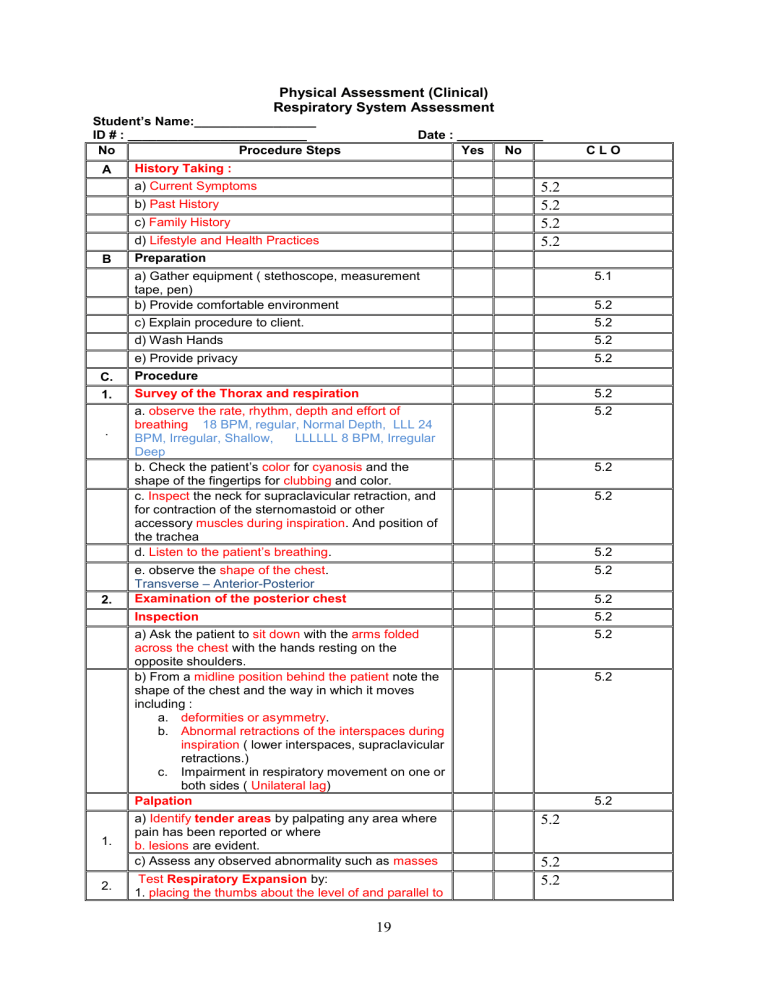
Physical Assessment (Clinical)
Respiratory System Assessment
Student’s Name:_________________
ID # : _________________________ Date : ____________
No Procedure Steps Yes No
A History Taking : a) Current Symptoms 5.2
5.2 b) Past History c) Family History d) Lifestyle and Health Practices
B Preparation a) Gather equipment ( stethoscope, measurement tape, pen) b) Provide comfortable environment c) Explain procedure to client. d) Wash Hands e) Provide privacy
C. Procedure
1. Survey of the Thorax and respiration
. a. observe the rate, rhythm, depth and effort of breathing 18 BPM, regular, Normal Depth, LLL 24
BPM, Irregular, Shallow, LLLLLL 8 BPM, Irregular
Deep b. Check the patient’s color for cyanosis and the shape of the fingertips for clubbing and color. c. Inspect the neck for supraclavicular retraction, and for contraction of the sternomastoid or other accessory muscles during inspiration . And position of the trachea d. Listen to the patient’s breathing . e. observe the shape of the chest .
Transverse – Anterior-Posterior
2. Examination of the posterior chest
Inspection
1.
2. a) Ask the patient to sit down with the arms folded across the chest with the hands resting on the opposite shoulders. b) From a midline position behind the patient note the shape of the chest and the way in which it moves including : a. deformities or asymmetry . b. Abnormal retractions of the interspaces during inspiration ( lower interspaces, supraclavicular retractions.) c. Impairment in respiratory movement on one or both sides ( Unilateral lag )
Palpation a) Identify tender areas by palpating any area where pain has been reported or where b. lesions are evident. c) Assess any observed abnormality such as masses
Test Respiratory Expansion by:
1. placing the thumbs about the level of and parallel to
5.2
5.2
5.2
5.2
5.2
C L O
5.2
5.2
5.2
5.2
5.2
5.2
5.2
5.2
5.2
5.1
5.2
5.2
5.2
5.2
5.2
5.2
19
the 10 th ribs and the hands Grasping the lateral rib cage, sliding them medially a bit in order to raise loose skin folds between thumbs and the spine
2. ask the patient to inhale deeply and watch for the
3. divergence of the thumbs during inspiration , feeling for the range & symmetry of the respiratory movements .
Feel for tactile fremitus ( palpable vibrations)
1. using either the ball of the or the ulnar surface of the hand .
2. Use both hands to compare sides
3. Ask the patient to repeat the words “ninety-nine” or
“ one, one, one .” If fremitus is faint ask the patient to speak more loudly.
4. palpate and compare symmetrical areas of the lungs .
Percussion a) Percuss the posterior chest while the patient keeps both arms crossed in front of the chest. b) press the distal interphalangeal joint of the middle finger ( pleximeter) firmly on the surfaces to be percussed avoiding surface contact by any other part of the hand.
c) with the middle finger of the other hand slightly flexed and relaxed strike over the pleximeter with a quick sharp but relaxed writ motion, using the tip of the flexor finger not the finger pad.
d) Percuss the thorax in symmetrical locations from the apices to the lung bases. twice in each location, compare two areas d. when percusing the lower posterior chest, stand somewhat to the side rather than directly behind the patient.
Identify the level of diaphragmatic dullness ( during quiet respiration) a. with the pleximeter finger held above and parallel to the expected level of the dullness, Percuss in progressive steps downward until dullness clearly replaces resonance. b. Check the level of this change near the middle of the hemithorax and also more laterally putting a point by a pen on each level.
Diaphragmatic excursion a. Ask the client to exhale fully then hold . b. Percuss for diaphragmatic dullness as above and put a point. c. Ask the client to take a deep breath and hold d. Again percuss for diaphragmatic dullness and put a point e. measure the distance between the two points
(5-6cm)
Auscultation
5.2
5.2
5.2
5.2
5.2
5.2
5.2
5.2
5.2
5.2
5.2
5.2
5.2
5.2
5.2
5.2
5.2
5.2
5.2
5.2
5.2
20
a. Listen to the breath sound with the diaphragm of the stethoscope after instructing the patient to breathe deeply through an open mouth .
1 . Bronchial: trachea.
2. Bronchovesicular: Primary Bronchi
3. Vesicular: Lungs.
1. Listen for Bronchovesicular sounds between
Scapulae
2. Listen for the Vesicular sounds over the lungs
3. Listen for any added sounds
5.2
5.2
5.2
5.2 b. If breath sounds heard located abnormally assess for transmitted voice sounds
1. Ask the patient to say “ ninety-nine” normally transmitted sound are muffled and indistinct .
2. Ask the patient to say “ee” normally a muffled long
E sound is heard.
3. Ask the patient to whisper “ninety-nine” or “one, two, three” . it will be heard faintly and indistinctly if heard at all.
Examination of the Anterior chest
3.
1
Inspection b.
Ask the patient to lie down into a supine position with the arms abducted .
if the patient has difficulty in breathing, he/she should be examined in the sitting position or with the head of the bed elevated at comfortable level.
Note the shape of the chest and the way in which it moves including : a. deformities or asymmetry . b. Abnormal retractions of the lower interspaces during inspiration c. Impairment in respiratory movement on one or both sides ( Unilateral lag )
Palpation
Identify tender areas by palpating any area where pain has been reported or where lesions are evident.
Assess any observed abnormality such as masses
5.2
5.2
5.2
5.2
5.2
5.2
5.2
5.2
5.2
5.2
5.2
5.2
2
Test Respiratory Expansion by:
1 . placing the thumbs along each costal margins with the hands along the lateral rib cage. As positioning the hands slide them medially a bit in order to raise loose skin folds between thumbs.
2. ask the patient to inhale deeply and watch for the divergence of the thumbs during inspiration, feeling for the range & symmetry of the respiratory movements .
3 Feel for tactile fremitus ( palpable vibrations)
21
5.2
5.2
1. using either the ball or the ulnar surface of the hand. Fremitus is decreased or absent over the pericardium .
2. Use both hands to compare sides
3. Ask the patient to repeat the words “ninety-nine” or
“ one, one, one .” If fremitus is faint ask the patient to speak more loudly.
4. palpate and compare symmetrical areas of the lungs.
Percussion c.
X a) Percuss the anterior and lateral chest while the patient keeps both arms abducted. press the distal interphalangeal joint of the middle finger ( pleximeter) firmly on the surfaces to be percussed avoiding surface contact by any other part of the hand b.
with the middle finger of the other hand slightly flexed and relaxed strike over the pleximeter with a quick sharp but relaxed wrist motion, using the tip of the flexor finger not the finger pad.
c. Percuss the thorax in symmetrical locations from the apices to the lung bases. twice in each location, compare two areas. In a woman to enhance percussion gently displace the breast with the left hand while percusing with the right d. when percusing the left chest, dullness will be heard at the level of the heart 3 rd to 5 th ICS, percussion of the left lung will be lateral to it.
Percuss for liver dullness with the pleximeter finger above and parallel to the expected upper border of the liver dullness, in progressive steps from resonance downward to the dullness in the right midclavicular line .
Auscultation a. Listen to the breath sound with the diaphragm of the stethoscope after instructing the patient to breathe deeply through an open mouth.
Listen for tracheal sounds at the suprasternal notch
Listen for bronchi al sounds over the manubrium
Listen for bronchovesicula r sounds at the 1 st and 2 nd interspaces. For primary bronchi
Listen for the Vesicular sounds over the lungs
Listen for any added sounds . b. If breath sounds heard located abnormally assess for transmitted voice sounds
D Procedure Termination
a) Put client in comfortable position according to health status b) Provide patient with reassurance c) Return back equipments d) Wash hands e) Document findings
5.2
5.2
5.2
5.2
5.2
5.2
5.2
5.2
5.2
5.2
5.2
5.2
5.2
5.2
5.2
5.2
5.2
5.2
5.2
5.2
5.2
5.2
5.2
5.2
22
23
