USE OF LOCAL ANESTHESIA, LECTURE-6
advertisement
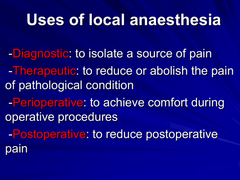
Uses of local anaesthesia -Diagnostic: to isolate a source of pain -Therapeutic: to reduce or abolish the pain of pathological condition -Perioperative: to achieve comfort during operative procedures -Postoperative: to reduce postoperative pain Diagnostic use Administration of local anaesthetic can be a useful way of finding the source of a patient's pain. An example of this is the pain of a pulpitis, which can be very difficult for both the patient and the dentist to isolate because of its tendency to be referred to other parts of the mouth or face Therapeutic use Local anaesthetics can, in themselves, constitute part of a treatment regimen for painful surgical conditions. The ability of the dentist to abolish pain for a patient. The use of a block technique to eliminate the pain of dry socket (localised osteitis) can be immensely helpful to the management of this very painful condition, particularly in the first few days. Inferior dental blocks of long-acting local anaesthetics such as bupivacaine can give total comfort for several hours, allowing patients to catch up on lost sleep and perhaps reduce the use of systemic analgesics to avoid overuse. Perioperative use • It reduces the arrhythmias, which are noted on electrocardiogram (ECG) during the surgery when significant afferent stimulation is taking place. This can be seen, for example, when a tooth is being elevated. It also provides local haemostasis to the operative site and provides immediate postoperative analgesia. Postoperative use After surgery with either local or general anaesthesia, the continuing effect of the anaesthetic is a most beneficial way of reducing patient discomfort. It helps to reduce or even eliminate the need for stronger (often narcotic) systemic analgesics. Basic Techniques of Local Anesthesia The various types of techniques used for deposition of these agents, in dentistry are as follows: (1) Surface or topical anesthesia, (2) Infiltration anesthesia, (3) Field block, and (4) Nerve block or conduction anesthesia . Surface or Topical Anesthesia By this method small terminal nerves in the surface area of the intact mucosa or the skin up to the depth of about 2 mm are anesthetised by application of a local anesthetic agent directly to the area . -Nerves Anesthetised Superficial nerve endings. -Indications i .Prior to the infiltration injection techniques or nerve blocks for making the insertion of the needle painless ii. Prior to carrying out incision and drainage of abscesses iii. Prior to removal of sutures . Spray I .The active in gradient is a suitable local anesthetic agent, such as 10% or 15% lignocaine hydrochloride in water base . II. Ethyl chloride spray: It produces anesthesia by refrigeration. When sprayed onto either mucous membrane or skin, it gets volatilised rapidly, and produces rapid anesthesia. Ointment It is used for similar purposes as spray. The active in gredient is a suitable local anesthetic agent, such as 5% lignocaine hydrochloride. Emulsion The active in gredient is a suitable local anesthetic agent, such as 2% lignocaine hydrochloride. Jet Injection Method: It is a technique by which a small amount of local anesthetic solution is expelled as a jet into submucosa without the use of a hypodermic needle. Specialised syringes are used for this technique. Infiltration Anesthesia or Local Infiltration This method is also known as terminal or peripheral anesthesia, as the induction of anesthesia is by the action of anesthetic agents on the terminal nerve fibers . Maxilla The maxilla has thin labial/buccal cortical plate; and moreover shows areas of porosity, and the compact bone presents numerous foramina which aid in absorption of local anesthetic solution. These factors, therefore, make the maxilla more favorable for infiltration anesthesia techniques . Mandible The bone is generally dense and has thicker cortical plates than maxilla, particularly in posterior region, more so in the region of external oblique ridge. Only the anterior part of mandible presents sufficient porosity, which is favorable for infiltration techniques. Advantages Easy and simple injection Very high success rate, and Good control of bleeding. Disadvantages The action is limited to a small area; hence considerable amount of solution has to be injected with multiple penetrations when large field is to be anesthetised. Indications This method is used when only the mucous membrane and the underlying connective tissues are to be anesthetised. Contraindications Presence of acute inflammation or infection at the site of injection. Applications Infiltration anesthesia helps in anesthetising (1) teeth as it affects dental nerves before they enter apical foramina; and (2) periodontal tissues . Other Applications Infiltration anesthesia is often used in conjunction with general anesthesia to reduce bleeding at the site of surgery; when vasoconstrictor is added to local anesthetic solution. Technique Needle: The recommended gauge is 25, 27 or 30; and the recommended length is 25 mm . Bevel of the needle: The bevel should be facing the bone . Point of insertion: It is in the middle of the area to be operated . Depth of penetration: It is beneath the mucous membrane into the connective tissue . This technique may require more than one needle insertions depending upon the extent of area to be anesthetised . Care should be taken to avoid injury to the tissues in the following ways : Avoid injecting the solution too rapidly. And cold solutions Avoid injecting too large a volume of the local anesthetic solution. Avoid injecting too superficially. And subperiostealy These situations will result in injury to the tissues in the form of pain at the time of injection, or persistent post-injection pain or sloughing of the overlying soft tissues . Types of Infiltration Anesthesia Submucosal or subcutaneous anesthesia Paraperiosteal or supraperiosteal anesthesia Subperiosteal anesthesia Intraligamentary (Periodontal ligament) anesthesia Intrapulpal anesthesia Intraosseous anesthesia Intraseptal anesthesia Palatal infiltration Technique: The local anesthetic solution is deposited in the immediate submucosal tissue layers. The solution diffuses through the interstitial tissues and reaches the terminal fibers of the nerve in the area of deposition of the local anesthetic solution . Procedure: The needle is inserted beneath the mucosal layers. Care should be exercised to avoid injecting too superficially. Excessive amounts injected superficially may lead to sloughing of the overlying tissues. Usually 0.25-0.5 ml of the local anesthetic solution is deposited. Submucosal Injection Paraperiosteal or Supraperiosteal Injection It is commonly called the local infiltration and is the most frequently used local anesthetic technique. The paraperiosteal injection is commonly used injection technique for obtaining anesthesia in the region of all maxillary teeth and mandibular anterior teeth because of thin cortical plates and abundant cancellous bone . Site of insertion: The needle is inserted through the mucosa, and the solution is deposited in close proximity to the periosteum or along the periosteum, in the vicinity of the apex of the tooth to be treated, as close to the bone as possible. Indications: This method is used for procedures in the entire maxilla and anterior mandible. In these areas, the cortical plates are thin, and there is abundant cancellous bone. The local anesthetic solution penetrates bone through Haversian canals. These canals are numerous near the apices of teeth near the surfaces. Pulpal anesthesia when treatment is limited to one or two teeth in maxilla, and anterior mandible. Soft tissue anesthesia for surgical procedures in a circumscribed area. Children and young adults. In children, this technique can be used in the posterior mandible to anesthetise deciduous molars as the cortical bone is thin in this region. Contraindications: Presence of acute inflammation or infection in the area of injection . Presence of dense bone covering the apices of teeth, as in maxillary first molar, because of overlying buttress of zygoma. Advantages: .High success rate .Technically easy injection .Usually atraumatic Disadvantages: The technique is not recommended for large areas because of: (i) need for multiple penetrations, (ii) the necessity to administer larger volumes of anesthetic solution, and (iii) satisfactory anesthesia cannot be always produced. Technique Needle: A 25 or 27 gauge short needle is recommended . Point of insertion: It is at the height of mucobuccal fold in the vicinity of the tooth to be anesthetised . Target area: The apical region or above the apex of the tooth to be anesthetised. Depth of insertion: Few millimeters. Bevel: The position of the bevel of the needle should be facing the bone. Landmarks: - Mucobuccal fold in the region of the tooth to be anesthetised. - Crown of the tooth. - Root contour of the tooth. -Position Procedure of the patient: The occlusal plane of maxillary teeth should be at an angle of 45° to the floor . -Position of the operator : i .For maxillary injections, for the right side, the operator stands by the side of the patient; and for the left side, the operator stands in front of the patient . ii. For mandibular injections, the operator stands by the side of the patient for the left side; and in front of the patient for the right side . -Preparation of the tissues at the site of injection with an antiseptic . -Application of topical anesthetic at the site of injection . -Retract the lip/cheek, pulling the tissues taut . -Take a preloaded syringe. Initially, hold it at an angle of 45° to the long axis of the tooth to be anesthetised, with the bevel of the needle facing the bone. Insert the needle at the height of mucobuccal fold, or a few millimeters away from the labial cortex . -Aspirate, if negative, deposit approximately 0.5 ml of the solution slowly over 20 seconds . -Depth of insertion: few millimeters . -Withdraw the syringe slowly . -Cover the needle . -Wait for 2-3 minutes, check for the signs and symptoms of anesthesia, and start the procedure.
