Bacterial Transformation Recombinant Selection (EXERCISE).doc
advertisement
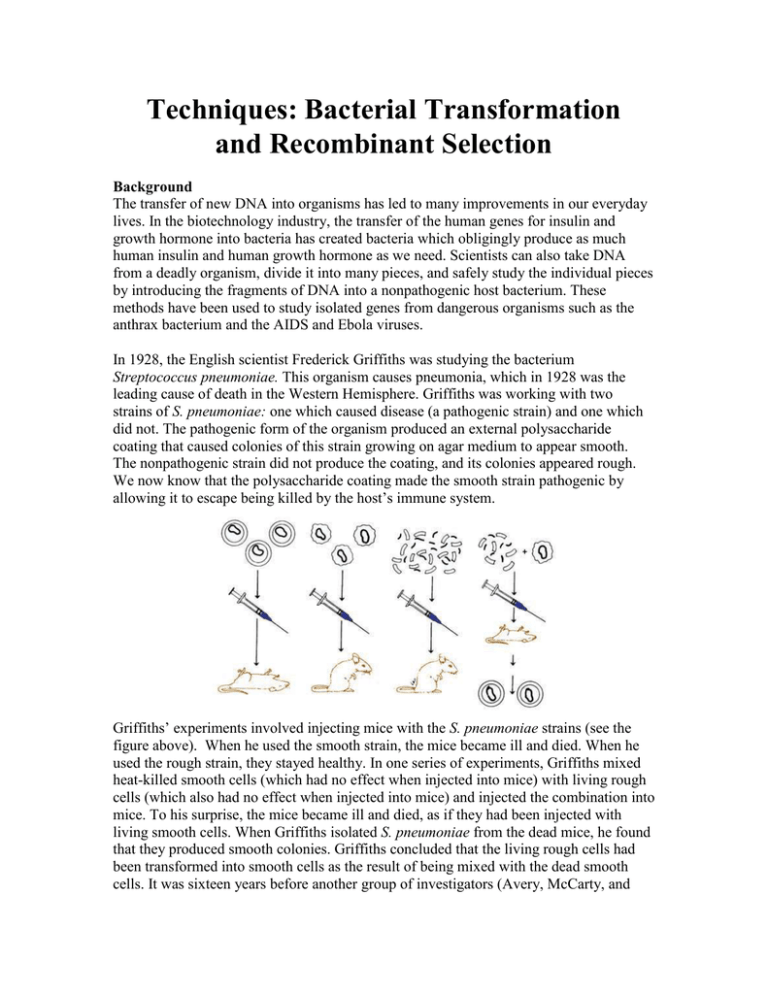
Techniques: Bacterial Transformation and Recombinant Selection Background The transfer of new DNA into organisms has led to many improvements in our everyday lives. In the biotechnology industry, the transfer of the human genes for insulin and growth hormone into bacteria has created bacteria which obligingly produce as much human insulin and human growth hormone as we need. Scientists can also take DNA from a deadly organism, divide it into many pieces, and safely study the individual pieces by introducing the fragments of DNA into a nonpathogenic host bacterium. These methods have been used to study isolated genes from dangerous organisms such as the anthrax bacterium and the AIDS and Ebola viruses. In 1928, the English scientist Frederick Griffiths was studying the bacterium Streptococcus pneumoniae. This organism causes pneumonia, which in 1928 was the leading cause of death in the Western Hemisphere. Griffiths was working with two strains of S. pneumoniae: one which caused disease (a pathogenic strain) and one which did not. The pathogenic form of the organism produced an external polysaccharide coating that caused colonies of this strain growing on agar medium to appear smooth. The nonpathogenic strain did not produce the coating, and its colonies appeared rough. We now know that the polysaccharide coating made the smooth strain pathogenic by allowing it to escape being killed by the host’s immune system. Griffiths’ experiments involved injecting mice with the S. pneumoniae strains (see the figure above). When he used the smooth strain, the mice became ill and died. When he used the rough strain, they stayed healthy. In one series of experiments, Griffiths mixed heat-killed smooth cells (which had no effect when injected into mice) with living rough cells (which also had no effect when injected into mice) and injected the combination into mice. To his surprise, the mice became ill and died, as if they had been injected with living smooth cells. When Griffiths isolated S. pneumoniae from the dead mice, he found that they produced smooth colonies. Griffiths concluded that the living rough cells had been transformed into smooth cells as the result of being mixed with the dead smooth cells. It was sixteen years before another group of investigators (Avery, McCarty, and MacLeod) showed that the “transforming principle,” the substance from the heat-killed smooth strain that caused the transformation, was DNA. Today, transformation is defined as the uptake and expression of free DNA by cells. Some bacteria undergo transformation naturally. Streptococcus pneumonia is one of these, as are Neisseria gonorrhea (the causative agent of gonorrhea) and Haemophilus influenza (the principle cause of meningitis in children under the age of 3). Each of these organisms has surface proteins that bind to DNA in the environment and transport it into the cell. Once inside the cell, the base sequence of the new DNA is compared to the bacterium’s DNA. If enough similarity in sequence exists, the new DNA can be substituted for the homologous region of the bacterium’s DNA. This is known as recombination. If the new DNA is not similar to the bacterium’s DNA, it is not incorporated into the genome and is broken down by intracellular enzymes. A model for DNA uptake in B. subtilis is shown below. Exogenous DNA binds to a select DNA receptor (discussed more below), ComEA, and perhaps to other protein(s) on the bacterial surface. The DNA undergoes endonucleolytic cleavage by NucA, leading to formation of new free termini. ComEA undergoes a conformational change, perhaps by bending at its hinge region, and delivers the DNA molecule to the channel formed by ComEC, which traverses the cytoplasmic membrane. The translocation of the DNA molecule to the cytosol as a single-stranded molecule is catalyzed by ComFA. The nontransported strand is degraded to acid soluble products by an unidentified nuclease. How do these organisms select for DNA that is likely to be beneficial to them? In Haemophilus and Neisseria, the DNA-binding proteins recognize and bind to particular base sequences, transporting in only DNA molecules containing those sequences. Each of these organisms has many copies of its recognition sequence in its genome. In Haemophilus, the recognition sequence is 11 bases long. One would expect this sequence to occur randomly once in 411 times, or once in about 5 million bases. Haemophilus, whose genome is about 5 million base pairs in size, has 600 copies of this sequence. The recognition sequences ensure that Haemophilus and Neisseria will mostly import DNA from members of their own species. Why would it be beneficial for a bacterium to bring in and use DNA from other members of its species? In Neisseria, transformation helps the organism to evade the immune system of its host (us!). Pathogenic Neisseria have stalk-like projections made of a protein called pilin on their surface. Our bodies’ immune system makes antibodies to the pilin protein, so we should be immune to reinfection by N. gonorrhea. But we are not. N. gonorrhea contains several versions of the pilin gene. In undergoing transformation by DNA containing different versions of the pilin gene, N. gonorrhea changes the version of pilin protein it synthesizes, evading recognition by the immune system’s antibodies. Natural transformations are not as rare as once thought. More and more often, scientists are discovering pathogenic organisms that transfer virulence genes between themselves. The major and minor pilin-like proteins in B. subtilis are color coded in the prior figure. Still, it is rare for most bacteria to take up DNA naturally from the environment. But by subjecting bacteria to certain artificial conditions, we can enable many of them to take up DNA. When cells are in a state in which they are able to take up DNA, they are referred to as competent. Making cells competent usually involves changing the ionic strength of the medium and heating the cells in the presence of positive ions (usually calcium). This treatment renders the cell membrane permeable to DNA. More recently, high voltage has also been used to render cells permeable to DNA in a process called electroporation. Once DNA is taken into a cell, the use of that DNA by the cell to make RNA and proteins is referred to as expression. In nature, the expression of the newly acquired DNA depends upon its being integrated into the DNA of the host cell. As discussed previously, the process of integration is known as recombination, and it requires that the new DNA be very similar in sequence to the host genome. However, researchers usually want to introduce into a cell DNA that is quite different from the existing genome. Such DNA would not be recombined into the genome and would be lost. To avoid this problem, scientists transform host cells with plasmid DNA. A plasmid is a small, circular piece of double-stranded DNA that has an origin of replication. An origin of replication is a sequence of bases at which DNA replication begins. Because they contain origins of replication, plasmids are copied by the host cell’s DNA replication enzymes, and each daughter cell receives copies of the plasmid upon cell division. Therefore, plasmids do not need to be recombined into the genome to be maintained and expressed. Additionally, since plasmids do not have to have DNA that is similar to the host cell’s DNA, DNA from other organisms can be maintained as a plasmid. Fortunately, it is relatively easy to introduce new DNA sequences into plasmids. Plasmids naturally occur in bacteria and yeast, and they are widely used as vehicles for introducing foreign DNA into these organisms. Thus far, no analogs of plasmids are known for higher plants and animals, which is one reason why genetic engineering is so much more difficult in higher organisms. In order to transform bacteria using plasmid DNA, biotechnologists must overcome two problems. Typically, cells that contain plasmid DNA have a disadvantage since cellular resources are diverted from normal cellular processes to replicate plasmid DNA and synthesize plasmid-encoded proteins. If a mixed population of cells with plasmids and cells without plasmids is grown together, then the cells without the plasmids grow faster. Therefore, there is always tremendous pressure on cells to get rid of their plasmids. To overcome this pressure, there has to be an advantage to the cells that have the plasmid. Additionally, we have to be able to determine which bacteria received the plasmid. We need a marker that lets us know that the bacterial colony we obtain at the end of our experiment was the result of a successful gene transfer. To accomplish both goals – making it advantageous for cells to retain plasmids and having a selectable marker so we can recognize when bacteria cells contain new DNA – we will use a system involving antibiotics and genes for resistance to antibiotics. You are probably already familiar with the terms antibiotic and antibiotic resistance from your own medical experiences (I am not making any inference to personal choices or lifestyles, here). The antibiotics used in transformation are very similar (or the same) as antibiotics used to treat bacterial infections in humans. In medical situations, the term antibiotic resistance has a very negative connotation since it indicates an infection that cannot be successfully treated with antibiotics. However, antibiotic resistance has a far more positive meaning in biotechnology, since it is the end result of a successful transformation experiment. In a typical transformation, billions of bacteria are treated and exposed to plasmid DNA. Only a fraction (usually fewer than 1 in 1000) will acquire the plasmid. Antibiotic resistance genes provide a means of finding the bacteria which acquired the plasmid DNA in the midst of all of those bacteria which did not. If the plasmid used to transform the DNA contains a gene for resistance to an antibiotic, then after transformation, bacteria that acquired the plasmid (transformants) can be distinguished from those that did not by plating the bacteria on a medium containing the antibiotic. Only the bacteria that acquired the plasmid will overcome the killing effect of the antibiotic and grow to form colonies on the plate. So the only colonies on an antibiotic plate after a transformation are the bacteria that acquired the plasmid. An individual colony is derived from one cell (which has divided by binary fission to produce the colony). This procedure accomplishes our two goals of giving an advantage to cells that have a plasmid so the plasmid is retained and of having a marker so we know our cells contain new DNA. Resistance to an antibiotic is known as a selectable marker; that is, we can select for cells that contain it. The selectable marker gene: beta-lactamase Ampicillin is a member of the penicillin family of antibiotics. The fungi that produce the antibiotics live in the soil, where they compete with soil bacteria. Ampicillin and the other penicillins help the fungi to compete by preventing the formation of the bacterial cell walls by inhibiting bacterial protein synthesis, thereby inhibiting bacterial growth. Ampicillin and the other penicillin antibiotics contain a chemical group called a betalactam ring. The ampicillin-resistance gene encodes beta-lactamase, an enzyme that destroys the activity of ampicillin by breaking down the beta-lactam ring. When a bacterium is transformed with a plasmid containing the beta-lactamase gene, it expresses the gene and synthesizes the beta-lactamase protein, which is secreted from the bacterium. As the ampicillin is broken down, the transformed bacterium retains its ability to form its cell wall and is able to replicate to form a colony. The colony continues to secrete betalactamase and forms a relatively ampicillin-free zone around it. After prolonged incubation, small satellite colonies of non-transformed bacteria that are still sensitive to ampicillin grow in these relatively ampicillin-free zones. This is why it is so important to plate your transformed bacteria to non-confluence and to not over-incubate your sample (in the figure below you can see the beta-galactosidase transformed “blue-colored” colonies surrounded by the non-transformed “clear-colored” satellite colonies). In the following figure on the next page, the ribbon structure (left) depicts a stimulated metallo-beta-lactamase. Two zinc ions (purple spheres) are in the enzyme's active site (with amino acids coordinating the metals represented as sticks). A beta-lactam antibiotic bound at the active site is represented as space-filled balls. The beta-lactamase gene is on the pUC19 plasmid employed as the vector DNA during the cloning of GFP. The presence of the beta-lactamase gene in the bacteria after they are transformed with the recombinant plasmid allows the bacteria to grow in ampicillincontaining media. The beta-lactamase gene is called a selectable marker because in the presence of ampicillin, it allows you to select for cells that have been successfully transformed with the recombinant plasmid. Bacteria cells that have not been transformed and do not contain the plasmid and its beta-lactamase gene will not be able to grow in the presence of ampicillin. Of course, there are color marker genes as well. An example of a color marker includes the lux genes, encoding the luciferase enzyme, which produces light in ocean bearing species. There are several genes involved in V. fischeri’s light production. The luciferase enzyme itself is composed of two different subunits encoded by two different genes. Synthesis of the aldehyde also requires the action of several genes, as does the regulation. Together, all of these genes required for the light production are called the lux genes. The group of lux genes from Vibrio fischeri has been placed in a plasmid. A word on laboratory safety… The bacterial host used in most molecular biology and teaching laboratories is Escherichia coli. Since E. coli is often associated with outbreaks of disease, concern may arise over its safety. Unfortunately, media reports on E. coli disease do not contain the background information necessary for understanding this issue. There are many naturally occurring strains of E. coli. They inhabit the lower intestinal tracts of many animals, including humans, cattle, and swine. The strains found in different animals vary genetically. The strain used in this lab is a weakened strain of the normal E. coli of the gut and does not cause disease. However, some genetic variants of E. coli do cause disease. These variants contain genes not found in the harmless organisms. These genes encode toxins and proteins that enable the organism to invade cells within the body. The nature of the disease genes varies; E. coli strains with different disease genes have been associated with several diseases. Some E. coli have genes for an enterotoxin, which causes the travelers’ diarrhea often called “Montezuma’s revenge.” The E. coli strain that causes the sometimes-fatal hemolyticuremic syndrome has genes that encode a toxin different from the travelers’ diarrhea toxin, and it also has genes that enable the bacteria to invade and disrupt cells lining the intestinal tract. Laboratory strains of E. coli used in molecular biology research do not contain any of these disease genes and are harmless under normal conditions. If introduced into a cut or into the eye, laboratory strains could conceivably cause infection, so standard safety precautions should be taken when handling the organisms. Every day, hundreds of scientists and their students handle these organisms (many in a rather cavalier manner) without any notable consequences. We do not recommend cavalier handling of any strain of E. coli, but the uneventful history of scientists with the organism should be reassuring. Purpose The purpose of this exercise is to provide you with information on gene transfer in E. coli. This includes a background on the history of transformation, a discussion of the science of transformation, an overview of plasmids available for transformation and an easy-to-follow procedure for transformation and recombinant selection. Materials per team (Bacterial Transformation – day 1) E. coli starter culture your pGFP cloned DNA sample LB liquid broth sterile 15 mL culturing tubes Bacterial Spreader sterile 1.5 mL microfuge tubes Beaker with ice Culture tube racks Water bath at 42°C 37oC incubator P20 pipetman yellow and blue pipetman tips (Recombinant Selection – day 2) Ultraviolet Light Box Agar plates from last period 50 mM CaCl2 LB agar plates LB agar Amp+ plates Parafilm P1000 pipetman P200 pipetman Procedures (Bacterial Transformation – day 1) – pGFP is transformed into bacteria and the recombinant bacteria are selected for on ampicillin laden Luria Broth Agar plates. 1. Mark one sterile 15 mL culturing tube as “+plasmid.” Mark another sterile 15 mL culturing tube as “–plasmid.” (Plasmid DNA will be added to the “+plasmid” tube; no plasmid DNA will be added to the “–plasmid” tube.) 2. Pre-chill the 15 mL culturing tubes by placing them both in ice. 3. A liquid culture of a transformation competent E. coli strain has been grown by your instructor (incubated at 37oC for 16 hr at 225 rpm). Carefully aliquot 1 mL of liquid culture into a 1.5 mL microfuge tube with a P1000 pipetman. 4. Place the 1.5 mL microfuge tube (containing the bacterial culture) into the centrifuge (be certain to balance the rotor with a tube containing an equal volume of water or another sample of bacteria from another group). 5. Centrifuge the sample at maximum speed (highest rpm setting) for 30 seconds. 6. Remove the sample from the centrifuge and carefully remove the supernatant (do not disturb the bacterial pellet) with a P1000 pipetman. 7. Gently resuspend the bacterial pellet in 500 uL of ice-cold calcium chloride (repeatedly pipetting in and out, about 5 times, with a P1000 pipetman). Examine the tube against light to confirm that no visible clumps of cells remain in the tube. The suspension should appear milky white. 8. Aliquot a 250 uL volume of resuspended cells to each 15 mL culturing tube (these are the big tubes pre-chilled in ice). Keep the 15 mL culturing tubes (with resuspended bacterial cells) in ice. 9. Use a P20 pipetman to add all of your plasmid DNA (20 L) to the “+plasmid” tube. Immerse the plasmid DNA directly into the cell suspension and quickly swirl the tube to mix the DNA with the cells. 10. Return the “+plasmid” tube to ice and incubate both tubes on ice for 15 minutes. 11. Following the 15 minute incubation on ice, “heat shock” the cells. Remove both tubes directly from ice and immediately immerse them in the 42°C water bath for 90 seconds. Gently agitate the tubes while they are in the water bath. Return both tubes directly to ice for at least 1 minute (the tubes stay on ice while you plate each of the samples). 12. Use a P1000 pipetman to add 250 μL Luria Broth (LB) to each tube. Gently tap the tubes with your finger to mix the LB with the cell suspension. Place the tubes in a test-tube rack on a rocker platform (with gentle agitation) at room temperature for a 15 minute recovery. 13. While the tubes are incubating, grab your labeled media plates (pre-heated in the 37oC incubator). Write your group lab name and date on them for identification. a. LB Amp+ plate “+plasmid.” This is an experimental plate. b. LB Amp+ plate “–plasmid.” This is a negative control. c. LB plate “+plasmid” and another LB plate “–plasmid.” These are positive controls to test the viability of the cells after they have gone through the transformation procedure. 14. Now you will remove some cells from each transformation tube and spread them on the plates. 15. Cells from the “–plasmid” tube should be spread on the “–plasmid” plates, and cells from the “+plasmid” tube should be spread on the “+plasmid” plates. 16. ALERT THE INSTRUCTOR YOU ARE READY TO PLATE THE CELLS. Use a P200 pipetman to add 100 μL of cells from the “–plasmid” transformation tube to each appropriate plate. Immediately spread the cells over the surface of the plate(s) using a bacterial spreader (THE INSTRUCTOR WILL SHOW YOU HOW TO APPLY THE CELLS USING ASEPTIC TECHNIQUE.) 17. Use a P200 pipetman to add 100 μL of cell suspension from the “+plasmid” tube to each appropriate plate. Immediately spread the cell suspension(s) as described in step 16. 18. Wrap the plates together with tape and place the plates upside down in the 37°C incubator and incubate. (Incubate them for approximately 24–36 hours in a 37°C incubator or 48–72 hours at room temperature.) 19. After the procedure, complete questions 1 through 12 of the worksheet. (Recombinant Selection – day 2) – Select for recombinants employing UV light. After the procedure, complete questions 13 through 20 of the worksheet. 1. Examine your plates and answer questions 13 through 20 of the worksheet. 2. Expose your LB Amp+ plate to Ultraviolet Light on the UV light box. Remember to affix the protective transparent cover, UV light is harmful to your eyes (UV light is incident upon you when standing in the Sunlight. It contains energy and can burn your tissues, i.e. sunburn or retinal cells of the eye.) WORKSHEET Bacterial Transformation and Recombinant Selection 1. In brief, describe an experiment in which you could show that the “transforming principle” is DNA based. _____________________________________________________________________ _____________________________________________________________________ _____________________________________________________________________ _____________________________________________________________________ _____________________________________________________________________ _____________________________________________________________________ _____________________________________________________________________ 2. Describe the model for DNA uptake in B. subtilis; elucidate the steps, not the specific proteins that are involved. _____________________________________________________________________ _____________________________________________________________________ _____________________________________________________________________ _____________________________________________________________________ _____________________________________________________________________ 3. How do organisms select for DNA that is likely to be beneficial to them? _____________________________________________________________________ _____________________________________________________________________ _____________________________________________________________________ _____________________________________________________________________ _____________________________________________________________________ 4. What are two popular and successful experimental processes available to render cells competent? How do these processes work? _____________________________________________________________________ _____________________________________________________________________ _____________________________________________________________________ _____________________________________________________________________ _____________________________________________________________________ 5. Why do plasmids not need to be recombined into the genome to be maintained and expressed? _____________________________________________________________________ _____________________________________________________________________ _____________________________________________________________________ _____________________________________________________________________ _____________________________________________________________________ 6. Why do scientists transform host cells with plasmid DNA? _____________________________________________________________________ _____________________________________________________________________ 7. Expression vectors produce a protein from a defined, genetic sequence inserted within its poly-linker cloning site. In general, why and how do expression vectors differ for prokaryotes (bacteria) and eukaryotes (human cells)? This is a tough question…think about it. (Hint: how do antibiotics selectively act against bacteria?) _____________________________________________________________________ _____________________________________________________________________ _____________________________________________________________________ _____________________________________________________________________ _____________________________________________________________________ 8. If a mixed population of cells with plasmids and cells without plasmids is grown together, the cells without the plasmids grow faster. Why? _____________________________________________________________________ _____________________________________________________________________ _____________________________________________________________________ _____________________________________________________________________ 9. Why would we use a system involving antibiotics and genes for resistance to antibiotics during recombinant bacterial selection? _____________________________________________________________________ _____________________________________________________________________ _____________________________________________________________________ _____________________________________________________________________ 10. Specifically, how do beta-lactam antibiotics work. What do they target in the cell? Are they effective in Eukaryotic (human) cells? Why or why not? (Hint: Google) _____________________________________________________________________ _____________________________________________________________________ _____________________________________________________________________ _____________________________________________________________________ _____________________________________________________________________ 11. Now I get to torture your mind. Why are two zinc ions (divalent cations) important for the function of the active site of the beta-lactamase enzyme? (Hint: Pulling Electrons) _____________________________________________________________________ _____________________________________________________________________ _____________________________________________________________________ _____________________________________________________________________ _____________________________________________________________________ _____________________________________________________________________ 12. In the chart below, predict your results. Write “yes” or “no,” depending on whether you think the plate will show growth. Give the reason(s) for your predictions 13. Observe the colonies through the petri plate lids. Do not open the plates. Record your observed results in the spaces above. If your observed results differed from your predictions, explain what you think may have occurred. 14. Count the number of individual colonies; mark each colony as it is counted (with a sharpie). If the cell growth is too dense to count individual colonies, record “lawn.” LB “+plasmid” LB “–plasmid” LB Amp+ “+plasmid” LB Amp+ “–plasmid” 15. Compare and contrast the number of colonies on each of the following pairs of plates. What does each pair of results tell you about the experiment? LB “+plasmid” & LB “–plasmid” LB Amp+ “–plasmid” & LB “–plasmid” LB Amp+ “+plasmid” & LB Amp+ “–plasmid” LB Amp+ “+plasmid” & LB “+plasmid” 16. What are you selecting for in this experiment? (i.e., what allows you to identify which bacteria have taken up the plasmid?) _____________________________________________________________________ _____________________________________________________________________ _____________________________________________________________________ _____________________________________________________________________ 17. What does the phenotype of the transformed colonies tell you? _____________________________________________________________________ _____________________________________________________________________ _____________________________________________________________________ _____________________________________________________________________ 18. What one plate would you first inspect to conclude that the transformation occurred successfully? Why? _____________________________________________________________________ _____________________________________________________________________ _____________________________________________________________________ _____________________________________________________________________ 19. Transformation efficiency is expressed as the number of antibiotic-resistant colonies per μg of plasmid DNA. The object is to determine the mass of plasmid that was spread on the experimental plate and that was, therefore, responsible for the transformants (the number of colonies) observed. Because transformation is limited to only those cells that are competent, increasing the amount of plasmid used does not necessarily increase the probability that a cell will be transformed. A sample of competent cells is usually saturated with the addition of a small amount of plasmid, and excess DNA may actually interfere with the transformation process. a. Determine the total mass (in μg) of plasmid used. Remember, you used 10 μL of plasmid at a concentration of 0.005 μg/μL. [total mass = volume × concentration] b. Calculate the total volume of cell suspension prepared. c. Now calculate the fraction of the total cell suspension that was spread on the plate. [volume suspension spread/total volume suspension = fraction spread] d. Determine the mass of plasmid in the cell suspension spread. [total mass plasmid (a) × fraction spread (c) = mass plasmid DNA spread] e. Determine the number of colonies per μg plasmid DNA. Express your answer in scientific notation. notation. [colonies observed/mass plasmid spread (d) = transformation efficiency] 20. What factors might influence transformation efficiency? Explain the effect of each factor you mention. _____________________________________________________________________ _____________________________________________________________________ _____________________________________________________________________ _____________________________________________________________________ _____________________________________________________________________

