ZOOL 409 Lab Week 3
advertisement
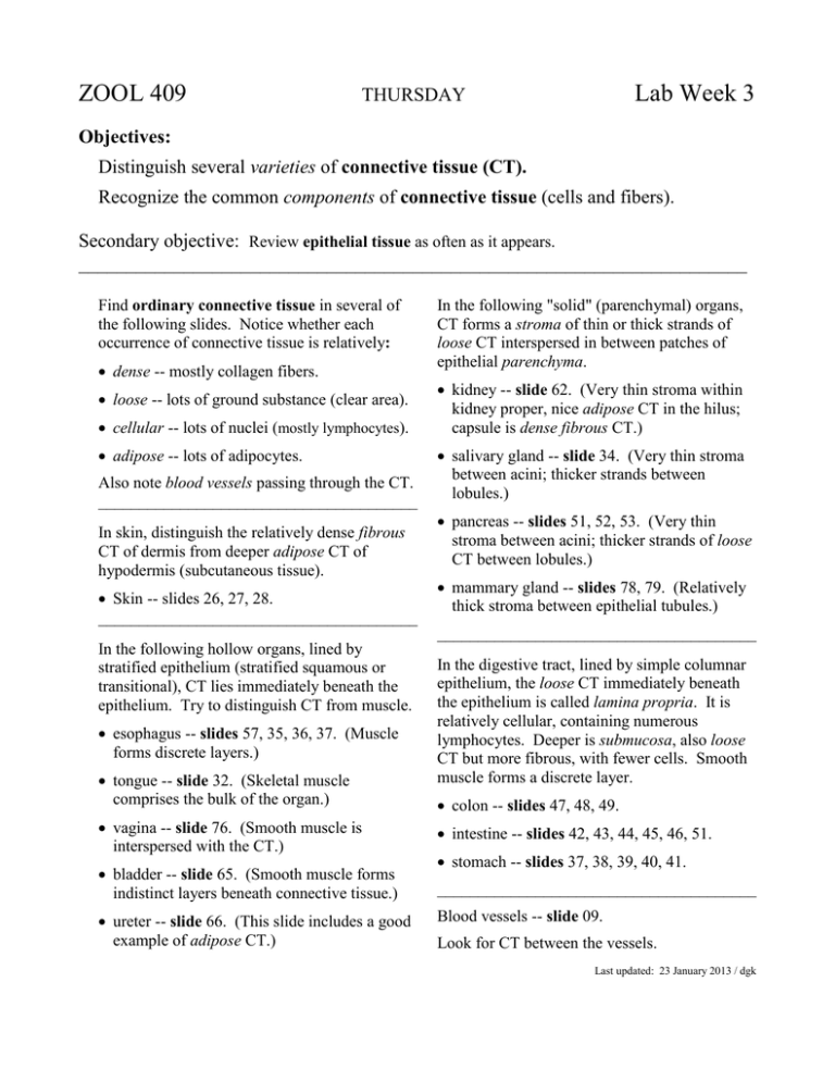
ZOOL 409 Lab Week 3 THURSDAY Objectives: Distinguish several varieties of connective tissue (CT). Recognize the common components of connective tissue (cells and fibers). Secondary objective: Review epithelial tissue as often as it appears. ______________________________________________________________________ Find ordinary connective tissue in several of the following slides. Notice whether each occurrence of connective tissue is relatively: dense -- mostly collagen fibers. loose -- lots of ground substance (clear area). cellular -- lots of nuclei (mostly lymphocytes). adipose -- lots of adipocytes. Also note blood vessels passing through the CT. _______________________________________ In skin, distinguish the relatively dense fibrous CT of dermis from deeper adipose CT of hypodermis (subcutaneous tissue). Skin -- slides 26, 27, 28. _______________________________________ In the following "solid" (parenchymal) organs, CT forms a stroma of thin or thick strands of loose CT interspersed in between patches of epithelial parenchyma. kidney -- slide 62. (Very thin stroma within kidney proper, nice adipose CT in the hilus; capsule is dense fibrous CT.) salivary gland -- slide 34. (Very thin stroma between acini; thicker strands between lobules.) pancreas -- slides 51, 52, 53. (Very thin stroma between acini; thicker strands of loose CT between lobules.) mammary gland -- slides 78, 79. (Relatively thick stroma between epithelial tubules.) _______________________________________ In the following hollow organs, lined by stratified epithelium (stratified squamous or transitional), CT lies immediately beneath the epithelium. Try to distinguish CT from muscle. esophagus -- slides 57, 35, 36, 37. (Muscle forms discrete layers.) tongue -- slide 32. (Skeletal muscle comprises the bulk of the organ.) In the digestive tract, lined by simple columnar epithelium, the loose CT immediately beneath the epithelium is called lamina propria. It is relatively cellular, containing numerous lymphocytes. Deeper is submucosa, also loose CT but more fibrous, with fewer cells. Smooth muscle forms a discrete layer. colon -- slides 47, 48, 49. vagina -- slide 76. (Smooth muscle is interspersed with the CT.) intestine -- slides 42, 43, 44, 45, 46, 51. bladder -- slide 65. (Smooth muscle forms indistinct layers beneath connective tissue.) _______________________________________ ureter -- slide 66. (This slide includes a good example of adipose CT.) stomach -- slides 37, 38, 39, 40, 41. Blood vessels -- slide 09. Look for CT between the vessels. Last updated: 23 January 2013 / dgk Connective Tissue Practice Quizzes To test/confirm your recognition of connective tissue, you are encouraged to take one or more ungraded quizzes. □ Please indicate when you are ready for a quiz. Your instructor will then provide a slide (see boxes at right) on which you should locate each of the listed structures. Each of the listed structures should be readily apparent in an appropriate region. Use the eyepiece pointer at low power to indicate layers or regions, higher power for cells and other small details. Do not call for a quiz until you feel confident of your ability to recognize the listed structures. You may also ask your instructor about any other structure that catches your interest. Note: As always, you are also encouraged to seek confirmation at any time for your recognition of structures on slides from your reference slide set -- particularly of any features not included in these quizzes. □ □ □ Connective tissues of tongue: ____lamina propria / CT papillae ____fibrous CT associated with muscle ____adipocytes ____blood vessel Connective tissues of salivary gland: ____lamina propria ____stromal CT ____adipocytes ____blood vessel Connective tissues of esophagus: ____lamina propria ____submucosa ____lymphoid CT ____blood vessel Connective tissues of stomach and duodenum: ____lamina propria ____submucosa ____lymphoid CT ____blood vessel Last updated: 18 January 2013 / dgk ZOOL 409 Lab Week 3 January 29, TUESDAY __________________________________________________________________________________ Visit a working laboratory and see how histological specimens are prepared. Meet in Room 2055 of Life Science 3 (immediately south of LSII). Objectives: Appreciate the technologies and skills involved in preparing histological specimens: Fixation Embedding Sectioning Mounting sections on slides Staining *** NOTE *** You may be given a "souvenir" slide during this histotechniques field trip. Keep this slide use later in the course. Navigation from LSII: Leave LSII at southwest corner, cross driveway southward to entrance of LS3. Elevator is to the left of the entry. On 2nd floor, from elevator turn left, left at cross-hallway, left again. Room 2055 is the fourth door on the left. [For stairway instead of elevator, take cross-hallway to left of entry, turn left (east) at end of cross-hallway. Stairway is second door on left. Turn left (east) at top of stairway. Room 2055 is the second door on the left.] This slide includes specimens from several organs, and displays some details more clearly than slides in our reference collections. As you learn to recognize various organs, look for and study them on this slide as well as on the slides available in laboratory. Navigation from parking lot entrance, south-west corner of LS3: Stairway is a few steps to the right of the entrance. Turn left (east) at top of stairway. Room 2055 is the second door on the left. Last updated: 20March 2013 / dgk Today is "field trip" day for ZOOL 409 Histology. Meet at Rm 2055 in LS III (the adjacent building to the south)


