ARGININE-GLYCINE-ASPARTIC ACID MOTIFS TURNIP YELLOW MOSAIC VIRUS COAT PROTEIN By: Jonathan Halama
advertisement
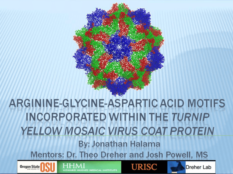
ARGININE-GLYCINE-ASPARTIC ACID MOTIFS INCORPORATED WITHIN THE TURNIP YELLOW MOSAIC VIRUS COAT PROTEIN By: Jonathan Halama Mentors: Dr. Theo Dreher and Josh Powell, MS Non-enveloped T=3 icosahedral symmetry of identical sequence. Single stranded positive sense RNA virus. Coat protein (CP) coded by sub-genomic strand. 180 coat proteins (CP), 60 as 1 of 3 quasi-equivalent sub-units. 60 A sub-units: pentameric capsomeres. 60 B sub-units and 60 C sub-units: hexameric capsomeres. RGD MOTIFS AND INTEGRIN Arginine-glycine-aspartic acid (RGD) A tripeptide motif used by Integrins as an attachment point. RGD motifs are found within viral proteins and facilitate cellular adhesion. Integrin (protein receptor) Integrin is a ligand used by some cells and viruses for adhesion and cell signaling. Stem cells up-regulate Integrin production. PURPOSE: STEM CELL GROWTH Utilize RGD motif as a binding site for Integrin attachment. Multiple binding sites on exterior capsid. Regulate stem cell adhesion. Control stem cell growth and differentiation. Mutant TYMV capsids are “vital for designing compatible biomaterials for tissue engineering purposes” (Gagandeep Kaur). HYPOTHESIS Arginine-Glycine-Aspartic acid (RGD) motifs can be engineered on the exterior of Turnip yellow mosaic virus (TYMV) coat proteins (CP) while maintaining the capsids: ability to assemble, structural stability, and allow binding of Integrin to the RGD motifs. COAT PROTEIN MONOMER RGD 44 Monomer COAT PROTEIN TRIMER RGD 44 Trimer ASSEMBLED CAPSID ASSEMBLED CAPSID INTEGRIN/RGD MOTIF INTERACTIONS INTEGRIN/RGD MOTIF INTERACTIONS INTEGRIN/RGD MOTIF INTERACTIONS MUTANTS Mutants Subcloning Agro infiltrated Capsid Extraction Successful Assembly RGD 44 Completed Completed Completed Yes RGD 55 Completed Completed Completed RGD 61 Completed Completed RGD 102 Completed Completed RGD 103 Completed Completed RGD 161 Completed Completed RGD 162 Completed Completed RGD 184 Incomplete Completed Completed SUB-CLONING AGROINFILTRATION AND RECOMBINATION VIRAL CAPSID EXTRACTION 10X NaPO4 Bentonite solution RGD TYMV deveined plant tissue samples MgSO4 2x Low Speed Spins: 13G High Speed Spin: 50K RPM Low Speed Spin: 13G High Speed Spin: 50K RPM Resuspend Isolated Capsids PROTEIN VISUALIZATION Coomassie gel Ladder Wild-type RGD 44 RGD 55 RGD 103 RGD 162 27 kDa 15 kDa Western blot Western blot prepared using: •SDS-PAGE. •Primary antibody: Rabbit anti-TYMV IgG. •Secondary antibody: Goat anti-rabbit IgG Horseradish Peroxidase (HRP) conjugate. Coomassie gel prepared using: •Sodium dodecyl sulfate polyarcylamide gel electrophoresis (SDS-PAGE). •Coomassie Brilliant Dye. 27 kDa 15 kDa CAPSID VISUALIZATION RGD 44 (60K) Electron Microscopy RGD 44 (125K) 50 nm Wild-type 28nm 50 nm CONCLUSIONS… RDG 44 expressed the mutant coat protein within the plant system, and self assembled forming capsids. RDG 55, 103, or 162 coat proteins were not detected within the plant system. Future work will determine if the protein was or was not being expressed. RDG 61, 62, 102, and 161 require further cloning techniques; transformation into Argobacterium, argoinfiltrated into the plant system, and capsid extraction to determine if coat protein can be expressed and self assemble within plant tissue. SPECIAL THANKS TO THE FOLLOWING… HHMI (Howard Hughes Medical Institute) URISC (Undergraduate Research, Innovation, Scholarship and Creativity) Dr. Kevin Ahern The Dreher Lab Special thanks to Josh Powell REFERENCES Kaur, G. / Valarmathi, M.T. / Potts, J.D. / Wang, Q. , Biomaterials, 29 (30), p.4074-4081,Oct 2008
