Development of Flexible Linker for Validation of Introduction
advertisement

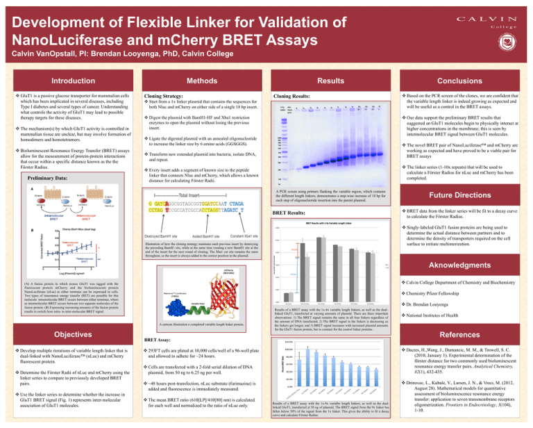
Development of Flexible Linker for Validation of NanoLuciferase and mCherry BRET Assays Calvin VanOpstall, PI: Brendan Looyenga, PhD, Calvin College Introduction v GluT1 is a passive glucose transporter for mammalian cells which has been implicated in several diseases, including Type I diabetes and several types of cancer. Understanding what controls the activity of GluT1 may lead to possible therapy targets for these diseases. v The mechanism(s) by which GluT1 activity is controlled in mammalian tissue are unclear, but may involve formation of homodimers and homotetramers. v Bioluminescent Resonance Energy Transfer (BRET) assays allow for the measurement of protein-protein interactions that occur within a specific distance known as the the Förster Radius. Preliminary Data: Methods Cloning Strategy: Results Cloning Results: v Start from a 1x linker plasmid that contains the sequences for both Nluc and mCherry on either side of a single 18 bp insert. v Digest the plasmid with BamH1-HF and Xba1 restriction enzymes to open the plasmid without losing the previous insert. v Based on the PCR screen of the clones, we are confident that the variable length linker is indeed growing as expected and will be useful as a control in the BRET assays. v Our data support the preliminary BRET results that suggested an GluT1 molecules begin to physically interact at higher concentrations in the membrane; this is seen by intermolecular BRET signal between GluT1 molecules. v Ligate the digested plasmid with an annealed oligonucleotide to increase the linker size by 6 amino acids (GGSGGS). v The novel BRET pair of NanoLuciferase™ and mCherry are working as expected and have proved to be a viable pair for BRET assays v Transform new extended plasmid into bacteria, isolate DNA, and repeat. v The linker series (1-10x repeats) that will be used to calculate a Förster Radius for nLuc and mCherry has been completed. v Every insert adds a segment of known size to the peptide linker that connects Nluc and mCherry, which allows a known distance for calculating Förster Radii. A Conclusions A PCR screen using primers flanking the variable region, which contains the different length linkers, demonstrates a step-wise increase of 18 bp for each step of oligonucleotide insertion into the parent plasmid. BRET Results: Future Directions v BRET data from the linker series will be fit to a decay curve to calculate the Förster Radius. v Singly-labeled GluT1 fusion proteins are being used to determine the actual distance between partners and to determine the density of transporters required on the cell surface to initiate multimerization. B Illustration of how the cloning strategy maintains each previous insert by destroying the preceding BamH1 site, while at the same time creating a new BamH1 site at the end of the insert for the next round of cloning. The Xba1 cut site remains the same throughout, so the insert is always added to the correct position in the plasmid. Aknowledgments v Calvin College Department of Chemistry and Biochemistry (A) A fusion protein in which mouse GluT1 was tagged with the fluorescent protein mCherry and the bioluminescent protein NanoLuciferase (nLuc) at either terminus can be expressed in cells. Two types of reasonance energy transfer (RET) are possible for this molecule: intramolecular BRET occurs between either terminus, where as intermolecular BRET occurs between two separate molecules of the fusion protein. (B) Expressing increasing amounts of the fusion protein results in swtich from intra- to inter-molecular BRET signal. v Chemistry Pfizer Fellowship A cartoon illustration a completed variable length linker protein. Objectives v Develop multiple iterations of variable length linker that is dual-linked with NanoLuciferase™ (nLuc) and mCherry fluorescent protein. v Determine the Förster Radii of nLuc and mCherry using the linker series to compare to previously developed BRET pairs. v Use the linker series to determine whether the increase in GluT1 BRET signal (Fig. 1) represents inter-molecular association of GluT1 molecules. v Dr. Brendan Looyenga Variable linker Results of a BRET assay with the 1x-4x variable length linkers, as well as the duallinked GluT1, transfected at varying amounts of plasmid. There are three important observations: 1) The BRET signal remains the same in all four linkers regardless of the amount of DNA transfected; 2) The BRET signal in the linkers is decreasing as the linkers get longer; and 3) BRET signal increases with increased plasmid amounts for the GluT1 fusion protein, but is constant for the control linker proteins . BRET Assay: References v Dacres, H.,Wang, J., Dumancic, M. M., & Trowell, S. C. (2010, January 1). Experimental determination of the förster distance for two commonly used bioluminescent resonance energy transfer pairs. Analytical Chemistry, 82(1), 432-435. v 293FT cells are plated at 10,000 cells/well of a 96-well plate and allowed to adhere for ~24 hours. v Cells are transfected with a 2-fold serial dilution of DNA plasmid, from 50 ng to 6.25 ng per well. v ~48 hours post-transfection, nLuc substrate (furimazine) is added and fluorescence is immediately measured. v The mean BRET ratio (610[LP]/410[80] nm) is calculated for each well and normalized to the ratio of nLuc only. v National Institutes of Health Results of a BRET assay with the 1x-9x varialbe length linkers, as well as the duallinked GluT1, transfected at 50 ng of plasmid. The BRET signal from the 9x linker has fallen below 50% of the signal from the 1x linker. This gives the ability to fit a decay curve and calculate Förster Radius v Drinovec, L., Kubale, V., Larsen, J. N., & Vreci, M. (2012, August 28). Mathematical models for quantitative assessment of bioluminescence resonance energy transfer: application to seven transmembrane receptors oligomerization. Frontiers in Endocrinology, 3(104), 1-10.
