Shuchi 2011-03-24 Visualizing the complete virus:
advertisement

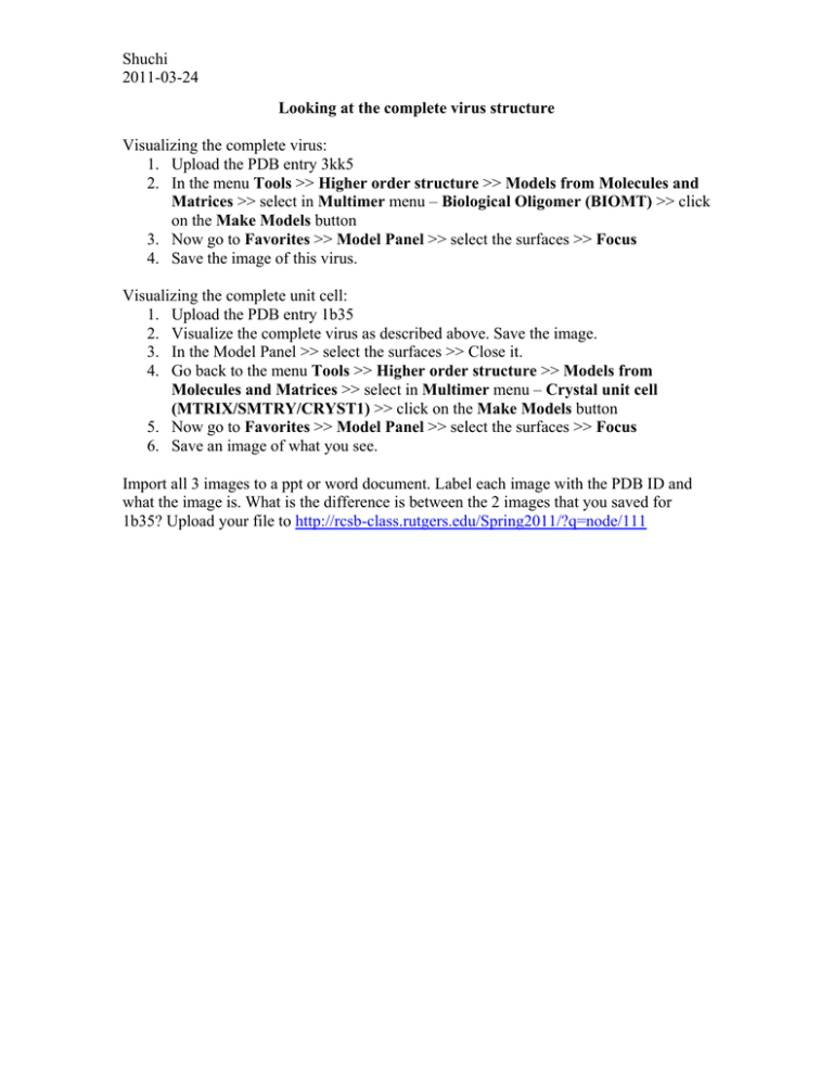
Shuchi 2011-03-24 Looking at the complete virus structure Visualizing the complete virus: 1. Upload the PDB entry 3kk5 2. In the menu Tools >> Higher order structure >> Models from Molecules and Matrices >> select in Multimer menu – Biological Oligomer (BIOMT) >> click on the Make Models button 3. Now go to Favorites >> Model Panel >> select the surfaces >> Focus 4. Save the image of this virus. Visualizing the complete unit cell: 1. Upload the PDB entry 1b35 2. Visualize the complete virus as described above. Save the image. 3. In the Model Panel >> select the surfaces >> Close it. 4. Go back to the menu Tools >> Higher order structure >> Models from Molecules and Matrices >> select in Multimer menu – Crystal unit cell (MTRIX/SMTRY/CRYST1) >> click on the Make Models button 5. Now go to Favorites >> Model Panel >> select the surfaces >> Focus 6. Save an image of what you see. Import all 3 images to a ppt or word document. Label each image with the PDB ID and what the image is. What is the difference is between the 2 images that you saved for 1b35? Upload your file to http://rcsb-class.rutgers.edu/Spring2011/?q=node/111
