MR in radiotherapy planning Presentation outline Why? 7/22/2014
advertisement

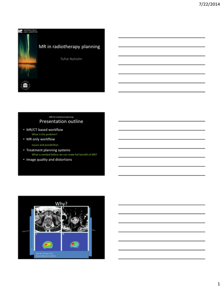
7/22/2014 MR in radiotherapy planning Tufve Nyholm MRI for treatment planning Presentation outline • MR/CT based workflow What is the problem? • MR only workflow Issues and possibilities • Treatment planning systems What is needed before we can make full benefit of MR? • Image quality and distortions Why? Functional imaging Van der Heide et al. Future Medicin (2011) 1 7/22/2014 Workflow of today Images Registration / Target definition Treatment planning Problem Registration RESULTS OFA MULTI-INSTITUTIONAL BENCHMARK TEST FOR CRANIAL CT/MR IMAGE REGISTRATION Ulin et al. Int. J. Radiation Oncology Biol. Phys., Vol. 77, No. 5, pp. 1584–1589, 2010 Method MR and CT examinations of head case were sent to 45 clinics for registration Result Standard deviation: 2.2 mm Switch from CT based to MR based workflow Imaging Target definition Treatment planning 2 7/22/2014 Which issues needs to be addressed Geometrical accuracy Which issues needs to be addressed Geometrical accuracy Imaging of markers Which issues needs to be addressed Geometrical accuracy Imaging of markers Imaging in fixation McJURY et al. BJR 2011 Hanvey et al. Phys Med Biol. 2011 3 7/22/2014 Which issues needs to be addressed Geometrical accuracy Imaging of markers Imaging in fixation Dose calculation Which issues needs to be addressed Geometrical accuracy Imaging of markers Imaging in fixation Dose calculation Positioning reference Which issues needs to be adressed Geometrical accuracy Imaging of markers Not specific for MR only radiotherapy Imaging in fixation Dose calculation Specific for MR only radiotherapy Positioning reference 4 7/22/2014 How? Dose calculation Positioning reference CT equivalent MR MR signal T2w CT UTE (Ultra short echo time) Manual segmentation and bulk densities 5 7/22/2014 Manual segmentation and bulk densities Segment all bone, air and lung on the MR images Assign bulk densities Advantage based on litterature • High dosimetric accuracy • Intuitive method Disadvantage • Very time demanding • Geometrical accuracy? Calculate the dose on the bulk CT and display on the MR Difference in calculated dose Registration based Registration based Advantage • High dosimetric accuracy • Automatic Disadvantage • Geometrical accuracy? Dowling JA, et al. Int J RadiatOncol Biol Phys 2012 6 7/22/2014 Automatic segmentation and bulk densities Air Air/soft tissue Soft tissue Bone Bone/soft tissue -1000 0 1000 HU 2000 Air Air/soft tissue TE: 3.76 ms Soft tissue Bone Bone/soft tissue TE: 0.07 ms 7 7/22/2014 Air Air/soft tissue Flip: 60 degrees Soft tissue Bone Bone/soft tissue Flip: 10 degrees Automatic segmentation and bulk densities T2 UTE1 UTE2 Fat Water Vessel Advantage • High dosimetric accuracy S-CT • High geometrical accuracy Disadvantage • Not intuitive – Need for QC CT • Not shown to work below the H&N region T1 Segmentation of Air Direct voxel-vise conversion 8 7/22/2014 Air Air/soft tissue Soft tissue Flip: 60 degrees Bone Bone/soft tissue Flip: 10 degrees Voxel by voxel esstimation Substitute CT (S-CT) CT MR 2 Uncertainty map 700 HU MR 1 0 HU Johansson et al. “CT substitute derived from MRI sequences with ultrashort echo time,” Med. Phys. 2011 Johansson et al. ” Voxel-wise uncertainty in CT substitute derived from MRI” Med. Phys. 2012 Gamma maps 3%-3mm Geometrical accuracy CT HU > 400 S-CT CT and S-CT Treatment planning of intracranial targets on MRI derived substitute CT data Jonsson et al. – Radiotherapy and oncology CT only S-CT only 9 7/22/2014 Comparison CT/substitute-CT Dose calculation DRR Advantage CT • High dosimetric accuracy • High geometrical accuracy Disadvantage • Not intuitive – Need for QC • Not shown to work below H&N region S-CT TPS and MRI Target delineation Dose calculations Today: Tomorrow: Image quality (some personal reflextions) The definition of the target volume and OAR’s are the most important steps of the radiotherapy workflow Point with MR: Image quality better possiblities to define relevant volumes So we should do what we can to maintain the image quality.. This could mean that RT needs to adopt to MR instead of the other way around 10 7/22/2014 Image quality (some personal reflextions) Distortions Patient dependent Gradient none-linearity Target defintion Dose calculation (MR only workflow) Registrations (CT/MR workflow) Distortions Patient dependent Chemical Shift Susceptibility Gradient none-linearity “Patient-related image distortions can be reduced by applying relatively strong slice selection and frequency-encoding gradients, for example, by setting the pixel bandwidth at twice the water-fat shift, all patient-related distortions are expected to be smaller than the pixel size.” 440 Hz at 1.5 T 880 Hz at 3 T 11 7/22/2014 Distortions Patient dependent Gradient none-linearity Distortions 1.5T 3T Umeå European capital of culture 2014 Thank You! 12


