LETTERS Mechanism of coupled folding and binding of an intrinsically disordered protein
advertisement
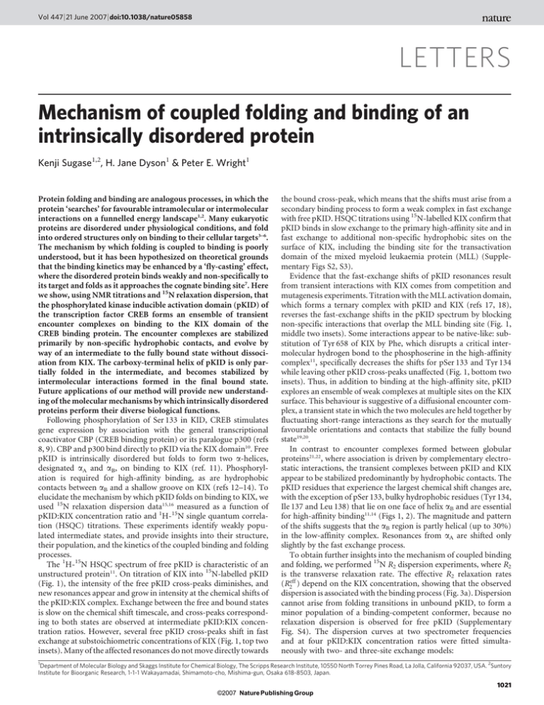
Vol 447 | 21 June 2007 | doi:10.1038/nature05858
LETTERS
Mechanism of coupled folding and binding of an
intrinsically disordered protein
Kenji Sugase1,2, H. Jane Dyson1 & Peter E. Wright1
Protein folding and binding are analogous processes, in which the
protein ‘searches’ for favourable intramolecular or intermolecular
interactions on a funnelled energy landscape1,2. Many eukaryotic
proteins are disordered under physiological conditions, and fold
into ordered structures only on binding to their cellular targets3–6.
The mechanism by which folding is coupled to binding is poorly
understood, but it has been hypothesized on theoretical grounds
that the binding kinetics may be enhanced by a ‘fly-casting’ effect,
where the disordered protein binds weakly and non-specifically to
its target and folds as it approaches the cognate binding site7. Here
we show, using NMR titrations and 15N relaxation dispersion, that
the phosphorylated kinase inducible activation domain (pKID) of
the transcription factor CREB forms an ensemble of transient
encounter complexes on binding to the KIX domain of the
CREB binding protein. The encounter complexes are stabilized
primarily by non-specific hydrophobic contacts, and evolve by
way of an intermediate to the fully bound state without dissociation from KIX. The carboxy-terminal helix of pKID is only partially folded in the intermediate, and becomes stabilized by
intermolecular interactions formed in the final bound state.
Future applications of our method will provide new understanding of the molecular mechanisms by which intrinsically disordered
proteins perform their diverse biological functions.
Following phosphorylation of Ser 133 in KID, CREB stimulates
gene expression by association with the general transcriptional
coactivator CBP (CREB binding protein) or its paralogue p300 (refs
8, 9). CBP and p300 bind directly to pKID via the KIX domain10. Free
pKID is intrinsically disordered but folds to form two a-helices,
designated aA and aB, on binding to KIX (ref. 11). Phosphorylation is required for high-affinity binding, as are hydrophobic
contacts between aB and a shallow groove on KIX (refs 12–14). To
elucidate the mechanism by which pKID folds on binding to KIX, we
used 15N relaxation dispersion data15,16 measured as a function of
pKID:KIX concentration ratio and 1H-15N single quantum correlation (HSQC) titrations. These experiments identify weakly populated intermediate states, and provide insights into their structure,
their population, and the kinetics of the coupled binding and folding
processes.
The 1H-15N HSQC spectrum of free pKID is characteristic of an
unstructured protein11. On titration of KIX into 15N-labelled pKID
(Fig. 1), the intensity of the free pKID cross-peaks diminishes, and
new resonances appear and grow in intensity at the chemical shifts of
the pKID:KIX complex. Exchange between the free and bound states
is slow on the chemical shift timescale, and cross-peaks corresponding to both states are observed at intermediate pKID:KIX concentration ratios. However, several free pKID cross-peaks shift in fast
exchange at substoichiometric concentrations of KIX (Fig. 1, top two
insets). Many of the affected resonances do not move directly towards
the bound cross-peak, which means that the shifts must arise from a
secondary binding process to form a weak complex in fast exchange
with free pKID. HSQC titrations using 15N-labelled KIX confirm that
pKID binds in slow exchange to the primary high-affinity site and in
fast exchange to additional non-specific hydrophobic sites on the
surface of KIX, including the binding site for the transactivation
domain of the mixed myeloid leukaemia protein (MLL) (Supplementary Figs S2, S3).
Evidence that the fast-exchange shifts of pKID resonances result
from transient interactions with KIX comes from competition and
mutagenesis experiments. Titration with the MLL activation domain,
which forms a ternary complex with pKID and KIX (refs 17, 18),
reverses the fast-exchange shifts in the pKID spectrum by blocking
non-specific interactions that overlap the MLL binding site (Fig. 1,
middle two insets). Some interactions appear to be native-like: substitution of Tyr 658 of KIX by Phe, which disrupts a critical intermolecular hydrogen bond to the phosphoserine in the high-affinity
complex11, specifically decreases the shifts for pSer 133 and Tyr 134
while leaving other pKID cross-peaks unaffected (Fig. 1, bottom two
insets). Thus, in addition to binding at the high-affinity site, pKID
explores an ensemble of weak complexes at multiple sites on the KIX
surface. This behaviour is suggestive of a diffusional encounter complex, a transient state in which the two molecules are held together by
fluctuating short-range interactions as they search for the mutually
favourable orientations and contacts that stabilize the fully bound
state19,20.
In contrast to encounter complexes formed between globular
proteins21,22, where association is driven by complementary electrostatic interactions, the transient complexes between pKID and KIX
appear to be stabilized predominantly by hydrophobic contacts. The
pKID residues that experience the largest chemical shift changes are,
with the exception of pSer 133, bulky hydrophobic residues (Tyr 134,
Ile 137 and Leu 138) that lie on one face of helix aB and are essential
for high-affinity binding11,14 (Figs 1, 2). The magnitude and pattern
of the shifts suggests that the aB region is partly helical (up to 30%)
in the low-affinity complex. Resonances from aA are shifted only
slightly by the fast exchange process.
To obtain further insights into the mechanism of coupled binding
and folding, we performed 15N R2 dispersion experiments, where R2
is the transverse relaxation rate. The effective R2 relaxation rates
(R2eff ) depend on the KIX concentration, showing that the observed
dispersion is associated with the binding process (Fig. 3a). Dispersion
cannot arise from folding transitions in unbound pKID, to form a
minor population of a binding-competent conformer, because no
relaxation dispersion is observed for free pKID (Supplementary
Fig. S4). The dispersion curves at two spectrometer frequencies
and at four pKID:KIX concentration ratios were fitted simultaneously with two- and three-site exchange models:
1
Department of Molecular Biology and Skaggs Institute for Chemical Biology, The Scripps Research Institute, 10550 North Torrey Pines Road, La Jolla, California 92037, USA. 2Suntory
Institute for Bioorganic Research, 1-1-1 Wakayamadai, Shimamoto-cho, Mishima-gun, Osaka 618-8503, Japan.
1021
©2007 Nature Publishing Group
LETTERS
NATURE | Vol 447 | 21 June 2007
½KIXkon
?
/
pKIDzKIX
pKID:KIX (twosite)
koff
pKIDzKIX
½KIXkon
?
/
kIB
pKID:KIX
? pKID:KIX (threesite)
/
koff
Free (F)
kBI
Intermediate (I)
Bound (B)
The two-site exchange model resulted in physically unrealistic
parameters; the effective microscopic dissociation constant KD
derived from the fits (41 mM for pSer 133) is inconsistent with the
macroscopic KD (1.3 mM) determined by isothermal titration calorimetry23. In contrast, the three-site exchange model fits better
(reduced x2 5 1.12 versus 1.60) and gives a mean KD (1.5 6
1.0 mM) that is in excellent agreement with isothermal titration
calorimetry (Table 1).
The dispersion curves for residues in the helical regions of bound
pKID were analysed in clusters, with the folding and unfolding rate
constants kIB and kBI fixed for all residues in each cluster. Three
clusters were found (Table 1); residues 124–128 in aA form a single
cluster (I), while there are two clusters in aB (clusters II and III,
comprising residues 133–139 and 140–144, respectively) which
appear to interact independently with KIX in both the encounter
complex and the intermediate.
We simulated 15N NMR spectra24 using the parameters of Table 1.
Even though the rate of exchange between bound and intermediate
states (kex,IB) is fast (kex,IB . DvIB, where DvIB is the chemical shift
difference between the intermediate and bound states), the overall
exchange process between free and fully bound states is slow (Fig. 3b),
1:0
as observed experimentally. Because exchange between I and B is fast,
the ‘bound’ shift (given by Dv*) is a population weighted average of
the chemical shifts in the I and B states. Comparison of the chemical
shift difference between the free and intermediate states, DvFI, to
Dv* provides a measure of the extent of folding of each residue in
the intermediate. For residues in cluster I, the average value of DvFI/
Dv* is greater than 0.9, suggesting that the aA helix is nearly fully
folded. Although aA residues (and Arg 131) make nearly native interactions in the intermediate, they experience additional millisecond
timescale fluctuations that contribute to relaxation dispersion. In
contrast, the average DvFI for residues 133–138 and 141 in the aB
helix is only about 70% of the fully bound shift, showing that this
helix is incompletely folded (Fig. 3c). Thus, in the intermediate, the
critical hydrophobic residues that anchor the pKID aB helix to the
KIX hydrophobic groove continue to search for the favourable intermolecular contacts that stabilize the final bound state.
To elucidate the mechanism of folding of aB, we looked for correlations between the chemical shifts determined from the dispersion
data and equilibrium chemical shift changes measured from HSQC
spectra, Dd (Fig. 3d). A linear correlation is observed between Dv*
and Dd. Importantly, Dv* correlates significantly better with the
differences in chemical shift between the bound state and the
ensemble of weak non-specific complexes, rather than with shift
differences relative to free pKID. This strongly suggests that the transient encounter complexes are essential for productive binding, and
evolve to the high-affinity complex without dissociation of pKID
from KIX.
As aB is incompletely folded in the intermediate identified from the
dispersion experiments, we conclude that this state is a pKID folding
1:1.2
110
S142
S129
15N
chemical shift (p.p.m.)
Concentration ratio (pKID:KIX)
pS133
115
L141
R135
S143
R124
L138
120
I137
Y134
R131
125
L138
10.0
9.5
9.0
8.0
8.5
1H chemical shift (p.p.m.)
Figure 1 | Chemical shift changes on pKID binding to KIX. 1H-15N HSQC
titration of [15N]-pKID with unlabelled KIX in which pKID:KIX
concentration ratios ranged from 1:0 to 1:1.2. The cross-peak colour changes
gradually from blue (free) to red (bound) according to the concentration
ratio. Exchange between the free and fully bound states of pKID is slow on
the chemical shift timescale. At substoichiometric concentrations of KIX,
several of the pKID cross-peaks shift away from their free positions in fast
exchange. The open circles (magenta) represent the fully bound chemical
shifts for the fast exchange process. These were estimated using the
dissociation constant (680 mM) determined by fitting the shifts observed for
KIX resonances in the presence of excess pKID (Supplementary Fig. S2).
Selected resonances are labelled. An expanded view of the central part of this
figure is shown in Supplementary Fig. S7. Insets show expanded views of the
7.5
7.0
cross-peaks for pSer 133 and Leu 138 at substoichiometric pKID:KIX ratios,
as follows. Top row, fast-exchange shift of the resonances of pSer 133 (left)
and Leu 138 (right) at pKID:KIX mole ratios of 1:0, 1:0.1, 1:0.2, 1:0.3, 1:0.4.
The contours are coloured according to the scale at the top of the figure.
Middle row, reversal of fast-exchange shifts for pSer 133 (left) and Leu 138
(right) on titration with MLL. The spectra correspond to pKID:KIX:MLL
mole ratios of 1:0:0 (dark blue contours), 1:0.3:0 (cyan), 1:0.3:0.1 (green),
1:0.3:0.2 (yellow) and 1:0.3:0.3 (red). Bottom row, HSQC cross-peaks of
pSer 133 (left) and Leu 138 (right) in the presence of wild-type KIX (cyan
contours) and the Tyr658Phe mutant (red) at a pKID:KIX mole ratio of
1:0.1. For reference, the cross-peaks for free pKID are shown in dark blue.
The scale is slightly expanded relative to the other insets.
1022
©2007 Nature Publishing Group
LETTERS
NATURE | Vol 447 | 21 June 2007
intermediate that differs significantly from the encounter complex.
Strictly, the coupled folding and binding process should be modelled
as four-site exchange (Free«Encounter«Intermediate«Bound).
However, exchange of pKID between the free state and the encounter
complex is fast and does not contribute to 15N R2 relaxation; dispersion arises from exchange between the encounter complex, the folding intermediate, and the fully bound state. At steady state, the steps
F«E«I (where E designates the encounter complex) can be replaced
by a single-step process with effective association and dissociation
rate constants kon
~kon kEI =(koff zkEI ) and koff
~koff kIE =(koff zkEI )
(ref. 25).
The values of kon
show that all residues of pKID except Arg 131
bind to KIX at similar rates (average kon
5 6.3 3 106 M21 s21) to
form the intermediate. The larger kon for Arg 131 suggests a possible
role for this residue in electrostatic steering26. In contrast, the effec
tive rate of dissociation from the intermediate, koff
, differs signifi
cantly for individual residues. Residues in aA (cluster I; average koff
is
21
12.7 s ) dissociate most slowly, whereas those in aB have more than
fivefold larger koff
values suggesting that they interact more weakly
a
αA
1.5
P
with KIX in the intermediate. The intermolecular contacts made by
aB residues are stabilized following evolution of the intermediate to
the final state. For the fully bound state, the effective microscopic
dissociation constants KD and the apparent dissociation rates koff are
the same for the residues in each cluster. Thus, the critical interfacial
hydrophobic residues in the aA and aB helices dissociate from the
fully bound state at the same rates (average koff is 6.5 s21 for Leu 128,
Tyr 134, Leu 138 and Leu 141). The increased apparent koff rates for
Asp 140 and Asp 144 cannot represent physical dissociation of pKID
from KIX because neighbouring residues have much smaller koff.
Rather, the dispersion data probably contain contributions from
additional exchange processes, reflecting either transient local distortions of this region of the aB helix or fluctuating electrostatic
interactions between the Asp side chains and His 602 and Lys 606
of KIX.
Our studies provide insights into the mechanism by which an
intrinsically disordered protein folds on binding to its target.
Application of our method to additional complexes will be needed
to establish whether coupled folding and binding is ubiquitous, or if
alternative mechanisms, such as conformational selection (Supplementary Fig. S1), also operate.
αB
b
pSer 133
pSer 133
30
0.5
0.0
120
125
130
135
Residue
140
145
R2eff (s –1
)
40
1.0
20
0
1.10
1,000
)
1.05
(mM
1/τ
2,000
on
1.00
cp (
rati
t
s –1
n
3,000 0.95
ce
)
con
KIX
c
3
15N
2
1
0
chemical shift (p.p.m.)
4 d
b
αA
αB
|∆ω*| (p.p.m.)
∆δav (p.p.m.)
a
L138
L141
L128
3
pS133
2
R125
1
R135
K136
R131
R124
Y134
S143
0
Figure 2 | Characteristics of the encounter complex. a, Chemical shift
differences for pKID backbone amide (1H and 15N) on binding to KIX,
calculated according to the formula Ddav 5 !((DdHN)2 1 (DdN/5)2), where
DdHN and DdN are the amide proton and nitrogen chemical shift differences,
respectively. The horizontal line corresponds to the average chemical shift
difference (0.131 p.p.m.) between the free and the fast-exchanging states.
Blue bars indicate residues that exhibit chemical shift deviations exceeding
the average by more than 1 s.d. (0.289 p.p.m.), and cyan and pale green bars
denote residues with intermediate and below average shift differences,
respectively. The open bars indicate chemical shift differences between free
pKID and fully bound pKID. The locations of the two helices aA and aB of
pKID and the phosphorylation site are shown at the top of the figure.
b, Structure of the high-affinity pKID:KIX complex11, showing a backbone
representation of pKID bound to the KIX surface. Chemical shift differences
between the free and fast-exchanging states are mapped onto the pKID
backbone using the same colour scheme as in a. The location of the pSer 133
side chain is shown. The figure was prepared using MOLMOL29.
0
1
2
3
4
|∆δ| (p.p.m.)
5
6
Figure 3 | Analysis of R2 dispersion experiments. a, 15N R2 relaxation
dispersion profile for pSer 133 of pKID recorded at 800 MHz (filled circles)
and 500 MHz (open circles). Dispersion curves for 1 mM [15N]-pKID in the
presence of 0.95, 1.00, 1.05 and 1.10 mM KIX are shown. b, Simulated 15N
NMR spectra of pSer 133 at a 15N resonance frequency of 81.1 MHz. The
spectra were simulated using the parameters obtained by fitting the R2
dispersion curves to the three-site exchange model. Chemical shifts in the
free form are set to zero. The pKID:KIX concentration ratios are depicted
using the same colour scheme as Fig. 1. c, Extent of folding in the
intermediate state as indicated by DvFI/Dv* ratios, displayed on the
structure of the high-affinity complex. Red, DvFI/Dv* . 0.9; orange,
0.5 , DvFI/Dv* , 0.9; yellow, DvFI/Dv* , 0.5. d, Correlation of 15N
chemical shift differences (Dv*) determined from the R2 dispersion
measurements with equilibrium shift differences (Dd). Chemical shift
differences between free pKID and the fully bound state (DdFB) are shown as
black squares, and between the encounter complex and fully bound state
(DdEB) are shown as red circles, with matching colours for the lines of best fit.
The scatter plots show a better correlation between Dv* and DdEB
(slope 5 0.95, R2 5 0.94) than between Dv* and DdFB (slope 5 0.79,
R2 5 0.79). This difference was shown to be statistically significant using an
F-test. The simulated and experimental chemical shift differences were
obtained from the simulated 15N NMR spectra (b) and from 1H-15N HSQC
spectra (Fig. 1), respectively. DdEB was calculated using the estimated fully
bound chemical shifts of the encounter complex (magenta circles in Fig. 1).
For resonances that undergo no fast-exchange shifts, the corresponding
DdFB values are used.
1023
©2007 Nature Publishing Group
LETTERS
NATURE | Vol 447 | 21 June 2007
Table 1 | R2 dispersion curve fitting with a three-site exchange model
Cluster
Residue
DvFI{
(p.p.m.)
DvFB{
(p.p.m.)
kon* 3 106
(M21 s21)
koff*
(s21)
I
I
I
I
Arg 124
Arg 125
Ile 127
Leu 128
1.33 (0.02){
1.17 (0.03)
1.47 (0.03)
2.31 (0.03)
1.71 (0.02){
0.84 (0.03)
1.87 (0.03)
2.79 (0.03)
6.5 (0.1){
5.0 (0.2)
7.3 (0.2)
8.1 (0.2)
13.2 (0.6){
15 (1)
13 (1)
9.9 (0.7)
Arg 130
Arg 131
2.12 (0.11)
1.77 (0.03)
4.01 (0.06)
3.25 (0.02)
5.4 (0.3)
15 (1)
II
II
II
II
II
II
pSer 133
Tyr 134
Arg 135
Lys 136
Leu 138
Asn 139
1.76 (0.04)
1.72 (0.05)
1.89 (0.05)
1.66 (0.03)
3.12 (0.05)
1.22 (0.05)
2.53 (0.04)
2.44 (0.06)
2.66 (0.05)
2.48 (0.03)
3.90 (0.05)
0.89 (0.03)
III
III
III
III
Asp 140
Leu 141
Ser 143
Asp 144
0.6 (0.1)
2.73 (0.05)
0.92 (0.06)
0.19 (0.06)
Ala 145
1.11 (0.01)
Apparent koff1
(s21)
kIB
(s21)
kBI
(s21)
KD* | |
(mM)
9.3
10.4
9.0
6.9
394 (8){
928 (10){
1.4
2.1
1.2
0.9
155 (21)
4.5 (0.3)
5.2
4.4
2241 (25)
34 (2)
78 (6)
957 (7)
1.0
0.3
5.5 (0.2)
6.4 (0.3)
8.9 (0.3)
7.1 (0.2)
7.3 (0.3)
4.2 (0.3)
54 (6)
56 (7)
65 (6)
55 (4)
48 (5)
137 (14)
6.2
6.5
7.5
6.4
5.6
15.8
1718 (10)
224 (10)
1.1
1.0
0.9
0.9
0.8
3.8
0.59 (0.01)
3.88 (0.06)
0.63 (0.03)
0.43 (0.02)
5.5 (0.5)
6.3 (0.3)
4.3 (0.2)
5.9 (0.4)
601 (43)
69 (6)
89 (12)
213 (33)
61.6
7.1
9.1
21.9
2852 (16)
326 (18)
11.3
1.1
2.1
3.7
0.81 (0.01)
7.62 (0.05)
16.9 (0.6)
10.1
340 (5)
511 (7)
1.3
See text for definitions of quantities in column headings.
{ Chemical shift differences DvFI and DvFB (in rad s21) were used in curve fitting; values shown here, in units of p.p.m., are calculated according to the formula: Dv(p.p.m.) 5 Dv(rad s21)/(2pB0),
where B0 is the 15N resonance frequency.
{ Values in parentheses are standard deviations.
1 Apparent koff are calculated as described in Methods.
| | Effective dissociation constants are calculated as described in Methods. Excluding the outlier Asp 140, the mean KD* obtained by fitting the dispersion data with a three-site exchange model is
1.5 6 1.0 mM.
6.
METHODS SUMMARY
15
N R2 relaxation rates were measured for [15N]-pKID:KIX at 1:0.95, 1:1.0, 1:1.05
and 1:1.10 concentration ratios using relaxation-compensated constant-time
Carr-Purcell-Meiboom-Gill pulse sequences15,27. Dispersion curves at all four
pKID:KIX concentration ratios and at two spectrometer frequencies were
fitted simultaneously for each residue using the program GLOVE28. Data were
fitted to two-site exchange (F«B) and three-site exchange (F«I«B) models.
The fitting parameters are a population-average intrinsic relaxation rate R20 ,
and koff
, folding and
effective association and dissociation rate constants kon
unfolding rate constants kIB and kBI, chemical shift differences between each pair
of states DvFI and DvFB, and total protein concentrations [pKID]0 and [KIX]0.
The parameters kIB, kBI and DvFI apply only to three-site exchange. The rate
constant for the binding process (FRB for two-site exchange and FRI for threesite exchange) depends on the concentration of free KIX, which is obtained from
the effective dissociation constant KD , determined independently for each
residue by fitting the dispersion data, and the total concentrations of pKID
and KIX:
qffiffiffiffiffiffiffiffiffiffiffiffiffiffiffiffiffiffiffiffiffiffiffiffiffiffiffiffiffiffiffiffiffiffiffiffiffiffiffiffiffiffiffiffiffiffiffiffiffiffiffiffiffiffiffiffiffiffiffiffiffiffiffiffiffiffiffiffiffiffiffiffiffiffiffiffiffiffiffiffi
2
1
½KIX~
{KD za½KIX0 {½pKID0 z KD {a½KIX0 z½pKID0 z4a½KIX0 KD ,
2
where a is the pKID:KIX concentration ratio (a 5 0.95, 1, 1.05 or 1.1). The
dispersion data at the different concentration ratios are related to each other
=kon
, while
through this equation. For the two-site exchange model, KD ~koff
=kon
|kBI =(kBI zkIB ), in which koff
is modified
for three-site exchange, KD ~koff
|kBI =(kBI zkIB )). In the
by the rate constants kBI and kIB (apparent koff ~koff
three-site exchange model, the intermediate is also a bound state and pKID can
is scaled by
dissociate from KIX only from the intermediate state. Therefore, koff
the population ratio: pI/(pI 1 pB) 5 kBI/(kBI 1 kIB).
Full Methods and any associated references are available in the online version of
the paper at www.nature.com/nature.
7.
8.
9.
10.
11.
12.
13.
14.
15.
16.
17.
Received 28 December 2006; accepted 18 April 2007.
Published online 23 May 2007.
1.
2.
3.
4.
5.
Tsai, C. J., Kumar, S., Ma, B. Y. & Nussinov, R. Folding funnels, binding funnels, and
protein function. Protein Sci. 8, 1181–1190 (1999).
Levy, Y., Cho, S. S., Onuchic, J. N. & Wolynes, P. G. A survey of flexible protein
binding mechanisms and their transition states using native topology based
energy landscapes. J. Mol. Biol. 346, 1121–1145 (2005).
Wright, P. E. & Dyson, H. J. Intrinsically unstructured proteins: Re-assessing the
protein structure-function paradigm. J. Mol. Biol. 293, 321–331 (1999).
Dyson, H. J. & Wright, P. E. Coupling of folding and binding for unstructured
proteins. Curr. Opin. Struct. Biol. 12, 54–60 (2002).
Dyson, H. J. & Wright, P. E. Intrinsically unstructured proteins and their functions.
Nature Rev. Mol. Cell Biol. 6, 197–208 (2005).
18.
19.
20.
21.
22.
Uversky, V. N. Natively unfolded proteins: A point where biology waits for physics.
Protein Sci. 11, 739–756 (2002).
Shoemaker, B. A., Portman, J. J. & Wolynes, P. G. Speeding molecular recognition
by using the folding funnel: The fly-casting mechanism. Proc. Natl Acad. Sci. USA
97, 8868–8873 (2000).
Chrivia, J. C. et al. Phosphorylated CREB binds specifically to nuclear protein CBP.
Nature 365, 855–859 (1993).
Kwok, R. P. S. et al. Nuclear protein CBP is a coactivator for the transcription factor
CREB. Nature 370, 223–226 (1994).
Parker, D. et al. Phosphorylation of CREB at Ser133 induces complex
formation with CPB via a direct mechanism. Mol. Cell. Biol. 16, 694–703
(1996).
Radhakrishnan, I. et al. Solution structure of the KIX domain of CBP bound to the
transactivation domain of CREB: A model for activator:coactivator interactions.
Cell 91, 741–752 (1997).
Shih, H. M. et al. A positive genetic selection for disrupting protein-protein
interactions: Identification of CREB mutations that prevent association with the
coactivator CBP. Proc. Natl Acad. Sci. USA 93, 13896–13901 (1996).
Shaywitz, A. J., Dove, S. L., Kornhauser, J. M., Hochschild, A. & Greenberg, M. E.
Magnitude of the CREB-dependent transcriptional response is determined by
the strength of the interaction between the kinase-inducible domain of CREB
and the KIX domain of CREB-binding protein. Mol. Cell. Biol. 20, 9409–9422
(2000).
Parker, D. et al. Analysis of an activator:coactivator complex reveals an essential
role for secondary structure in transcriptional activation. Mol. Cell 2, 353–359
(1998).
Loria, J. P., Rance, M. & Palmer, A. G. A relaxation-compensated Carr-PurcellMeiboom-Gill sequence for characterizing chemical exchange by NMR
spectroscopy. J. Am. Chem. Soc. 121, 2331–2332 (1999).
Mulder, F. A., Mittermaier, A., Hon, B., Dahlquist, F. W. & Kay, L. E. Studying
excited states of proteins by NMR spectroscopy. Nature Struct. Biol. 8, 932–935
(2001).
Goto, N. K., Zor, T., Martinez-Yamout, M., Dyson, H. J. & Wright, P. E.
Cooperativity in transcription factor binding to the coactivator CREB-binding
protein (CBP). The mixed lineage leukemia protein (MLL) activation domain
binds to an allosteric site on the KIX domain. J. Biol. Chem. 277, 43168–43174
(2002).
De Guzman, R. N., Goto, N. K., Dyson, H. J. & Wright, P. E. Structural basis for
cooperative transcription factor binding to the CBP coactivator. J. Mol. Biol. 355,
1005–1013 (2006).
Gabdoulline, R. R. & Wade, R. C. Biomolecular diffusional association. Curr. Opin.
Struct. Biol. 12, 204–213 (2002).
Berg, O. G. & von Hippel, P. H. Diffusion-controlled macromolecular interactions.
Annu. Rev. Biophys. Biophys. Chem. 14, 131–160 (1985).
Tang, C., Iwahara, J. & Clore, G. M. Visualization of transient encounter complexes
in protein-protein association. Nature 444, 383–386 (2006).
Volkov, A. N., Worrall, J. A. R., Holtzmann, E. & Ubbink, M. Solution structure and
dynamics of the complex between cytochrome c and cytochrome c peroxidase
1024
©2007 Nature Publishing Group
LETTERS
NATURE | Vol 447 | 21 June 2007
23.
24.
25.
26.
27.
28.
determined by paramagnetic NMR. Proc. Natl Acad. Sci. USA 103, 18945–18950
(2006).
Zor, T., De Guzman, R. N., Dyson, H. J. & Wright, P. E. Solution structure of the KIX
domain of CBP bound to the transactivation domain of c-Myb. J. Mol. Biol. 337,
521–534 (2004).
Johnson, C. S. Jr & Moreland, C. G. The calculation of NMR spectra for many-site
exchange problems. J. Chem. Ed. 50, 477–483 (1973).
Shoup, D. & Szabo, A. Role of diffusion in ligand binding to macromolecules and
cell-bound receptors. Biophys. J. 40, 33–39 (1982).
Selzer, T. & Schreiber, G. New insights into the mechanism of protein-protein
association. Proteins Struct. Funct. Genet. 45, 190–198 (2001).
Tollinger, M., Skrynnikov, N. R., Mulder, F. A., Forman-Kay, J. D. & Kay, L. E. Slow
dynamics in folded and unfolded states of an SH3 domain. J. Am. Chem. Soc. 123,
11341–11352 (2001).
McElheny, D., Schnell, J. R., Lansing, J. C., Dyson, H. J. & Wright, P. E. Defining the
role of active-site loop fluctuations in dihydrofolate reductase catalysis. Proc. Natl
Acad. Sci. USA 102, 5032–5037 (2005).
29. Koradi, R., Billeter, M. & Wüthrich, K. MOLMOL: A program for
display and analysis of macromolecular structures. J. Mol. Graph. 14, 51–55
(1996).
Supplementary Information is linked to the online version of the paper at
www.nature.com/nature.
Acknowledgements We thank M. Martinez-Yamout for assistance with NMR
titrations. This work was supported by the National Institutes of Health and the
Skaggs Institute for Chemical Biology.
Author Contributions K.S. and P.E.W. designed the experiments, and K.S., P.E.W.
and H.J.D. analysed the data and wrote the manuscript.
Author Information Reprints and permissions information is available at
www.nature.com/reprints. The authors declare no competing financial interests.
Correspondence and requests for materials should be addressed to P.E.W.
(wright@scripps.edu).
1025
©2007 Nature Publishing Group
doi:10.1038/nature05858
METHODS
Sample preparation. Uniformly 15N-labelled KID domain (residues 116–147) of
rat CREB was expressed as a GB1 fusion protein30 in BL21-DE3 cells in M9
minimal medium; Ser 133 phosphorylation was accomplished in Escherichia coli
by coexpression with protein kinase A. The GB1 fusion protein was cleaved with
Factor Xa and pKID was purified by reverse-phase HPLC. Unlabelled KIX
domain (residues 586–672) of mouse CBP was prepared as described11. The
proteins were dissolved separately in NMR buffer (90% H2O/10% D2O,
20 mM Tris-d11-acetate-d4 (pH 7.0 at 30 uC), 50 mM NaCl, 2 mM NaN3) and
concentrated. Samples of the [15N]-pKID:KIX complex for the R2 dispersion
experiments were prepared from a single concentrated solution of each protein
to make the concentration ratios accurate; the pKID concentration was kept at
1.0 mM while KIX concentration was 0.95, 1.0, 1.05 or 1.1 mM. The concentration was determined from the absorbance at 280 nm, using extinction coefficients of 12.95 mM21 cm21 for KIX and 1.49 mM21 cm21 for pKID.
NMR methods. 15N R2 relaxation rates were measured on Bruker 500 and 800
MHz spectrometers at a temperature of 30 uC using relaxation-compensated
constant-time Carr–Purcell–Meiboom–Gill (CPMG) pulse sequences15,27. R2
dispersion spectra were acquired as two-dimensional data sets with a constant
relaxation delay of 40 or 60 ms. Four data points, including a reference spectrum
acquired with the CPMG blocks omitted, were collected in duplicate and were
used to estimate the absolute uncertainties and the signal-to-noise ratio of each
spectrum. The KD (680 mM) for the fast exchanging complex was estimated by
fitting the shifts of KIX resonances induced by excess pKID.
Fitting of R2 dispersion profiles. Dispersion curves were fitted to two-site
exchange (F«B) and three-site exchange (F«I«B) models using the in-house
program GLOVE28. The fitting parameters are a population-average intrinsic
relaxation rate R20 , effective association and dissociation rate constants kon
and
koff
, folding and unfolding rate constants kIB and kBI, chemical shift differences
between each pair of states DvFI and DvFB, and total protein concentrations
[pKID]0 and [KIX]0. The fitting parameters kIB, kBI and DvFI apply only to the
three-site exchange model. If the kinetic rate constants are much faster than the
differences in intrinsic relaxation rate between the states, the intrinsic relaxation
rate for each state cannot be derived31. The rate constant for the binding process
(FRB for the two-site exchange model and FRI for the three-site exchange
model) depends on the concentration of free KIX, which is obtained from the
effective dissociation constant KD , determined independently for each residue by
fitting the dispersion data, and the total concentrations of pKID and KIX:
½KIX~
qffiffiffiffiffiffiffiffiffiffiffiffiffiffiffiffiffiffiffiffiffiffiffiffiffiffiffiffiffiffiffiffiffiffiffiffiffiffiffiffiffiffiffiffiffiffiffiffiffiffiffiffiffiffiffiffiffiffiffiffiffiffiffiffiffiffiffiffiffiffiffiffiffiffiffiffiffiffiffiffi
2
1
{KD za½KIX0 {½pKID0 z KD {a½KIX0 z½pKID0 z4a½KIX0 KD ð1Þ
2
where a is the pKID:KIX concentration ratio (a 5 0.95, 1, 1.05 or 1.1). For each
residue, the dispersion data at the different concentration ratios are related to
each other through this equation. For the two-site exchange model,
KD ~koff
=kon
, while for three-site exchange, KD ~kof
f =kon |kBI =(kBI zkIB ), in
which koff is modified by the rate constants kBI and kIB (apparent
|kBI =(kBI zkIB )). In the three-site exchange model, the intermediate
koff ~koff
state is also a bound form and pKID can dissociate from KIX only from the
intermediate state. Therefore, koff
must be scaled by the population ratio: pI/
(pI 1 pB) 5 kBI/(kBI 1 kIB).
The R2 dispersion curves at all four pKID:KIX concentration ratios and at the
two spectrometer frequencies were fitted simultaneously for each residue. Fits
were initially performed for each individual residue and the goodness of fit was
assessed by the reduced x2 value (x2 divided by the degree of freedom, designated
x2r,individual ). However, it is reasonable to assume that the folding and unfolding
processes for residues in a local element of secondary structure will be correlated;
neighbouring residues within a given structural element are likely to fold and
unfold at similar rates. The R2 dispersion curves of pKID were therefore fitted in
clusters; the folding and unfolding rate constants kIB and kBI were treated as
global parameters for all residues in the cluster and the goodness of fit was
assessed from x2r,cluster values. The two pKID helices were treated separately in
the cluster analysis. The initial cluster in each helix consisted of two neighbouring residues; additional residues were added to extend each cluster in subsequent
rounds of data fitting. For each cluster, the goodness of fit was assessed by
comparing the reduced x2 value to that obtained by fitting the constituent
residues individually. The fitting quality does not change for a correctly identified cluster, but the reduced x2 value decreases with the global parameters
because the degree of freedom increases. The cluster analysis was therefore
performed by increasing the size of each cluster one residue at a time until the
ratio of the reduced x2 values (x2r,cluster /x2r,individual ) increased and became greater
than 1. At this point, the added residue was omitted and new clusters were tested.
Residues that lie outside the pKID helices were not included in the cluster
analysis but their dispersion curves were fitted individually.
Exchange models. For the two-site exchange model, the following analytical
equation32 can be used:
1
1
½KIXkon
R2eff ~R20 z
zkoff
{ cosh{1 Dz cosh gz {D{ cosðg{ Þ
2
tcp
"
#
2
1
Yz2DvFB
ffi ,
where D+ ~ +1z pffiffiffiffiffiffiffiffiffiffiffiffiffiffiffi
2
Y 2 zj2
sffiffiffiffiffiffiffiffiffiffiffiffiffiffiffiffiffiffiffiffiffiffiffiffiffiffiffiffiffiffiffiffiffiffiffiffiffiffiffiffiffiffiffiffiffiffi
ð2Þ
qffiffiffiffiffiffiffiffiffiffiffiffiffiffiffiffi
1
+Yz Y 2 zj2 ,
g+ ~tcp
2
2
zkoff
{Dv2FB z4½KIXkon
koff ,
Y~ ½KIXkon
j~2DvFB ½KIXkon {koff
where tcp is the interval between 180u pulses in CPMG. In the case of a three-site
exchange model, R2eff can be calculated according to33:
R2eff ~R20 {
1
lnðl1 Þ
tcp
ð3Þ
Under the experimentally accessible condition, R2eff is dominated by the largest
eigenvalue l1 of the matrix
!
t t t t I exp {A 2cp exp {A 2cp
R exp {A 2cp exp {A 2cp
ð4Þ
t t t t {R exp {A 2cp exp {A 2cp
I exp {A 2cp exp {A 2cp
where R[ ] and I[ ] are functions to extract the real or imaginary elements,
respectively, of the complex matrix. As the matrix A is a 3-by-3 evolution matrix
as given below (A* is its complex conjugate) for the three-site exchange model,
the matrix size shown as (4) is 6-by-6. If the kinetic rate constants are much faster
than differences in intrinsic relaxation rate between the sites,
0
1
{koff
0
½KIXkon
B
C
{kBI A
ð5Þ
A~@ {½KIXkon koff zkIB {iDvFI
{kIB
0
kBI {iDvFB
Effective dissociation constant for the three-site exchange model. In our
model, the intermediate state is one of the bound forms. Therefore, the effective
dissociation constant KD is given by
KD ~
½pKID½KIX
½pKID : KIXI z½pKID : KIXB
ð6Þ
where [pKID], [KIX], [pKID:KIX]I and [pKID:KIX]B are the concentrations of
free pKID, free KIX, complex in the intermediate state, and complex in the fully
bound state, respectively. When both numerator and denominator are divided
by the total pKID concentration, KD can be expressed by the populations of the
free, intermediate and bound states together with [KIX],
KD ~
pF ½KIX
pI zpB
ð7Þ
All populations can be derived from the kinetic rate constants using the
condition of microscopic reversibility,
pF ~
koff
kBI
koff kBI zkon ½KIXðkIB zkBI Þ
pI ~
kon
½KIXkBI
½KIXðk zk Þ
koff kBI zkon
IB
BI
pB ~
kon
½KIXkIB
k zk ½KIXðk zk Þ
koff
BI
IB
BI
on
ð8Þ
Substitution of equation (8) into (7) gives
KD ~
koff
kBI
|
kon
kBI zkIB
ð9Þ
Calculation of NMR spectra for multiple-site exchange. Excited magnetization
at each resonance frequency M(v) in the presence of multiple-site exchange can
be calculated by24:
M(v) 5 2v M0R[P?(At)21?1]
(10)
where M0 is the equilibrium magnetization, which only scales the calculated
NMR spectrum and therefore an arbitrary value can be used unless noise is taken
into account. R[ ] is the function to extract the real part of the complex number.
©2007 Nature Publishing Group
doi:10.1038/nature05858
P is a row vector whose elements are the population in each state. A is the KuboSack matrix24. 1 is a column vector whose elements are all 1. In the case of our
three site-exchange model,
P~ð pF
0
B
AðvÞ~B
@
pI
pB Þ
0
{½KIXkon
ziv
{R2F
koff
½KIXkon
0
{R2B
{koff
{kIB {iðDvFI {vÞ
kBI
kIB
0
{R2B
{kBI {iðDvFB {vÞ
0
0 1
1
B C
C
1~B
@1A
1
0
1
C
C
A
ð11Þ
The free KIX concentration at each pKID:KIX concentration ratio used in the
dispersion measurements was calculated by equation (1) and populations were
then derived using equation (8). The intrinsic relaxation rates in the free form
0
) were experimentally determined by measuring 15N R2 dispersions with
(R2F
pKID in the free form. The R20 rates obtained with the 1:1 concentration ratio
0
.
sample were used as R2B
30. Huth, J. R. et al. Design of an expression system for detecting folded protein
domains and mapping macromolecular interactions by NMR. Protein Sci. 6,
2359–2364 (1997).
31. Grey, M. J., Wang, C. & Palmer, A. G. III. Disulfide bond isomerization in basic
pancreatic trypsin inhibitor: Multisite chemical exchange quantified by CPMG
relaxation dispersion and chemical shift modeling. J. Am. Chem. Soc. 125,
14324–14335 (2003).
32. Carver, J. P. & Richards, R. E. General 2-site solution for chemical exchange
produced dependence of T2 upon Carr-Purcell pulse separation. J. Magn. Reson. 6,
89–105 (1972).
33. Allerhand, A. & Gutowsky, H. S. Spin-echo studies of chemical exchange. II.
Closed formulas for two sites. J. Chem. Phys. 42, 1587–1599 (1965).
©2007 Nature Publishing Group
doi: 10.1038/nature05858
SUPPLEMENTARY INFORMATION
Figure S1. Schematic diagram illustrating the problem that is addressed in this paper. How unstructured proteins
bind to their targets is poorly understood. In the case of the binding of the phosphorylated kinase inducible domain of
CREB (pKID) to the KIX domain of CBP, we find that the mechanism more nearly approximates the lower of these
two possibilities, with the formation of unstructured encounter complexes and an intermediate, partly folded complex
before formation of the final, fully folded complex.
www.nature.com/nature
1
doi: 10.1038/nature05858
SUPPLEMENTARY INFORMATION
Figure S2. 1H-15N HSQC titration of [15N]-KIX with unlabeled pKID over a KIX :pKID concentration ratios ranging
from 1:0 to 1:4. The cross peaks are color coded from blue (free KIX) through yellow at 1:1 ratio of KIX:pKID, then
from yellow through red and magenta to black as pKID is added up to a 4-fold excess. Exchange between free KIX
and the stoichiometric (1:1) complex is slow on the chemical shift time scale. Addition of further pKID beyond 1:1
stoichiometry causes many of the KIX resonances to shift in fast exchange away from their positions in the 1:1
KIX:pKID complex.
www.nature.com/nature
2
doi: 10.1038/nature05858
SUPPLEMENTARY INFORMATION
Figure S3 Orthogonal views of the pKID:KIX complex2 showing pKID bound in the high affinity binding groove on
the surface of KIX. The KIX surface is colored green to indicate the location of residues whose HSQC cross peaks
undergo additional chemical shift changes upon titration of excess pKID into the 1:1 KIX:pKID complex (Fig. S2).
The location of the MLL binding site is indicated.
www.nature.com/nature
3
doi: 10.1038/nature05858
SUPPLEMENTARY INFORMATION
Figure S4. R2 dispersion data measured for 15N-labeled pKID in the free form at 800 MHz.
www.nature.com/nature
4
doi: 10.1038/nature05858
SUPPLEMENTARY INFORMATION
Figure S5. KIX concentration dependence of 15N R2 dispersion curves recorded at 800 MHz (filled circles) and 500
MHz (open circles). Dispersion curves for 1mM [15N]-pKID in the presence of 0.95, 1.00, 1.05, and 1.10mM KIX are
shown.
www.nature.com/nature
5
doi: 10.1038/nature05858
www.nature.com/nature
SUPPLEMENTARY INFORMATION
6
doi: 10.1038/nature05858
www.nature.com/nature
SUPPLEMENTARY INFORMATION
7
doi: 10.1038/nature05858
www.nature.com/nature
SUPPLEMENTARY INFORMATION
8
doi: 10.1038/nature05858
www.nature.com/nature
SUPPLEMENTARY INFORMATION
9
doi: 10.1038/nature05858
www.nature.com/nature
SUPPLEMENTARY INFORMATION
10
doi: 10.1038/nature05858
SUPPLEMENTARY INFORMATION
Figure S6.Simulated 15N NMR spectra at a 15N resonance frequency of 81.1 MHz. The spectra were simulated using
the parameters obtained by fitting the R2 dispersion curves to the three-site exchange model. Chemical shifts in the
free form are set to zero. The pKID:KIX concentration ratios are depicted using the same color scheme as Figure 1.
www.nature.com/nature
11
doi: 10.1038/nature05858
www.nature.com/nature
SUPPLEMENTARY INFORMATION
12
doi: 10.1038/nature05858
www.nature.com/nature
SUPPLEMENTARY INFORMATION
13
doi: 10.1038/nature05858
www.nature.com/nature
SUPPLEMENTARY INFORMATION
14
doi: 10.1038/nature05858
SUPPLEMENTARY INFORMATION
Figure S7.Expanded view of the central region of Figure 1, labelled to show all resonances that are shifted in the
titration of pKID with KIX. Slow-exchange pairs that do not show the additional fast-exchange shift are connected by
dotted lines.
www.nature.com/nature
15
