Rotavirus nonstructural proteins: a structural perspective K. Suguna
advertisement
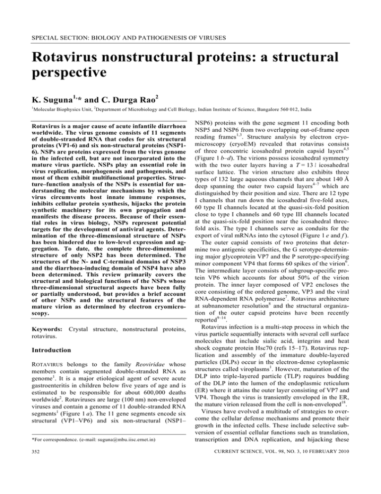
SPECIAL SECTION: BIOLOGY AND PATHOGENESIS OF VIRUSES Rotavirus nonstructural proteins: a structural perspective K. Suguna1,* and C. Durga Rao2 1 Molecular Biophysics Unit, 2Department of Microbiology and Cell Biology, Indian Institute of Science, Bangalore 560 012, India Rotavirus is a major cause of acute infantile diarrhoea worldwide. The virus genome consists of 11 segments of double-stranded RNA that codes for six structural proteins (VP1-6) and six non-structural proteins (NSP16). NSPs are proteins expressed from the virus genome in the infected cell, but are not incorporated into the mature virus particle. NSPs play an essential role in virus replication, morphogenesis and pathogenesis, and most of them exhibit multifunctional properties. Structure–function analysis of the NSPs is essential for understanding the molecular mechanisms by which the virus circumvents host innate immune responses, inhibits cellular protein synthesis, hijacks the protein synthetic machinery for its own propagation and manifests the disease process. Because of their essential roles in virus biology, NSPs represent potential targets for the development of antiviral agents. Determination of the three-dimensional structure of NSPs has been hindered due to low-level expression and aggregation. To date, the complete three-dimensional structure of only NSP2 has been determined. The structures of the N- and C-terminal domains of NSP3 and the diarrhoea-inducing domain of NSP4 have also been determined. This review primarily covers the structural and biological functions of the NSPs whose three-dimensional structural aspects have been fully or partially understood, but provides a brief account of other NSPs and the structural features of the mature virion as determined by electron cryomicroscopy. Keywords: rotavirus. Crystal structure, nonstructural proteins, Introduction ROTAVIRUS belongs to the family Reoviridae whose members contain segmented double-stranded RNA as genome1. It is a major etiological agent of severe acute gastroenteritis in children below five years of age and is estimated to be responsible for about 600,000 deaths worldwide2. Rotaviruses are large (100 nm) non-enveloped viruses and contain a genome of 11 double-stranded RNA segments1 (Figure 1 a). The 11 gene segments encode six structural (VP1–VP6) and six non-structural (NSP1– *For correspondence. (e-mail: suguna@mbu.iisc.ernet.in) 352 NSP6) proteins with the gene segment 11 encoding both NSP5 and NSP6 from two overlapping out-of-frame open reading frames1,3. Structure analysis by electron cryomicroscopy (cryoEM) revealed that rotavirus consists of three concentric icosahedral protein capsid layers4,5 (Figure 1 b–d). The virions possess icosahedral symmetry with the two outer layers having a T = 13 l icosahedral surface lattice. The virion structure also exhibits three types of 132 large aqueous channels that are about 140 Å deep spanning the outer two capsid layers4–7 which are distinguished by their position and size. There are 12 type I channels that run down the icosahedral five-fold axes, 60 type II channels located at the quasi-six-fold position close to type I channels and 60 type III channels located at the quasi-six-fold position near the icosahedral threefold axis. The type I channels serve as conduits for the export of viral mRNAs into the cytosol (Figure 1 e and f ). The outer capsid consists of two proteins that determine two antigenic specificities, the G serotype-determining major glycoprotein VP7 and the P serotype-specifying minor component VP4 that forms 60 spikes of the virion6. The intermediate layer consists of subgroup-specific protein VP6 which accounts for about 50% of the virion protein. The inner layer composed of VP2 encloses the core consisting of the ordered genome, VP3 and the viral RNA-dependent RNA polymerase7. Rotavirus architecture at subnanometer resolution8 and the structural organization of the outer capsid proteins have been recently reported9–14. Rotavirus infection is a multi-step process in which the virus particle sequentially interacts with several cell surface molecules that include sialic acid, integrins and heat shock cognate protein Hsc70 (refs 15–17). Rotavirus replication and assembly of the immature double-layered particles (DLPs) occur in the electron-dense cytoplasmic structures called viroplasms1. However, maturation of the DLP into triple-layered particle (TLP) requires budding of the DLP into the lumen of the endoplasmic reticulum (ER) where it attains the outer layer consisting of VP7 and VP4. Though the virus is transiently enveloped in the ER, the mature virion released from the cell is non-enveloped18. Viruses have evolved a multitude of strategies to overcome the cellular defense mechanisms and promote their growth in the infected cells. These include selective subversion of essential cellular functions such as translation, transcription and DNA replication, and hijacking these CURRENT SCIENCE, VOL. 98, NO. 3, 10 FEBRUARY 2010 SPECIAL SECTION: BIOLOGY AND PATHOGENESIS OF VIRUSES Figure 1. Rotavirus architecture as determined by electron cryomicroscopy. a, Polyacrylamide gel showing the 11 dsRNA segments and their protein coding assignments. b, Cryo-EM reconstruction of rotavirus triple-layered particle (TLP). The outer capsid proteins VP4 and VP7 are shown in orange and yellow respectively. c, A cutaway representation of the TLP showing the three capsid layers. The intermediate layer consisting of VP6 is shown in blue and the inner layer of VP2 is shown in green. d, Schematic depiction of the core showing the RNA as conical spirals surrounding the transcriptase (red) anchored to the VP2 layer at the five-fold axes. d and e, Cryo-EM reconstruction model of the transcriptionally active DLP showing release of the transcribed RNA through channels located at the five-fold vertex. Figure reproduced from Jayaram et al. (2004) with permission from B. V. Prasad. processes for its own advantage by manipulation of key cellular factors by proteolytic cleavage or modification, suppression of antiviral innate immune responses and altering the intracellular environment conducive for virus growth. Structural proteins determine the virion architecture, host and cellular tropism, antigenic specificities and pathogenicity whereas the non-structural proteins play crucial roles in selective modification and directing essential cellular processes for viral protein synthesis and replication leading to pathogenesis. Understanding the structure and function of NSPs is important as they could serve as potential targets for intervention against viral diseases. Of the six non-structural proteins encoded by the viral genome, the complete three-dimensional structure of only one protein NSP2 has been determined. Whereas the structure of NSP3 has been determined in two parts; the central tetrameric coiled coil region of NSP4 containing the diarrhoea-inducing, VP4- and Ca2+-binding domains has been determined. Poor expression, aggregation and insolubility of the recombinant NSPs hindered their crystallization. In this review, we primarily focus on the structural and functional aspects of the rotavirus NSPs CURRENT SCIENCE, VOL. 98, NO. 3, 10 FEBRUARY 2010 whose three-dimensional structural features have been fully or partially understood, but provide only a brief description of the properties of other NSPs. NSP1, encoded by genome segment 5, is least conserved among the strains and contains cysteine–histidinerich zinc-binding domain19. It was recently shown to function as E3 ubiquitin ligase and mediate suppression of cellular innate immune responses by targeting interferon regulatory factors IRF3, IRF5 and IRF7 to proteasome-mediated degradation20–23. It also inhibits NFκB activation by preventing the degradation of inhibitor of κB (κBα) through proteasome-mediated degradation of the β-transducin repeat containing protein (β-TrCP), a component of the E3 ubiquitin ligase complex23. NSP1 from different strains differ in their abilities to suppress innate immune responses and hence influence the replication efficiency of the virus in the infected cell24. Thus, mutants of NSP1 are viable but replicate slowly25. NSP5, the major product coded by segment 11, is a 20 kDa polypeptide that undergoes phosphorylation and glycosylation yielding isoforms ranging in size from 26 to 37 kDa26–29. It is a serine–threonine-rich protein and 353 SPECIAL SECTION: BIOLOGY AND PATHOGENESIS OF VIRUSES contains target sites for multiple cellular kinases. However, the cellular kinases that are required for its phosphorylation have yet to be identified. NSP5 appears to undergo hyperphosphorylation and dephosphorylation, and the hyperphosphorylated forms are found in the insoluble viroplasm fractions30,31. Its phosphorylation is modulated by interaction with other non-structural protein NSP2 (ref. 31). It also possesses ATPase activity32. NSP5 interacts with NSP2 and the two proteins are the major components of the viroplasm structures in the infected cell33. It also interacts with NSP6 (ref. 34), the viral RNAdependent RNA polymerase VP1 (ref. 35) and the inner capsid protein VP2 (ref. 36). Using RNA interference, NSP5 was shown to be essential for virus replication and encapsidation37,38. The C-terminal region binds calcium and the C-terminal 68 residues appear to direct NSP5 hyperphosphorylation, insolubility and calcium-dependent viroplasm-like structure formation39. NSP6 is a minor product encoded by an alternative open reading frame in gene segment 11 which is out-offrame with that of NSP5 (ref. 3). NSP6 interacts with NSP5 (ref. 34). The biological function of NSP6 in the infected cell is yet to be elucidated. NSP3 Efficient initiation of translation of most cellular mRNAs requires circularization of the mRNA mediated by bringing together of the translation initiation factor eIF-4E bound to the cap structure at the 5′ end and the poly(A)binding protein (PABP) bound to the poly(A) tail at the 3′ end by the scaffold protein eIF-4G40–43. Rotavirus mRNAs lack a poly(A) tail but instead contain a consensus sequence at their 3′ ends1. NSP3 is a sequence-specific RNA binding protein and binds the tetranucleotide sequence that is conserved at the 3′ ends of all rotaviral mRNAs44,45. NSP3 interacts at the same site on eIF-4GI as that is used by PABP46,47. NSP3 also interacts with RoXaN, a cellular protein of unknown function, and mediates nuclear localization of PABP-C1 in rotavirusinfected cells48,49. This competition between NSP3 and PABP for eIF-4G1 was believed to facilitate virus RNA translation and shut-off of host protein synthesis46,50,51. However, recent studies indicate that NSP3 is not required for viral RNA translation, but could be important for inhibition of cellular mRNA translation52. NSP3 has also been implicated in the extraintestinal spread of the virus in the infected host53,54, but the mechanism is not understood. NSP3 is a 36 kDa protein encoded by genome segment 7 (refs 55, 56). The protein occurs as a dimer and consists of two separable domains with the N-terminal RNAbinding domain consisting of residues 1–170 and the eIF4G1-interacting domain formed by the last 107 residues of the C-terminus. These two domains are separated by the 354 dimerization domain lying between amino acids 150 and 206 (ref. 47). The crystal structures of the RNA-binding and eIF-4G1-interacting domains of NSP3 from Simian rotavirus strain SA11 have been determined separately57,58 and that of the region corresponding to residues 165–205 of the dimerization domain is yet to be determined. Crystal structure of the N-terminal RNA-binding domain of NSP3 (NSP3-N) was determined at a resolution of 2.45 Ǻ in complex with a hexanucleotide57 (Figure 2). The heart-shaped homodimer of NSP3-N (residues 4– 164) is formed by intertwining of the two subunits with structurally similar N-terminal segments but rather dissimilar C-terminal segments. The crystal structure revealed that the mRNA binding takes place in the deep cleft formed at the extensive dimeric interface which is largely polar in nature. Five bases (5′-U-G-A-C-C-3′) are visible in the electron density. One end of the cleft where the 3′-terminal nucleotide binds is completely blocked thus preventing any internal mRNA binding. NSP3-N is involved in extensive hydrogen bonds, van der Waals contacts, salt bridges, stacking and water bridges with the consensus tetranucleotide segment at the 3′ end of the ligand. Involvement of all the 2′-hydroxyl groups of these four nucleotides in hydrogen bonds with the protein suggests the preference of RNA sequence to that of DNA. Though NSP3-N exists in dimeric state both in native and mRNA-bound forms, mRNA binding was shown to increase its stability. The structure of the C-terminal domain of NSP3 (NSP3C) in complex with a 30 residue fragment of elF-4G (residues 132–160) that also interacts with PABP was determined at 2.38 Ǻ resolution58. NSP3-C (residues 206–315) is a symmetric homodimer with a rod-like structure having two identical binding sites for elF-4G at the dimeric interface (Figure 3). The binding pockets have predominantly hydrophobic character. Each subunit has 3α helices and a terminal β-strand which forms a 3-stranded Figure 2. Dimer of NSP3-N with bound RNA (pdb code: 1knz). CURRENT SCIENCE, VOL. 98, NO. 3, 10 FEBRUARY 2010 SPECIAL SECTION: BIOLOGY AND PATHOGENESIS OF VIRUSES β-sheet with the two N-terminal strands of the elF-4G fragment. Both NSP3-N and NSP3-C use dimerization as a strategy for the creation of a highly specific binding sites with high affinity. The structure of these complexes provides valuable insights into the mechanism by which NSP3 preferentially recognizes the conserved sequence at the 3′ end of the viral mRNA and competes with PABP for binding to the same site on the translational initiation factor eIF-4G leading to hijacking of the host cell translation machinery for selective translation of viral mRNAs51. NSP2 The 35-kDa NSP2 is a major component of virus replicase complexes59 and along with NSP5 forms viroplasm structures that are sites of viral RNA replication, packaging and assembly1. Co-expression of NSP2 and NSP5 in uninfected cells results in upregulation of NSP5 hyperphosphorylation and formation of viroplasm-like structures31,60. NSP2 possesses single-stranded RNA binding61, nucleic acid helix destabilizing and Mg2+-dependent nucleoside triphosphatase (NTPase)62, nucleoside diphosphate kinase (NDP kinase)63 and RNA triphosphatase (RTPase)64 properties. NSP2 can catalyse the removal of Figure 3. Dimer of NSP3-C (cyan and green) in complex with elF-4G fragment (pink and yellow) (pdb code: 1lj2). CURRENT SCIENCE, VOL. 98, NO. 3, 10 FEBRUARY 2010 the γ phosphate from either RNA or NTP, but RNA is a better substrate. It is proposed that NTPase and the associated NDP kinase activities promote the conversion of spent NDPs into NTPs within the viroplasms64. In presence of RNA, because of the low Km value of its RTPase activity, it is thought to switch to an alternative activity linked to RNA replication, translocation and packaging. The biological unit of this highly basic protein is a doughnut-shaped octamer formed by tail–tail interaction of two tetramers. In the crystal structure of NSP2 from group A and group C rotaviruses determined at a resolution of 2.6 Å and 2.2 Å, the octamer is formed by the crystallographic 422 symmetry65,66 (Figure 4). Each monomer in the asymmetric unit consists of two domains: the N-terminal domain with a new fold and the C-terminal domain with a histidine-triad (HIT)-like fold observed in the active site of cellular nucleotidyl hydrolases (Figure 5). A 25 Å-deep and 30 Å-wide cleft lies between these two domains which are separated by a 10 residue loop. Nucleoside triphosphate-binding sites are located on either side of the groove (Figure 5). The C-terminal domain of NSP2, though structurally similar to the HIT family proteins, lacks the typical signature HIT motif (HxHxHxx: the second H is the catalytic residue and x is a hydrophobic residue), but exhibits a HIT-like motif (H221-G-(K/H)-X-H225-X-R) responsible for binding and hydrolysis of NTPs64,65. Other striking differences are that HIT proteins function as dimers and hydrolyse at the α-phosphate, whereas NSP2 is an octamer and hydrolyses at the γ-phosphate. His225 of NSP2 has been identified as the catalytic residue by a comparison with the structure of PKCI (protein kinase C Figure 4. NSP2 octamer viewed down the four-fold axis. Subunits are shown in different colours (pdb code: 1l9v). 355 SPECIAL SECTION: BIOLOGY AND PATHOGENESIS OF VIRUSES interacting protein). This study suggests that the hydrolytic mechanism of NSP2 is significantly different from that of cellular enzymes. The four grooves lined predominantly with basic residues that extend across the octamer surface function as the ssRNA-binding regions65,67. Structural studies on the complexes of NSP2 and the H225A mutant with nucleotide analogues not only provided many interesting details of the mode of nucleotide binding and catalytic mechanism but also revealed the nucleoside diphosphate kinase-like activity of NSP2 which was not reported before63. NSP4 NSP4, encoded by genome segment 10, is a 175 amino acid long-multifunctional protein, and is essential for virus morphogenesis and pathogenesis1. It is the first virus-encoded enterotoxin to be identified68. It exists in multiple forms in the infected cell. The full-length protein is anchored in the ER through the N-terminal hydrophobic domains and the C-terminal region of about 131 residues attains cytoplasmic orientation69,70 (Figure 6). It contains an uncleaved signal sequence at the N-terminus followed by three hydrophobic domains H1–H3 (Figures 6 and 7). The region from residue 45 to 175 constitutes the cytoplasmic tail (CT) that exhibits all the known important biological properties associated with the protein Figure 5. A subunit of NSP2. N- and C-terminal domains are shown in different colours (pdb code: 1l9v). 356 which include intracellular calcium mobilization68,71, membrane permeabilization72,73, Ca2+- and VP4-binding1, double-layered particle-binding74–77 and diarrhoea induction in newborn mouse pups68,78,79 (Figure 7). The region encompassing H3 to residue 137 exists as an α-helical tetrameric coiled coil domain (CCD) whereas the C-terminal region is highly flexible77,79. Though NSP4 was considered to be a tetramer1,80, our studies demonstrated that the CT forms highly ordered multimeric structures with both the N-terminal amphipathic helix (residues 73–85) and the extreme C-terminus being important for multimerization79. Our results also showed that the multimerizing abilities differed widely among NSP4s from different strains (unpublished results). The flexible C-terminus is also an important determinant of the biological functions of the protein81. A secreted form of NSP4 containing residue 112–175 has also been identified that exhibited diarrhoea-inducing ability similar to that of full-length protein82. A synthetic peptide corresponding to residues 114–135 also exhibited diarrhoeainducing activity, but was about 100-fold less efficient than the full-length protein68. Because of the multimerization properties of the CT and the highly flexible nature of the C-terminus, the complete CT was not amenable for crystallization81,83,84. Solution of the structure of the cytoplasmic domain of NSP4 should throw light on its diverse functions. Though the structure of a synthetic peptide that contains residues 95–137 of NSP4 was reported85, we were successful in crystallizing the 95–145 residue segment of the recombinant protein from a number of different viral strains83,84 (Figure 8). Two of these structures showed a tetrameric coiled-coil, calcium-binding domain similar to the one observed in the synthetic peptide84. The domain is formed by four parallel α-helices with a calcium ion located inside the helical bundle coordinating with four Gln and two Glu side chains. The extra C-terminal residues corresponding to the inter-species variable region could not be located in the electron density in spite of the resolution of one of the structures being 1.7 Å, indicating the Figure 6. Schematic representation of the ER-anchored NSP4 and its role in the budding of DLP into the ER lumen during morphogenesis of DLP into mature triple-layered particle. CURRENT SCIENCE, VOL. 98, NO. 3, 10 FEBRUARY 2010 SPECIAL SECTION: BIOLOGY AND PATHOGENESIS OF VIRUSES Figure 7. Schematic representation of the different domains of rotavirus NSP4. Figure 8. Two views of the tetrameric coiled-coil structure of NSP4 (pdb code:2o1k). The two dimers (one shown in grey and the other in colour) are related by a two-fold rotation. inherent flexibility of this region. The tetramer is formed by a two-fold rotation of a dimer having two chains related by an angle of rotation of 81°. Differences between the structures of the recombinant protein segments and the synthetic peptide are confined to a few residues at the C-terminus of the helix and in crystal packing owing to the extra residues present in the recombinant proteins84,85. Our recent studies on NSP4 from other strains indicate considerable variation in their oligomeric states and it would be of interest to investigate if the structural differences correlate with differences in biological activities. 1. Estes, M. K. and Kapikian, A. Z., Rotaviruses. In Fields Virology (eds Knipe, D. M. and Howley, P. E.), Wolters Kluwer-Lippincott Williams and Wilkins, Philadelphia, 2007, 5th edn, pp. 1917– 1974. 2. Parashar, U. D., Gibson, C. J., Breese, J. S. and Glass, R. I., Rotavirus and severe childhood diarrhea. Emerg. Infect. Dis., 2006, 12, 304–306. 3. Mattion, N. M., Mitchell, D. B., Both, G. W. and Estes, M. K., Expression of rotavirus proteins encoded by alternative open reading frames of genome segment 11. Virology, 1991, 181, 295– 304. CURRENT SCIENCE, VOL. 98, NO. 3, 10 FEBRUARY 2010 4. Prasad, B. V., Wang, G. J., Clerx, J. P. and Chiu, W., Threedimensional structure of rotavirus. J. Mol. Biol., 1988, 199, 269– 275. 5. Jayaram, H., Estes, M. K. and Prasad, B. V., Emerging themes in rotavirus cell entry, genome organization, transcription and replication. Virus Res., 2004, 101, 67–81. 6. Shaw, A. L., Rothnagel, R., Chen, D., Ramig, R. F., Chiu, W. and Prasad, B. V., Three-dimensional visualization of the rotavirus hemagglutinin structure. Cell, 1993, 74, 693–701. 7. Prasad, B. V., Rothnagel, R., Zeng, C. Q.-Y., Jakana, J., Lawton, J. A., Chou, W. and Estes, M. K., Visualization of ordered genomic RNA and localization of transcriptional complexes in rotavirus. Nature, 1996, 382, 471–473. 8. Li, Z., Baker, M. L., Jiang, W., Estes, M. K. and Prasad, B. V., Rotavirus architecture at subnanometer resolution. J. Virol., 2009, 83, 1754–1766. 9. Dormitzer, P. R., Nason, E. B., Prasad, B. V. and Harrison, S. C., Structural rearrangements in the membrane penetration protein of a non-enveloped virus. Nature, 2004, 430, 1053–1058. 10. Aoki, S. T., Settembre, E. C., Trask, S. D., Greenberg, H. B., Harrison, S. C. and Dormitzer, P. R., Structure of rotavirus outerlayer protein VP7 bound with a neutralizing Fab. Science, 2009, 324, 1444–1447. 11. Lepault, J. et al., Structural polymorphism of the major capsid protein of rotavirus. EMBO J., 2001, 20, 1498–1507. 12. Greig, S. L., Berriman, J. A., O’Brien, J. A., Taylor, J. A., Bellamy, A. R., Yeager, M. J. and Mitra, A. K., Structural determinants of rotavirus subgroup specificity mapped by cryo-electron microscopy. J. Mol. Biol., 2006, 356, 209–221. 13. Lawton, J. A., Zeng, C. Q.-Y., Mukherjee, S. K., Cohen, J., Estes, M. K. and Prasad, B. V., Three-dimensional structural analysis of recombinant rotavirus-like particles with intact and aminoterminal-deleted VP2: implications for the architecture of the VP2 capsid layer. J. Virol., 1997, 71, 7353–7360. 14. Lu, X. et al., Mechanism for coordinated RNA packaging and genome replication by rotavirus polymerase VP1. Structure, 2008, 16, 1678–1688. 15. Guerrero, C. A. et al., Heat shock cognate protein 70 is involved in rotavirus cell entry. J. Virol., 2002, 76, 4096–4102. 16. Guerrero, C. A., Mendez, E., Zarate, S., Isa, P., Lopez, S. and Arias, C. F., Integrin αvβ3 mediates rotavirus cell entry. Proc. Natl. Acad. Sci. USA, 2000, 97, 14644–14649. 17. Lopez, S. and Arias, C. F., Multistep entry of rotavirus into cells: a Versaillesque dance. Trend. Microbiol., 2004, 12, 271–278. 18. Lopez, T., Camacho, M., Zayas, M., Najera, R., Sanchez, R., Arias, C. F. and Lopez, S., Silencing the morphogenesis of rotavirus. J. Virol., 2005, 79, 184–192. 19. Hua, J. and Patton, J. T., The carboxyl-half of the rotavirus nonstructural protein NS53 (NSP1) is not required for virus replication. Virology, 1994, 198, 567–576. 20. Barro, M. and Patton, J. T., Rotavirus NSP1 inhibits expression of type I interferon by antagonizing the function of interferon regulatory factors IRF3, IRF5, and IRF7. Proc. Natl. Acad. Sci. USA, 2005, 102, 4114–4119. 21. Graff, J. W., Ewen, J., Ettayebi, K. and Hardy, M. E., Zincbinding domain of rotavirus NSP1 is required for proteasomedependent degradation of IRF3 and autoregulatory NSP1 stability. J. Gen. Virol., 2007, 88, 613–620. 22. Barro, M. and Patton, J. T., Rotavirus NSP1 inhibits expression of type I interferon by antagonizing the function of interferon regulatory factors IRF3, IRF5, and IRF7. J. Virol., 2007, 81, 4473–4481. 23. Graff, J. W., Ettayebi, K. and Hardy, M. E., Rotavirus NSP1 inhibits NFκB activation by inducing proteasome-dependent degradation of β-TrCP: a novel mechanism of IFN antagonism. PLoS Pathogens, 2009, 5, 1–12. 24. Feng, N., Sen, A., Nguyen, H., Vo, P., Hoshino, Y., Deal, E. M. and Greenberg, H. B., Variation in antagonism of the interferon 357 SPECIAL SECTION: BIOLOGY AND PATHOGENESIS OF VIRUSES 25. 26. 27. 28. 29. 30. 31. 32. 33. 34. 35. 36. 37. 38. 39. 40. 41. 42. 43. 358 response to rotavirus NSP1 results in differential infectivity in mouse embryonic fibroblasts. J. Virol., 2009, 83, 6987–6994. Patton, J. T. et al., Effect of intragenic rearrangement and changes in the 3′ consensus sequence on NSP1 expression and rotavirus replication. J. Virol., 2001, 75, 2076–2086. Afrikanova, I., Miozzo, M. C., Giambiagi, S. and Burrone, O., Phosphorylation generates different forms of rotavirus NSP5. J. Gen. Virol., 1996, 77, 2059–2065. Gonzalez, S. A. and Burrone, O. R., Rotavirus NS26 is modified by addition of single O-linked residues of N-acetylglucosamine. Virology, 1991, 182, 8–16. Blackhall, J., Munoz, M., Fuentes, A. and Magnusson, G., Analysis of rotavirus nonstructural protein NSP5 phosphorylation. J. Virol., 1998, 72, 6398–6405. Poncet, D., Lindenbaum, P., L’Haridon, R. and Cohen, J., In vivo and in vitro phosphorylation of rotavirus NSP5 correlates with its localization in viroplasms. J. Virol., 1997, 71, 34–41. Sen, A., Agresti, D. and Mackow, E. R., Hyperphosphorylation of the rotavirus NSP5 protein is independent of serine 67 or NSP2, and the intrinsic insolubility of NSP5 is regulated by cellular phosphatases. J. Virol., 2006, 80, 1807–1816. Afrikanova, I., Fabbretti, E., Miozzo, M. C. and Burrone, O. R., Rotavirus NSP5 phosphorylation is up-regulated by interaction with NSP2. J. Gen. Virol., 1998, 79, 2679–2686. Bar-Magen, T., Spencer, E. and Patton, J. T., An ATPase activity associated with the rotavirus phosphoprotein NSP5. Virology, 2007, 369, 389–399. Eichwald, C., Rodriguez, J. F. and Burrone, O. R., Characterization of rotavirus NSP2/NSP5 interactions and the dynamics of viroplasm formation. J. Gen. Virol., 2004, 85, 625–634. Torres-Vega, M. A., Gonzalez, R. A., Duarte, M., Poncet, D., Lopez, S. and Arias, C. F., The C-terminal domain of rotavirus NSP5 is essential for its multimerization, hyperphosphorylation and interaction with NSP6. J. Gen. Virol., 2000, 81, 821–830. Arnoldi, F., Campagna, M., Eichwald, C., Desselberger, U. and Burrone, O. R., Interaction of rotavirus polymerase VP1 with nonstructural protein NSP5 is stronger than that with NSP2. J. Virol., 2007, 81, 2128–2137. Berois, M., Sapin, C., Erk, I., Poncet, D. and Cohen, J., Rotavirus nonstructural protein NSP5 interacts with major core protein VP2. J. Virol., 2003, 77, 1757–1763. Campagna, M., Eichwald, C., Vascotto, F. and Burrone, O. R., RNA interference of rotavirus segment 11 mRNA reveals the essential role of NSP5 in the virus replicative cycle. J. Gen. Virol., 2005, 86, 1481–1487. Lopez, T., Rojas, M., Ayala-Breton, C., Lopez, S. and Arias, C. F., Reduced expression of the rotavirus NSP5 gene has a pleiotropic effect on virus replication. J. Gen. Virol., 2005, 86, 1609–1617. Sen, A., Sen, N. and Mackow, E. R., The formation of viroplasmlike structures by the rotavirus NSP5 protein is calcium regulated and directed by a C-terminal helical domain. J. Virol., 2007, 81, 11758–11767. Tarun, S. Z., Wells, S. E., Deardorff, V. A. and Sachs, A. B., Translation initiation factor eIF4G mediates in vitro poly(A) taildependent translation. Proc. Natl. Acad. Sci. USA, 1997, 94, 9046–9051. Wells, S. E., Hillner, P. E., Vale, R. D. and Sachs, A. B., Circularization of mRNA by eukaryotic translation initiation factors. Mol. Cell, 1998, 2, 135–140. Sachs, A., Physical and functional interactions between the mRNA cap structure and the poly(A) tail. In Translational Control of Gene Expression (eds Sonenberg, N., Hershey, J. W. B. and Mathews, M. B.), Cold Spring Harbor Laboratory Press, Cold Spring Harbor, New York, 2000, pp. 447–467. Preiss, T. and Hentze, M. W., Dual function of the messenger RNA cap structure in poly(A)-tail-promoted translation in yeast. Nature, 1998, 392, 516–520. 44. Poncet, D., Laurent, S. and Cohen, J., Four nucleotides are the minimal requirement for RNA recognition by rotavirus nonstructural protein NSP3. EMBO J., 1994, 13, 4165–4173. 45. Chizhikov, V. and Patton, J. T., A four-nucleotide translation enhancer in the 3′-terminal consensus sequence of the nonpolyadenylated mRNAs of rotavirus. RNA, 2000, 6, 814–825. 46. Piron, M., Vende, P., Cohen, J. and Poncet, D., Rotavirus RNAbinding protein NSP3 interacts with eIF4GI and evicts the poly(A) binding protein from eIF4F. EMBO J., 1998, 17, 5811–5821. 47. Piron, M., Delaunay, T., Grosclaude, J. and Poncet, D., Identification of the RNA-binding, dimerization, and eIF4GI-binding domains of rotavirus nonstructural protein NSP3. J. Virol., 1999, 73, 5411–5421. 48. Vitour, D., Lindenbaum, P., Vende, P., Becker, M. M. and Poncet, D., RoXaN, a novel cellular protein containing TPR, LD, and zinc finger motifs, forms a ternary complex with eukaryotic initiation factor 4G and rotavirus NSP3. J. Virol., 2004, 78, 3851–3862. 49. Harb, M. et al., Nuclear localization of cytoplasmic poly(A)binding protein upon rotavirus infection involves the interaction of NSP3 with eIF4G and RoXaN. J. Virol., 2008, 82, 11283– 11293. 50. Padilla-Noriega, M., Paniagua, O. and Guzman-Leon, S., Rotavirus protein NSP3 shuts off host cell protein synthesis. Virology, 2002, 298, 1–7. 51. Varani, G. and Allain, F. H.-T., How a rotavirus hijacks the human protein synthesis machinery. Nature Struct. Biol., 2002, 9, 158–160. 52. Montero, H., Arias, C. F. and Lopez, S., Rotavirus nonstructural protein NSP3 is not required for viral protein synthesis. J. Virol., 2006, 80, 9031–9038. 53. Mossel, E. C. and Ramig, R. F., Rotavirus genome segment 7 (NSP3) is a determinant of extraintestinal spread in the neonatal mouse. J. Virol., 2002, 76, 6502–6509. 54. Mossel, E. C. and Ramig, R. F., A lymphatic mechanism of rotavirus extraintestinal spread in the neonatal mouse. J. Virol., 2003, 77, 12352–12356. 55. Mattion, N. M., Cohen, J., Aponte, C. and Estes, M. K., Characterization of an oligomerization domain and RNA-binding properties on rotavirus nonstructural protein NS34. Virology, 1992, 190, 68–83. 56. Rao, C. D., Das, M., Ilango, P., Lalwani, R., Rao, B. S. and Gowda, K., Comparative nucleotide and amino acid sequence analysis of the sequence-specific RNA-binding rotavirus nonstructural protein NSP3. Virology, 1995, 207, 327–333. 57. Deo, R. C., Groft, C. M., Rajashankar, K. R. and Burley, S. K., Recognition of the rotavirus mRNA 3′ consensus by an asymmetric NSP3 homodimer. Cell, 2002, 108, 71–81. 58. Groft, C. M. and Burley, S. K., Recognition of eIF4G by rotavirus NSP3 reveals a basis for mRNA circularization. Mol. Cell, 2002, 9, 1273–1283. 59. Gallegos, C. O. and Patton, J. T., Characterization of rotavirus replication intermediates: a model for the assembly of singleshelled particles. Virology, 1989, 172, 616–627. 60. Fabbretti, E., Afrikanova, I., Vascotto, F. and Burrone, O. R., Two non-structural rotavirus proteins, NSP2 and NSP5, form viroplasm-like structures in vivo. J. Gen. Virol., 1999, 80, 333– 339. 61. Taraporewala, Z. F., Chen, D. and Patton, J. T., Multimers formed by the rotavirus nonstructural protein NSP2 bind to RNA and have nucleoside triphosphatase activity. J. Virol., 1999, 73, 9934– 9943. 62. Taraporewala, Z. F. and Patton, J. T., Identification and characterization of the helix-destabilizing activity of rotavirus nonstructural protein NSP2. J. Virol., 2001, 75, 4519–4527. 63. Kumar, M. et al., Crystallographic and biochemical analysis of rotavirus NSP2 with nucleotides reveals a nucleoside diphosphate kinase-like activity. J. Virol., 2007, 81, 12272–12284. CURRENT SCIENCE, VOL. 98, NO. 3, 10 FEBRUARY 2010 SPECIAL SECTION: BIOLOGY AND PATHOGENESIS OF VIRUSES 64. Caprio, R. V., Gonzalez-Nilo, F. D., Riadi, G., Taraporewala, Z. F. and Patton, J. T., Histidine triad-like motif of the rotavirus NSP2 octamer mediates both RTPase and NTPase activities. J. Mol. Biol., 2006, 362, 539–554. 65. Jayaram, H., Taraporewala, Z., Patton, J. T. and Venkataram Prasad, B. V., Rotavirus protein involved in genome replication and packaging exhibits a HIT-like fold. Nature, 2002, 417, 311–315. 66. Taraporewala, Z. F., Jiang, X., Caprio, R. V., Jayaram, H., Prasad, B. V. and Patton, J. T., Structure–function analysis of rotavirus NSP2 octamer by using a novel complementation system. J. Virol., 2006, 80, 7984–7994. 67. Jiang, X., Jayaram, H., Kumar, M., Ludtke, S. J., Estes, M. K. and Prasad, B. V., Cryoelectron microscopy structures of rotavirus NSP2-NSP5 and NSP2-RNA complexes: implications for genome replication. J. Virol., 2006, 80, 10829–10835. 68. Ball, J. M., Tian, P., Zeng, C. Q.-Y., Morris, A. P. and Estes, M. K., Age-dependent diarrhea induced by a rotaviral nonstructural glycoprotein. Science, 1996, 272, 101–104. 69. Bergmann, C. C., Maass, D., Poruchynsky, M., Atkinson, P. H. and Bellamy, A. R., Topology of the non-structural rotavirus receptor glycoprotein NS28 in the rough endoplasmic reticulum. EMBO J., 1989, 8, 1695–1703. 70. Chan, W.-K., Au, K. S. and Estes, M. K., Topography of the simian rotavirus nonstructural glycoprotein (NS28) in the endoplasmic reticulum membrane. Virology, 1988, 164, 435–442. 71. Dong, Y., Zeng, C. Q.-Y., Ball, J. M., Estes, M. K. and Morris, A. P., The rotavirus enterotoxin NSP4 mobilizes intracellular calcium in human intestinal cells by stimulating phospholipase C-mediated inositol 1,4,5-trisphosphate production. Proc. Natl. Acad. Sci. USA, 1997, 94, 3960–3965. 72. Tian, P., Ball, J. M., Zeng, C. Q.-Y. and Estes, M. K., The rotavirus nonstructural glycoprotein NSP4 possesses membrane destabilization activity. J. Virol., 1996, 70, 6973–6981. 73. Newton, K., Meyer, J. C., Bellamy, A. R. and Taylor, J. A., Rotavirus nonstructural glycoprotein NSP4 alters plasma membrane permeability in mammalian cells. J. Virol., 1997, 71, 9458– 9465. 74. Au, K. S., Chan, W. K., Burns, J. W. and Estes, M. K., Receptor activity of rotavirus nonstructural glycoprotein NS28. J. Virol., 1989, 63, 4553–4562. CURRENT SCIENCE, VOL. 98, NO. 3, 10 FEBRUARY 2010 75. Au, K. S., Mattion, N. M. and Estes, M. K., A subviral particle binding domain on the rotavirus nonstructural glycoprotein NS28. Virology, 1993, 194, 665–673. 76. Meyer, J. C., Bergmann, C. C. and Bellamy, A. R., Interaction of rotavirus cores with the nonstructural glycoprotein NS28. Virology, 1989, 171, 98–107. 77. O’Brien, J. A., Taylor, J. A. and Bellamy, A. R., Probing the structure of rotavirus NSP4: a short sequence at the extreme C terminus mediates binding to the inner capsid particle. J. Virol., 2000, 74, 5388–5394. 78. Horie, Y. et al., Diarrhea induction by rotavirus NSP4 in the homologous mouse model system. Virology, 1999, 262, 398–407. 79. Jagannath, M. R., Kesavulu, M. M., Deepa, R., Sastri, P. N., Kumar, S. S., Suguna, K. and Rao, C. D., N- and C-terminal cooperation in rotavirus enterotoxin: novel mechanism of modulation of the properties of a multifunctional protein by a structurally and functionally overlapping conformational domain. J. Virol., 2006, 80, 412–425. 80. Taylor, J. A., O’Brien, J. A. and Yeager, M., The cytoplasmic tail of NSP4, the endoplasmic reticulum-localized non-structural glycoprotein of rotavirus, contains distinct virus binding and coiled coil domains. EMBO J., 1996, 15, 4469–4476. 81. Deepa, R., Sastri, P. N., Jagannath, M. R., Indi, S. S., Kiranmayee, P., Suguna, K. and Rao, C. D., The flexible C terminus of the rotavirus non-structural protein NSP4 is an important determinant of its biological properties. J. Gen. Virol., 2008, 89, 1485–1496. 82. Zhang, M., Zeng, C. Q.-Y., Morris, A. P. and Estes, M. K., A functional NSP4 enterotoxin peptide secreted from rotavirusinfected cells. J. Virol., 2000, 74, 11663–11670. 83. Deepa, R., Jagannath, M. R., Kesavulu, M. M., Rao, C. D. and Suguna, K., Expression, purification, crystallization and preliminary crystallographic analysis of the diarrhoea-causing and virulence-determining region of rotaviral nonstructural protein NSP4. Acta Crystallogr., 2004, D60, 135–136. 84. Deepa, R., Durga Rao, C. and Suguna, K., Structure of the extended diarrhea-inducing domain of rotavirus enterotoxigenic protein NSP4. Arch. Virol., 2007, 152, 847–859. 85. Bowman, G. D. et al., Crystal structure of the oligomerization domain of NSP4 from rotavirus reveals a core metal-binding site. J. Mol. Biol., 2000, 304, 861–887. 359
