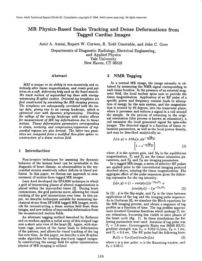
From: AAAI Technical Report SS-94-05. Compilation copyright © 1994, AAAI (www.aaai.org). All rights reserved.
MR Physics-Based
Amir A. Amini,
Snake Tracking and Dense Deformations
Tagged Cardiac Images
Rupert
W. Curwen,
R. Todd Constable,
from
and John C. Gore
Departmentsof Diagnostic Radiology, Electrical Engineering,
and Applied Physics
Yale University
New Haven, CT 06510
Abstract
2
MRIis unique in its ability to non-invasively and selectively alter tissue magnetization, and create grid patterns on a soft, deforming body such as the heart muscle.
We track motion of myocardial tag lines with energy.
minimizing B-spline snakes. Oriented tag templates are
first constructed by simulating the MRimaging process.
The templates are subsequently correlated with the image data, giving rise to an energy landscape, which is
optimized over with dynamic programming. Tracking
the valleys of the energy landscape with snakes allows
for measurement of MRtag deformations due to tissue
motion. Tissue deformation parameters corresponding
to strain, vorticity, and compression/expansion of myocardial regions are also derived. The latter two quantities are computedfrom a modified thin-plate spline reconstruction of a dense motion field.
1
Introduction
Non-invasive techniques for assessing the dynamic
behavior of the human heart can be invaluable in the
diagnosis of heart disease, as abnormalities in the myocardial motion sensitively reflect deficits in blood perfusion. In this paper, we discuss our approach to measurement of motion from tagged MRimages.
Leon Axel developed the SPAMM
technique in which
a grid of intersecting planes of altered magnetization is
placed within the myocardial tissue [2]. During heart
contractions, the grid patterns move,allowing for visual
tracking of the grid intersections over time. In this paper, we describe techniques suitable for measuring mechanical strain from SPAMM
tagged MRimages, methods for reconstructing a dense motion field from these
images, and new indices of myocardial deformation from
the reconstructed motion fields.
An alternate tagging method described by Zerhouni
and co-workers applies a radial array of thin striped tags
over a short axis view of the heart [8]. Again, with starburst tags, motion of the tissue leads to deformation
of the pattern, and allows for visual tracking of the tag
line over time. In this paper, we describe a new B-spline
snake algorithm for tracking star-burst tagged images.
In constructing the energy field for spline optimization
physics of MRimaging is utilized.
126
NMR Tagging
In a normal MRimage, the image intensity is obtained by measuring the NMRsignal corresponding to
each tissue location. In the presence of an external magnetic field, the local nuclear spins sum to provide the
tissue magnetization. Application of an RF pulse of a
specific power and frequency content leads to absorption of energy by the spin system, and the magnetization is rotated by 90 degrees, into the transverse plane,
where it precesses and induces a signal in a coil around
the sample. In the process of returning to the original orientation (this process is knownas relaxation),
coil measures the local generated signal for spin-echo
images. The detected signal is a function of tissue relaxation parameters, as well as the local proton density,
and may be described analytically as:
fl(z,
Y) = AMo(x, y)e-
where A is the system gain, and M0is the equilibrium
magnetization. 7"1 and 7"2 are the tissue relaxation parameters, and TE and TR are imaging parameters.
In a tagged MRimage, a series of selective RF pulses
are applied prior to the conventional imaging protocol
decribed above, rotating the tissue magnetization. The
aggregate effect of the pulse sequences gives the following expression for the tag intensity:
f2(z, y) = (1 cos(¢(z)))e -TMT’ +
(COS(¢(x)) -- -T°/r’
(2)
In (2) , ¢ is the flip-angle, and To is the time between
application of the tag and the imaging pulse sequence.
As in Guttman [6], we simulate the Bloch equations for
the MRimaging process, and obtain a sequence of tag
profiles as a function of time. The tag profiles approximate inverted Gaussian functions which undergo Ta tissue relaxation, becomingless visible in later phases of
the heart cycle (fig. I). In these simulations the following parameters were used: duration of tag pulse was
T, = 1.8 msec, Gyromagnetic Ratio was 26800 HZ/G,
gradient strength was G~ = 0.052 G/ram, Tn = 1 sec,
and Ta = 0.5 see. The RF pulse had the following form:
B,(t) = Vov(t)w(t)cos2,qot
(3)
where v is a sinc pulse, w is the Hammingwindow, and
V0 = 0.09 G.
3
Energy
Field
The simulation returns a predicted tag profile in the
direction perpendicular to image tag lines as a function of time and is normalized to lie between zero and
one. To create an energy field, the following approach
is taken. For points within tag lines in the the vertical orientation, a set of profiles are concatenated along
the vertical axis to create a correlation kernel, g*. The
correlation kernel is then successively rotated to create kernels along other orientations. Additional energy
fields are constructed using correlation kernels for tag
endpoints. Let the image be represented by g2. The
normalized correlation, p(z, Y)
{ffg~(x
f f n (x + 6~, y + 6~)g2(x, y)dx@
+5~,y+5~)dzdyf f g2(z,y)dzdy}
5
1/~ (4)
with 0 _< p _< 1, and with p = 1 when 91 is a constant multiple of 92. In order to increase the discrimination power of the technique the energy field is set
to -pn(z, y), where n is a positive integer less than 10.
Endpoint energy fields are termed p,, and are obtained
~rom correlating endpoint masks with the tag data. For
~ach tag endpoint, a correlation mask is generated, and
:s used to construct endpoint energy images for subsequent frames. These kernels essentially have half of the
~indow filled with tag profiles, and depending on the
;ag line, have half filled with zero intensities, or intensi;ies from surrounding organs. Currently, tag endpoint
:oordinates are specified in the first frame. All subsetuent endpoints are determined by the algorithm.
t
B-Spline
Snakes
with
for i > 2, and SI(pl,p2) = minp0 Eo(po,pt,p2). In general, for an order k B-spline, Si is a function of k control
points. Also, note that the minimization yields the optimal open spline, as is the case for a tag line. In order to
find a closed snake, one performs Mapplications of the
above recurrence, where M is the number of possible
choices for the endpoint pi, and for each optimization
fixing the end point to be one of the M choices, repeating for all Mpossibilities, and finally choosing the
minimum. Figure 2 shows the initial star-burst image
and results from the DP algorithm. Figure 3 shows the
energy field for a diagonal tag line, and for one endpoint
of the same tag line.
Dense
Tissue
Velocity
Field
Estimation
Deformation
Indices
and
In the case of SPAMM
grids, from following grid line
intersections, we obtain displacement information at a
set of loci in the image. In the case of star-burst images,
tracking the tag lines will yield displacement information at points where the tag lines intersect the heart
wall. Clearly, it is desirable to obtain a dense field of
displacement vectors so that myocardial motion can be
inspected at all points falling within the myocardium.
As with the case of tag lines, we perform spline approximation of the data [5, 4] for providing dense motion information. Twovariational principles are posed
which we numerically solve, yielding components of
the displacement vector field for all pixels in the LV
myocardiumh
DP
(i,j)ED
Weuse B-spline snakes [7, 3] to represent image tag
ines. The spline is given by the following expression:
o~(u) -- uT/vIP
(5)
vhere u is a column vector of powers of u, the spline
mrameter, P is a sequence of control points, and Mis
L matrix which blends the control points. The approach
s to minimize the following expression along tags:
E,o,°,
= -(/p"(+Cu))au
+
(6)
n equation (6), the first term maximizes pn along the
ength of the snake, and second and third terms attract
he snake to the endpoint of tag lines.
The discrete form of Etot~t, for a quadric spline may
,e written as:
~total
= Eo(Po,pl,p2)
+""-I- EN-s(PN-3,PN-2,pN-1)
vhere p/ are the B-spline control points. DynamicPro:ramming (DP) [1] may be used to optimize the curve
n the control point space to minimize EtotaZ using the
ollowing recurrence
Si(pi,pi+l)
= minEi-l(pi-l,pi,pi+,)
Pt--I
(7)
127
In the above expression, d is used to denote the u, or
v component of displacement, A is in generM a nonnegative function of x and y and controls the degree
of smoothing, and D is the set of points with known
displacement information. Successive Over Relaxation
(SOR)is used to solve the resulting discrete linear systems of equations to obtain dense displacement information. This method calculates LV displacement at all
points of a 2D tagged cardiac MRimage utilizing the
tracked image tag information. Results of dense motion estimation from 2 consecutive frames of a SPAMM
tagged MRimage sequence are shown in figure 4.
5.1
Measures
traction,
for Tissue
Expansion,
and Circulation
Con-
In previous work, authors have reported measuring
such quantities as torsion, and rotational motion of the
tissue. However, with previous techniques, this information could only be obtained at a specific set of points
within the myocardium. With the methods described,
we can obtain displacement vectors on all parts of the
* We would like to perform the spline
approximation
without
knowledge of LV boundary.
One idea which is currently
being
pursued is to normalize the derivative
component of the integrand
with respect
to components of the image gradient
of an untagged
MR cardiac
scan performed
at the same time point
of the ECG
wave.
of figure 5 is shown in figure 6 where the maximum
and minimumprincipal strain directions and values are
shown. It should be pointed out that extension of quantities obtained from equations (8), (9), (10), and (11)
3D is possible with tagging. Naturally, there are three
principal strain directions in 3D.
myocardium. From this information, we obtain differential vector quantities which describe local rotations
and expansions.
Tissue
Expansion and Contraction
Expansion or contraction of the myocardiumin an arbitrary area within the LVwall between the endocardial
and epicardial surfaces maybe written as:
6
(9)
~r17 . ~ds = L E7 .17 dA
wherethe integral on the left. is a line integral computed
on a curve F which bounds the myocardial mass of interest, ~ is the normal to F, and I7 is a dense displacement
vector field. The integral on the right is over the area
boundedby F, and E7’ 17 is the divergence of the vector
field. This provides an easy way to compute a quantitative measure of tissue expansion. The strength of
this measureis that it is invariant to rigid body motion,
and so can be used as a measure of compressibility, of
non-rigid deformation, or of tissue expansion, or contraction.
Circulation
Torsion has been described to be of major significance in
the study of LV. Wecan evaluate circulation accurately
around any contour bounding the LV myocardium:
f
r17 " i’ds = ~A(~7 × V)ndA
Conclusions
In conclusion, we have described new computer vision
algorithms, which utilize the physics of medical imaging for tracking with energy-minimizing snakes. From a
clinical standpoint, the computer vision reconstruction
algorithm described here maybe useful as an initial step
in measurement of local myocardial deformation. Mechanical strain can also be measured with suitable accuracy from tagged images.
References
[1] A. A. Amini, T. E. Weymouth, and R. C. Jain.
Using dynamic programming for solving variational
problems in vision. IEEE Transactions on Pattern
Analysis and Machine Intelligence,
12(9):855-867,
1990.
[2] L. Axel, R. Goncalves, and D. Bloomgarden. Regional heart wall motion: Two-dimensional analysis
and functional imaging with mr imaging. Radiology,
183:745-750, 1992.
(10)
where F is a planar contour, t is the tangent to such a
curve, n is the binormal of the curve, and the integral
is evaluated around r. (~7 × 17)n is the component
curl of 1~ perpendicular to the plane of F describing infinitesimal circulation of the displacement vector field
at a point. It is important to note that this information can be evaluated around any planar contour which
passes through the myocardium.
[3] A. Blake, R. Curwen, and A. Zisserman. A framework for spatio-temporal control in the tracking of
visual contours. Internalional Journal of Computer
Vision, 11(2):127-145, 1993.
5.2
[5] W. Grimson. An implementation of a computational
theory of visual surface interpolation.
Computer
Vision, Graphics, and Image Processing, 22:39-69,
1983.
[4] F. Bookstein. Principal warps: Thin-plate splines
and the decomposition
of deformations.
IEEE
Transactions on Pattern Analysis and Machine Intelligence, 11(6):567-585, 1989.
Strain
Strain is a measure of local deformation of a line element due to tissue motion and is independent of the
rigid motion of the LV. To compute the local 2D strain
for a given triangle, correspondence of the 3 vertices
with a later time is sufficient [2]. With such information
known, an affine map F is completely determined. By
the polar decomposition theorem, F can be decomposed
as F = R. U, where R is a rotation matrix, and U is
a symmetric matrix representing the 2D strain. Under
the linear motion assumption, strain in the direction of
a vector £ can be expressed as
1( Wxlu 1)
[6] M. Guttman, J. Prince, and E. McVeigh. Tag and
contour detection in tagged mr images of the left.
ventricle. In IEEE Transactions on Medical Imaging, in press.
[7] S. Menet, P. Saint-Marc, and G. Medioni. B-snakes:
Implementation and application to stereo. In Proceedings of Third International Conference on Computer Vision, pages 720-726, 1990.
[8] E. Zerhouni, D. Parish, W. Rogers, A. Yang. and
E. Shapiro. Humanheart: Tagging with mr imaging
- a method for noninvasive assessment of myocardial
motion. Radiology, 169:59-63, 1988.
(ll)
Twodirections within each such triangle are of particular interest, namely, the directions of principal
strain, representing the maximumand minimumstretch
within a triangle, and corresponding to the eigenvectors
of U. The results of strain analysis done on images
128
1
09
oB
0.7
0.6
~G
~’ ".’,
~..:;.
"’,’,’.
"’.... ’.
.’;’’.~ 160ros
.’.’ .~ 240ms
’." .~ 320ms..
/’..’..:
400ms....
05
04
0.3
0.2
Ol
o
-8
¯6
-4
-2
0
2
4
6
Perpendicular
dostanco
tr~)rn tag center (me)
B
Figure 1: Simulated tag profiles for successive time inslanls.
Figure 4: Reconstruction of dense u and v components
of displacement from 2 frames of a SPAMM
tagged image.
Figure 2: Results from tracking with DP snakes. The
initial,
unoptimized placement of B-Spline snakes is
shown in top left. Tag lines in subsequent frames are
focalized and tracked automatically.
Pig, ure 3: E.ergy tield for frame ] in Ihe previous figure.
,I’ and p~" are displayed for a diagonal tag It,e, and o,e
"ndpoiud..
129
Figure 5: Two consecut.ive frames of SPAMM
tagged
cardiac images. Hand-t.raced tag lines are superimposed.
Figure 6: Strain measurement made from figure 5: principal straixl values and directions.


