From: AAAI Technical Report SS-94-05. Compilation copyright © 1994, AAAI (www.aaai.org). All rights reserved.
Deformable
Models for Biomedical
Demetri
Terzopoulos
Department
of Computer Science
University
of Toronto,
Toronto, Canada,
Introduction
The rapid development of computer and noninvasive
sensor technologies is revolutionizing medicine, offering
scientists and physicians powerful new investigative and
diagnostic tools. Computer-based medical image analysis is an especially challenging task which has not kept
pace with the ability to acquire digital images in various modalities. In response to this challenge, we have
been developing several new segmentation, registration,
shape reconstruction, and motion tracking techniques
for multidimensional medical image analysl~s. Our work
exploits deformable models.
Deformable models offer a fundamentally dynamic
approach to nonrigid shape and motion analysis based
on computational physics. Typically, the models are
governed by the Lagrangian mechanics of elastic media, and they are coupled to various data sets (and
they may be manipulated interactively) thorough force
fields. Although they were originally developed in the
context of computer vision [1] and computer graphics
[2], deformable models are also naturally applicable to
biomedical data analysis. This is because the human
body is a complex, highly deformable structure.
In this summary, I will not attempt to survey the
growing cavalcade of research on deformable models in
medical image analysis (see, e.g., the forthcoming collection [3]). I will, however, review someof our recent
work which applies deformable models to a variety of
static and time varying medical data sets from scales
on the macroscopic to the microscopic. The subsequent
sections focus on the extraction of 3D models from serial microscopy, opthalmic image analysis, dynamic 3D
cardiac image analysis, and tracking for biomechanics.
The details are available in the references.
3D Models
from Serial
Microscopy
In [4, 5] we propose a new approach to the segmentation, reconstruction, and visualization of 3D models
from serial microscopy. Wehave developed an interactive system which exploits recent computer graphics
and computer vision techniques to significantly reduce
the time required to build such models. The key ingredients of the system are a digital "blink comparator" for section registration, deformable contour models
(the popular "snakes") for semi-automated cell segmentation, and voxel-based techniques for 3D reconstruc-
118
Data Analysis
M5S 1A4
tion and visualization of complex volumes with internal structures. To date, the system has been applied
to serial EMimages of neuronal tissue and images of
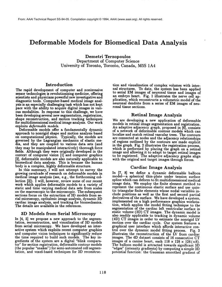
an embryo heart. Fig. 1 illustrates
the nerve cell application, which reconstructs a volumetric model of the
neuronal dendrite from a series of EMimages of neuronal tissue sections.
Retinal
Image Analysis
We are developing a new application of deformable
models in retinal image segmentation and registration.
An adaptive adjacency graph, proposed in [6], consists
of a network of deformable contour models which can
localize and match retinal vascular trees. The contours
are connected at nodes and the adjacency relationships
of regions outlined by the contours are made explicit
in the graph. Fig. 2 illustrates the registration process.
which is performed by placing the graph on a retinal
image and allowing it to adapt to target retinal images
to be registered. The adaptive adjacency graphs align
with the original and target images through forces.
Cardiac
Image Analysis
In [7, 8] we define a dynamic deformable balloon
model--a spherical thin-plate under tension surface
spline which can deform to fit multidimensional medical
image data. We employ the finite element method to
represent the continuous elastic surface and use quintic triangular finite elements whose nodal variables include positions as well as the first and second partial
derivatives of the surface. Wehave developed a system,
implemented on a high performance grap]lics workstation, which applies the model fitting technique to the
segmentation of the cardiac left ventricular surface in
static volume (3D) CT images. The dynamic model
also readily applicable to tracking in dynamic volume
(4D) CT images in order to estimate the nonrigid
motion over the cardiac cycle. Our system feat’ures a
graphical user interface which affords interactive control over the dynamic model fitting process. Fig. 3
illustrates
the reconstruction of the LV from cardiac
images. The 4D dataset consists of 16 consecutive CT
images of a canine heart, each 118 x 128 × 128(x8).
The balloon model is attracted towards significant 3D
"edges" (intensity gradients) by computing a simple
potential function: the Gaussian smoothed gradient of
¯(a)
(a)
._..
(c)
(b)
(b)
Figure 2: Detection and matching of vascular tree on
retinal image with adaptive adjacency graph
(a) Initial position of AAG(white). (b) Matched
the image. The final LV reconstruction is shown in
Fig. 3(f). Finally, Fig. 4 illustrates the dynamicsurface
model tracking the LVduring the cardiac cycle.
(d)
ire h Segmentation, reconstruction, and visualizaof neuronal dendrite.
A serial tissue section image with deformable con" model (white) adhering to cell membrane.(b)
atially segmentedcell interiors stacked in 3D. (c)
Jme rendered view of translucent dendrite revealin$
rnal structures. (d) Shaded surface of reconstructed
Jendrite model.
119
Tracking
for Biomechanics
In [9] we apply continuous nonlinear Kalman filtering
theory to devise new recursive shape and motion estimators. The estimators employ the Lagrange equations
of deformable superquadric models as Kalman system
models. To produce nonstationary shape and motion
estimates from time-varying visual data, the system
model continually synthesizes nonrigid motions in response to generalized forces arising from the inconsistency between the incoming observations and the estimated model state. The observation forces account
formally for instantaneous uncertainties and incomplete
information. A Riccati procedure updates a covariance
(4)
(6)
Ca)
(8)
(lO)
(e)
(f)
Figure3: Intensity
andprocessed
CT sliceof LV.
(a)Intensity
imageXZ planeslice91.(b)Edgedetected
image.
(c)Crosssection
of initial
model.
(d)-(e)
sectionof modeldeforming
to leftventricle.
(f)
reconstruction
ofleftventricle.
(12)
matrixwhichtransforms
theforcesin accordance
with
thesystemdynamics
andthepriorobservation
history.
(14)
Thetransformed
forcesinducechanges
in thetranslational,
rotational,
anddeformational
statevariables
of
thedeformable
superquadric
systemmodelin orderto
increase
itsconsistency
withthedata.
Theestimation
technique
generalizes
to constrained
multibody
modelscomprising
deformable
superquadric
parts.Fig.5 showsshapeandmotionestimation
of an
articulated
multipartupperbodymodelcomposedof
(16)
5 dynamicsuperquadrics
connected
by 4 point-to-point
constraints
usingdatacollected
fromtheraisingand
flexingmotionof the armsof a humansubject.The
data was collectedusing WATSMART,a commercial
non-contact, 3D motion digitizing and analysis system.
(a)
(b)
The system employs multiple optoelectric measurement
cameras and infrared light emitting diode markers that
Figure4: Tracking
of LV overa cardiac
cycle
may be attached to various body parts of a moving
(a)Sagittal
sliceof successive
CT volumes
overone
Carsubject. The 3D coordinate data were collected using
diaccycle.
(b)Tracked
LV surface
(notto scale).
120
/
4 cameras and 32 markers at a 50 Hz samplin~ rate,
yielding 120 time frames. Fig. 5(a) shows a vzew
the data points and the initial part models. Figs. 5(b)
showthe models fitted to the data. The fitting process
is driven by constraint forces and data forces from the
first frame of the motion sequence. Figs. 5(c) and (d)
show two frames of the multibody model tracking the
articulated motion of the arms.
/
\\
Acknowledgements
This summarydescribed joint work with I. Carlbom{Digital, CambridgeResearch Lab), K. Harris (Harvard Medical
School), P. Jasiobedzki (Univ. of Toronto, whosupplied
the images for Fig. /refiig:retina), T. McInerney(Univ.
Toronto), and D. Metaxas(Univ. of Pennsylvania). I thank
N. Ayache(INRIA)for providing 3Dedge filtering software,
D. Goldgof(Univ. S. Florida) for providing cardiac images,
and J. Stevens (Eye Research Institute of Canada)for providingretinal images, and S. Tupling (Univ. of Toronto) for
WATSMART
data. The research reported herein was supported by Digital EquipmentCorporation, the Information
TechnologyResearch Center of Ontario, and the National
Research Council of Canada. The author is a Fellow of the
CanadianInstitute for AdvancedResearch.
(a)
References
[1] D. Terzopoulos, A. Witldn, and M. Kass. Constraints on
deformable models: Recovering 3D shape and nonrigid
motion.Artificial lntelligence, 36(1):91-123,1988.
[2] D. Terzopoulos and K. Fhischer. Deformable morals.
The Visual Computer,4(6):306-331, 1988.
A.
Singh, D. Goldgof, and D. Terzopoulos. Deformable
[3]
Models in Medical Image Analysis. IEEEComputerSociety Press, Los Alamitos, CA,1994. To appear.
[4] I. Carlbom, D. Terzopoulos, and K. M. Harris. Reconstructing and visualizing modelsof neuronal dendrites.
In N. M. Patrikalakis, editor, Scientific Visualization
~q~ePhysical
Phenomena,
pages 623-638.Springer-Verlag,
w York, 1991.
(b)
[5] I. Carlbom, D. Terzopoulos, and K. Harris. Computerassisted registration, segmentation, and 3Dreconstruction from Images of neuronal tissue sections. IEEE
Transactions on MedicalImaging, 1994. In press.
P.
Jasiobedzki. Registration of retinal images using
[6]
adaptive adjacency graphs. In Proc. glth IEEE Symposium on ComputerBased Medical Systems, pages 40-45,
AnnArbor, MI, June 1993.
[7] T. McInerney and D. Terzopoulos. A finite element
model for 3D shape reconstruction and nonrigid motion
tracking. In Fourth International Conference on Computer Vision (Iccg’93), pages 33-37, Berlin, Germany,
May1993. IEEE ComputerSociety Press.
[8] T. McInerney and D. Terzopoulos. A dynamic finite
element surface model for segmentation and tracldng
in multidimensional medical images with application to
cardiac 4D image analysis. IEEETransactions on Computerized MedicalImagingand Graphics, 1994. [n press.
[9] D. Metaxasand D. Terzopoulos. Shape and nonrigid motion estimation through physics-based synthesis. IEEE
Transactions on Pattern Analysis and MachineIntelligence, 15(6):580-591,1993.
(d)
ure 5: Tracking of articulated humanarm motion.
Data points and initial configuration of deformable
-~rquadrie parts. (b) Reconstructed multibody
hi of torso and arms. (c-d) Tracking of arm motion.
121


