From: AAAI Technical Report SS-94-05. Compilation copyright © 1994, AAAI (www.aaai.org). All rights reserved.
Planning for Image-GuidedRadiosurgery
** John R. Adler t Jean-Claude
*t Rhea Tombropoulos
Latombe
¯ Robotics Laboratory,
Department of Computer Science
t Department of Neurosurgery
¯ Section on Medical Informatics,
School of Medicine
University,
Stanford, CA 94305, USA, {as [ rhea}@flamingo.stanford.edu
Achim Schweikard
Stanford
Abstract: In radiosurgery a moving beam of radiation is
used as an ablative surgical instrument to destroy brain tu.
roots. Classical radiosurgical systems rely on rigid skeletal
fixation of the anatomicregion to be treated. This fixation
procedureis very pain]ui for the patient andlimits radiosurgical proceduresto brain lesions. Furthermore,due to the
necessity of rigid fixation, radiosurgicaltreatmentwith classical systems cannot be fractionated. A new camera-guided
system capable of tracking patient motion during treatment
has been built to overcomethese problems. The radiation
source is movedby a six degree-of-freedom robotic arm. In
addition to offering a morecost effective, less invasive, and
less painful treatment, the robotic gantry allows for arbitrary
spatial motionof the radiation source. Basedon this feature
we can treat non-spherical lesions with accuracies unachievable with classical radiosurgical systems. The system introduces a newclass of radiosurgicai procedures, called nonstereotactic, or image-guidedradiosurgery. At the heart of
these procedures are algorithms for planning both a treatment and the corresponding beam motion, given the geometric description of lesion shapesand relative locations in
the particular case.
Acknowledgments:
This research is funded in part by the
Sheik EnanyFund, Lorraine Ulshafer Fundand by the National
Library of Medicine(LM-05305and: LM-07033).The authors
thank MohanBodduluri, Joe Depp, Martin Murphyand Bo
Preising of AccurayCorp. for comments
and suggestions.
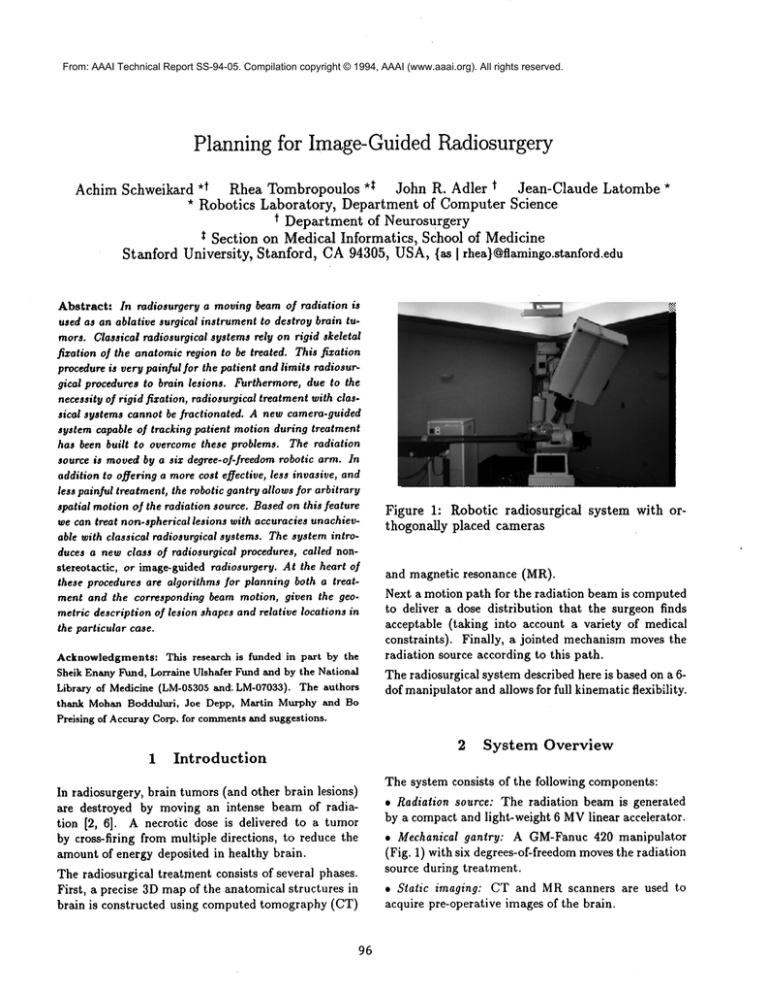
Figure 1: Robotic radiosurgical
thogonally placed cameras
system with or-
and magnetic resonance (MR).
Next a motion path for the radiation beam is computed
to deliver a dose distribution that the surgeon finds
acceptable (taking into account a variety of medical
constraints). Finally, a jointed mechanism moves the
radiation source according to this path.
The radiosurgical system described here is based on a 6dof manipulator and allows for full kinematic flexibility.
2
1
*
System
Overview
Introduction
In radiosurgery, brain tumors (and other brain lesions)
are destroyed by moving an intense beam of radiation [2, 6]. A necrotic dose is delivered to a tumor
by cross-firing from multiple directions, to reduce the
amount of energy deposited in healthy brain.
The radiosurgical treatment consists of several phases.
First, a precise 3D map of the anatomical structures in
brain is constructed using computed tomography (CT)
96
The system consists of the following components:
¯ Radiation source: The radiation beam is generated
by a compact and light-weight 6 MVlinear accelerator.
¯ Mechanical gantry: A GM-Fanuc 420 manipulator
(Fig. 1) with six degrees-of-freedom movesthe radiation
source during treatment.
¯ Static imaging: CT and MRscanners are used to
acquire pre-operative images of the brain.
~reatment planning: At the heart of the new system
tre geometric planning algorithms. Given constraints
)laced by the desired dose distribution, a beam path
s computed. The beam path must observe additional
:onstraints, i.e. the path must be collision-free and
nust not obstruct the surveillance and on-line vision
;ubsystems.
, Direct dosimetry: A dosimetry program simulates the
~lanned treatment and displays the dose distribution
vhich will be generated by a computed plan.
On-line positioning: A vision system with two orthog,nal x-ray camerasacquires imagesof the patient’s skull
wice every second and computes the patient’s position
,y correlating the images to precomputed radiographs.
:mall movements of the head are compensated for by
¯ corresponding motion of the manipulator arm, while
¯ rger movementscause the radiation process to be inerrupted.
)uring treatment, the 6-dof arm moves in point-tooint modethrough a series of configurations. At each
onfiguration, the beam is activated for a small time
aterval 6t, while the arm is held still.
~urrently, the beam has a circular cross-section, the
¯ dius of which remains constant throughout a given
peration. However, this radius initially can be set
etween 5mmand 40mmby selecting an appropriate
~llimator for focusing the beam.
’he new system offers the following advantages over
¯r]ier radiosurgical and radiotherapeutical systems
~eee.g. [7, 8, 9]):
* The tissue does not have to be fixed in space. In
earlier radiosurgical systems this was done with
stereotaxic frames¯ Besides being very painful for
the patient, this fixation was only possible for the
lesions in the head, so the use of radiosurgical
methods was limited.
¯ Treatment can be fractionated, i.e. the total dose
can be delivered in a series of 2-30 treatments,
where only a small dose is delivered during each
treatment¯ Fractionation has turned out to be
highly effective in radiation therapy, but could not
be used in radiosurgery due to the necessity of
rigidly fixing the tissue in space.
¯ Based on geometric planning algorithms the radiation dose can be focussed within the lesion with
high accuracy so that healthy tissue can be protected, and side effects of radiation can be reduced
dramatically¯
97
a)
Figure 2: Cross-firing
3
Geometric
Treatment
b)
at a tumor
Planning
The objective of treatment planning is to find a motion
suitable for treating the particular condition presented
by a patient. This planning problem is illustrated in
Fig. 2. The figure schematically depicts an axial crosssection of the brain with a circular tumor T. Weassume that the beam is cylindrical, i.e. generated by a
cylindrical collimator. If T is irradiated from only one
direction rl, the healthy tissue along the beam absorbs
approximately the same dose as the tumor. If, instead,
we use two directions, rl and r2, the dose deposited in
the tumor is approximately twice the dose in healthy
tissue (Fig. 2-a). Using more beam directions can lead
to further improvementsof the dose distribution, and a
very sharp drop-off of the dose in the tissue surrounding the (spherical) tumor region can be achieved.
this type of treatment, the axes of all beams cross a
single point in space, called the isocenter. However,
for tumors of non-spherical shape this single-isocenter
treatment procedure is problematic (Fig. 2-b).
Treatment planning has to take into account the following constraints.
¯ Sharp dose drop-off in the healthy tissue surrounding the tumor should be achieved to protect healthy tissue. The region receiving high dose
should match the tumor region.
¯ Certain structures in the anatomy (especially the
brain) are very critical and/or radiation sensitive.
The dose in such critical regions should be very
small.
¯ Dose homogeneity;for certain lesions, it is essential that the dose inside the lesion be uniform. It
is also desirable that the dose in the healthy noncritical structures be uniform.
Our methodfor finding a suitable treatment plan proceeds in two steps:
¯ Beamselection: beamsare selected according to
the given lesion shape¯Theselection uses a reachability modelof the manipulatorworkspace.
¯ Beamweighting: an intensity or weight is assigned
to each beam.The weight is given by the duration
of beamactivation.
4
Selecting
X
Beam Configurations
Spherical Tumors: First assume the tumor is spherical, with center p and radius r. Weset the radius of
all beamsto r. The beamconfigurations are then selected in the followingway.Theaxes of all beamscross
the tumorcenter p. (p is the isocenter.) Directions for
the beamscould then be chosen by placing the axis of
each beamthrough a vertex of a regular Plato polyhedron centered at p. However,the orientation of such a
polyhedronis not defined, and the choice of this orientation becomesproblematicif there are critical healthy
structures in the tumor vicinity. Furthermorethere is
no regular Plato polyhedron with more than 12 vertices. A moreevendistribution is obtained using mo.re
directions. For n > 12 the methods in [13] provide
point grids on spheres whichare adequate for our purposes. Thuseach beamaxis crosses the isocenter p and
a point on a spherical grid centered at p. Our planning
methoduses a large set of beams(typically n > 300)
for a single sphere, and addressesthe presence of critical regions not during the beamselection phase, but
during the beamweighting phase.
Ellipsoidal Shapes: While the schemein the previous
paragraph gives both very sharp dose drop-off around
a sphere-shapedlesion and sufficient homogeneity,generalizations of this schemefor treating non-spherical
tumors are not obvious.
Afirst attempt is to use two spheres instead of a single sphere, whereeach sphere is treated independently.
However,it is generally difficult to cover a tumorwith
arbitrary shape with spheres, such that the volumeof
overlap with the tumor environmentis small, and the
entire shape is covered. This is aggravatedby the fact
that the two spheres should not intersect, since this
wouldyield very inhomogeneous
dose distributions, i.e.
high dose (so-called hot spots) in the intersection region of the spheres and cold spots outside such regions.
Studies on complicationrates showthat hot spots, even
inside the tumor,shouldbe avoidedif at all possible.
98
Figure 3: Isodosic surface, beam directions. Region inside surface absorbs 50%or more of maximum dose
However,a simple generalization of the above singlesphere methodis obtained in the following way.
Instead of using a single isocenter, weplace a series of
isocenters evenly spaced along a line segment. Each
such isocenter is treated in the same wayas a single
isocenter. Central to this approachis the fact that each
sphere receives very low dose, and manyspheres are
overlaid. In this way we can avoid hot spots in the
intersection of two spheres.
The dose distribution resulting from such a motion as
well as isocenter points and beamdirections for generating this distribution are shownin figure 3. Thefigure
showsan 50%isodosic surface, i.e. the region inside the
surface receives 50%or more of the maximum
dose.
There are several problemswith this method:
1. If weuse the sameset of directions for all isocenters thus chosen, manyof the beamswill be parallel. Accumulateddose fall-out from such beams
will cause dose inhomogeneityin the surrounding
tissue.
2. Dose inhomogeneity in the tumor mayoccur. The
dose in the tumorcenter will be muchhigher than
the dose along the tumor boundary. This inhomogeneityis in large part due to the cylindrical
collimator shape.
To address the first problem, we choose rotated beam
sets for each isocenter. Hencethe beamsfor different
isocenters do not line up along a certain direction, and
accumulationof fall-off is avoided.In practice, rotating
~he entire beam set aimed at one isocenter by a fixed
~mount turns out to be equivalent to choosing each
ndividual beam direction from a finer spherical point
;rid. Furthermore, instead of using several beams for
inch isocenter, we can use a larger set of isocenters,
Lgain evenly spaced on a line segment and a single
)earn (from the finer sphere grid) for each such isocen.er. Thus we place a single beam through each isocenter
)oint along the line segment. Each beam direction is
.hen randomly chosen from a fine grid of points on the
mit sphere.
[’he second problem can be addressed in several ways.
kn obvious wayis t ° use a collimator of non-cylindrical
hape. For example, we can use a collimator focusing
he beam to a rectangular shape. Using beam direcions as in figure 3 in combination with a rectangular
ollimator provides very high homogeneityand does not
equire additional hardware or safety features for movo
ag parts within the beam collimator. For these tenons, rectangular collimators have been added to the
ew system.
~eneral Shapes: The above approach is restricted
the pseudo-cylindrical shapes in figure 3 and shapes
btained from bending such shapes. A direct general:ation of this scheme is the following. With the alorithm given in [13], we compute a grid of n points
n the tnmor surface. Each such point is now treated
s an isocenter, again placing one beam with direction
aosen as above through each isocenter. In experiments
turns out that this scheme addresses both the first
nd the second of the mentioned problems: AccumuLtion along certain principal directions is avoided by
sing a fine grid. Accumulation of high dose in the
1mot center is avoided by placing isocenters on the
lmor surface. To achieve homogeneity, the point grid
a the input surface can be expandedor retracted inter=tively. A path thus generated is refined by computing
earn weights.
5
Computing Beam Weights
uring the second planning step weights are assigned to
De previously selected beams. Let CI,...,Cn denote
te cylindrical beamsat the n selected configurations.
Figure 4: Regions T, H, minimal and maximal cell
~mln resp. Igrnax.
tion provides a coarse model for photon beam characteristics. Refinements described in [11] allow for a more
accurate representation of these characteristics.)
Weconsider two disjoint regions T (for tumor) and
(for healthy tissue), with the following constraints: the
dose delivered at each point in T must be larger than
some value a, while the dose at each point of H must
be below f~ (/~ < a). The n cylinders, T, and H define
an arrangement of cells in space. Each cell is defined
as a maximal connected set not containing any piece
of the boundaries of regions T and H or the cylinders.
For each cell we compute a label. A cell ~ in cylinders
CQ,..., Cih has label l = {il,. ¯., ik}.
The calculation of wl,..., wn reduces to finding a point
in the intersection of two n-dimensional polyhedral sets:
If ~ is in T and labeled by {it,..., it } then ~: determines
the inequality:
o~ < wi~ + ... + wi~.
A cell ~ labeled by {jl,...,
jk’} in H gives:
# > wj, +...+ w~,.
The inequalities for all cells in T determine a convex
polyhedral set Pa. Similarly, the inequalities derived
from the cells in H determine a polyhedron P#. If Pa
and P~ intersect, any point (Wl,..., w,) in the intersection gives a dose distribution that satisfies the given
constraints.
Otherwise, the problem admits no solution.
More generally, we can specify several healthy critical
or non-critical regions HI,...,Hq marked by distinct
maximal doses ~l,...,f~q.
We then obtain polyhedra
Pa, Pa~,..., Pa~. Any point (wl,..., Wn) in the intersection of these polyhedra determines a dose distribution that satisfies the input constraints.
The intersection of the polyhedral sets Pa, Pa~, ¯ ¯., P#,
is another n-dimensional convex polyhedral set. Extreme points of this set can be computed, allowing us
ur method will assign a dose value wi > 0 to every
i. Each wi is a factor specifying activation duration at
te particular (static) beamconfiguration. The values
1,-.., wn determine a distribution D, defined as folws. Ifp is a point and C1,.-., Ck are all the cylinders
~ntaining p, then D(p) = wl +... + wk. (This defini-
99
(a)
(c)
(d)
(e)
)
(k)
(f)
Ca)
(m)
(n)
(o)
Figure 6: Phantom study for sample case.
~c implies the inequality given by ~’, since all wi are
positive or null. A T-cell is called minimal,if its label is
not a superset of any other T-label (figure 4). Similarly,
a cell in-Hi is called maximal,if its label is not a subset
of any other label in Hi. Thus, in the above polyhedral
intersection test, we need only to consider minimal Tcells and maximal H-cells.
6
(d)
Implementation
A treatment planning system with 3D graphical user
interface based on the above beam selection and weighting methods is the kernel of the new system.
Figure 5: Sample case (Stanford Medical Center).
(a) Axial CT with tumor delineated in black (b)
3D reconstruction of tumor (c) Isodosic surface (d)
Isodosic surface overlaid on tumor reconstruction
to deal directly with criteria for optimization (e.g., in
addition to satisfying the input constraints defined by
a, ~x,..., ~q, minimize the dose delivered to some region Hi).
Reducing the number of inequalities:
We consider
two inequalities deriving from two cells ~ and td in T.
Let L - {il,...,ik}
and L~ - {jl,-..,J~’} be the labels
of ~¢ and ~. If L C L~, then the inequality given by
I00
Inputs: The tumor and critical tissues are described
as polyhedra. This is done as follows: the regions of
interest are delineated by polygons in the MRimages
showing axial, sagital, or coronal cross-sections of the
brain. These polygons are thickened between two crosssections, and a 3D reconstruction of the tumor shape
is formed.
The radius r of the (cylindrical) beam and the number
n of beam configurations are given as input.
To compute beam weights, our implementation uses a
modified simplex algorithm to find a point in the intersection of k n-dimensional polyhedra.
1 example is shownin figure 5 a...d. Figure a shows
axial cross-section of the brain for a 69 year old
.tient presenting a large non-spherical tumor of the
x cerebri, delineated in the cross-section. Figure 5 b
~ws the 3D reconstruction of the tumor outline. A
;ular grid of 600 points was expandedon a surface renstruction, giving isocenter points. Beamdirections
re chosen from a fine spherical grid describing reachle points in the work space. Figure 5 c shows the
% isodosic surface resulting from the computed mo.n. Figure 5 d overlays the input shape with this
:face to illustrate
the matching between input and
tput shapes. For verification, the motion was exceed exposing a photographic film phantom (Fig. 6).
test series for the new system was carried out to
duate the possibilities of generating prespecified dose
tributions. In this test series, cases treated during a
nparison period with an earlier system for frame~ed stereotactic radiosurgery were considered. In
:h case plan data for the actual treatment plan permed were retrieved and plans were recomputed with
: new planner. The comparison is based on phot.phic film phantom studies in water, dose-volume
tograms, as well as 2D and 3D dose visualization
¯ adigms. First results of the test series and variants
~he above methods are discussed in [11].
7
Conclusion
)rototype of the new system has been installed at
.nford Medical Center. Several systems will soon be
~alled at major medical research institutions.
r methods divide treatment planning into two steps:
Selection of beam configurations. (2) Computation
lose weights for these configurations. For the secI step, we proposed a general theoretical approach.
.~ evaluation of the planning methods gives very enraging results. The design of new collimator methand corresponding generalizations of the planning
adigms is under way. The planning method relies on
ghted arrangements in 3D space. This is not limited
:ylinders, and seems appropriate for treatment plang in conventional radiation therapy as well.
likely that the newsystems will allow for radiosurd treatment of tumors outside the brain, including
mrs for which no treatment is currently available. In
ticular, extensions are desirable for very radiationstant tumors and cases in which conventional radiah therapy would cause too much damage to healthy
ue and is not applicable.
101
References
[1] Barth, N. H. An Inverse Problem in Radiation Therapy. Intern. J. Radiation Oncology
Biol. Phys., 18:425-431, 1990.
[2] Betty, O. O., Munari, C., and Rosler, R. Stereotactic Radiosurgery with the Linear Accelerator:
Treatment of Arteriovenous Malformations. Neurosurgery, 24(3):311-321, 1989.
[3] Brahme, A. Optimization of stationary and moving beam radiation therapy techniques. Radiother. Oncol., 12:127-140, 1988.
[4] Chazelle, B., Edelsbrunner, H., Guibas, L., and
Sharir. M. A singly-exponential
stratification
scheme for semi-algebraic varieties and its applications. In Proc. 16th Int. Colloq. Automata
Lang. Programm., Lecture Notes in Computer Science, Springer Verlag, 372:179-192, 1989.
[5] Halperin, D. and Yap, C.K. Combinatorial Complexity of Translating a Box in Polyhedral 3-Space
9th Annual ACMSyrup. on Computational Geometry, 29-37, 1993.
[6] Larsson, B. et at. The High Energy Proton Beam
as a Neurosurgical Tool. Nature, 182:1222-1223,
1958.
[7] Lutz, W., Winston, K. R., and Maleki, N. A System for stereotactic radiosurgery with a linear accelerator. Int. J. Radiation OncologyBiol. Phys.,
14:373-381, 1988.
[8] Podgorsak, E. B., et al. Dynamic Stereotactic Radiosurgery Intern. J. Radiation Oncology
Biol. Phys., 14:115-126, 1988.
[9] Rice, R. K., et al. Measurements of Dose Distributions
in Small Beams of 6 MeVX-Rays.
Phys. Meg. Biol., 32:1087-1099, 1987.
[10] Schweikard, A., Adler, J. R., and Latombe, J.
C., Motion Planning in Stereotaxic Radiosurgery.
Proc. 1EEE lnt. Conf. Robotics and Automation,
Atlanta, GA, May 1993, 1909-1916. (Extended
version to appear in 1EEE Tr. Robotics and Automation.)
[11] Schweikard, A., Tombropoulos, R. Z., Adler, J.
R., and Latombe, J. C., Treatment Planning for
a radiosurgical System with General Kinematics.
to appear: Proc. IEEE Int. Conf. Robotics and
Automation, 1994.
[12] Troccaz, J. et al. Conformal External Radiotherapy of Prostatic
Carcinoma: Requirements and
Preliminary Results, Rep. No. 9121, TIMC-IMAG,
Facult~ de M~decine, Grenoble, 1993.
[13] Turk, G. Re-Tiling Polygonal Surfaces. Computer
Graphics, 26(3):55-64, 1992.


