Document 13759527
advertisement
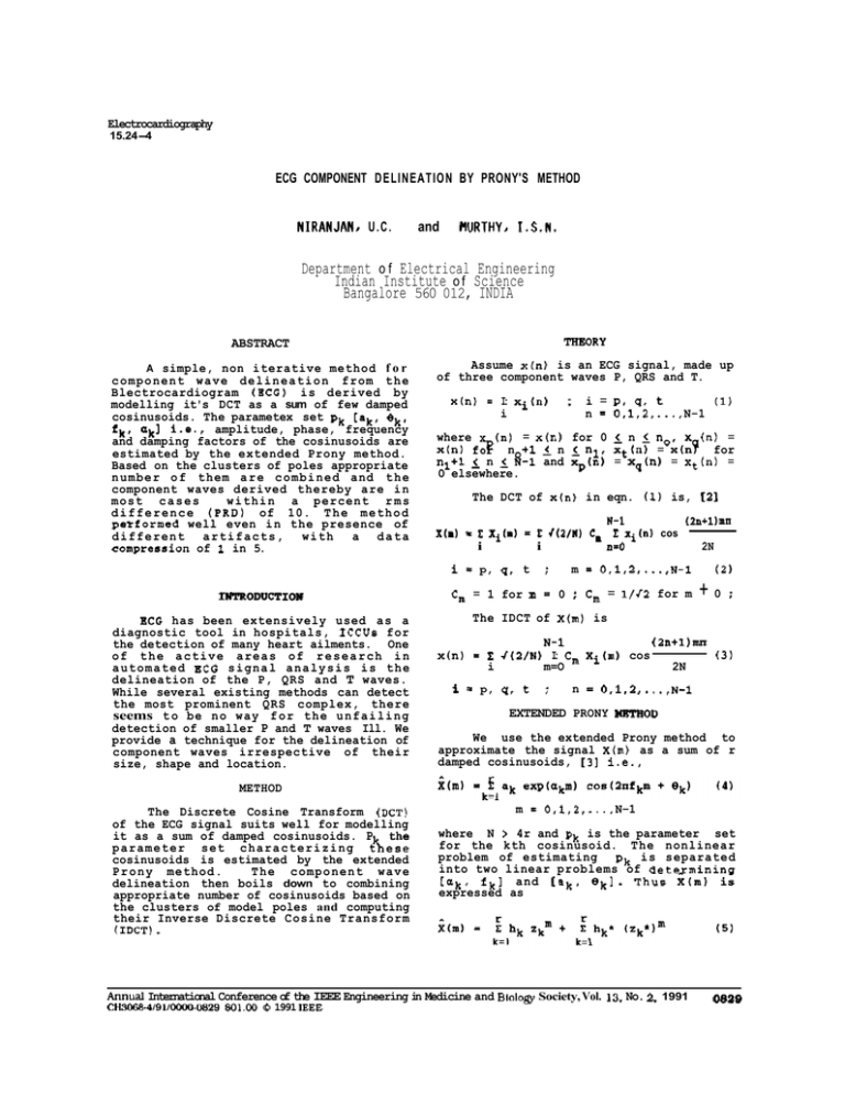
Electrocardiography 15.24-4 ECG COMPONENT DELINEATION BY PRONY'S METHOD NIRANJAN, U . C . and HURTHY, I . S . N . Department o f Electrical Engineering Indian Institute o f Science Bangalore 560 012, INDIA ABSTRACT THEORY A simple, non iterative method f o r c o m p o n e n t w a v e d e l i n e a t i o n from t h e Blectrocardiogram (ECG) is derived by modelling it's DCT as a sum of few damped cosinusoids. The parametex set pk [ak, 8 k # fk' akl i.e., amplitude, phase, frequency and damping factors of the cosinusoids are estimated by the extended Prony method. Based on the clusters of poles appropriate n u m b e r o f them a r e c o m b i n e d and t h e component waves derived thereby are in most c a s e s w i t h i n a percent r m s d i f f e r e n c e ( P R D ) of 10. T h e m e t h o d petformed well even in the presence of different artifacts, with a data compression of 1 in 5. Assume x(n) is an ECG signal, made up of three component waves P, QRS and T. x(n) = I: xi(") i BCG has been extensively used as a diagnostic tool in hospitals, ICCUs for the detection of many heart ailments. One of t h e a c t i v e a r e a s of r e s e a r c h i n a u t o m a t e d ECG s i g n a l a n a l y s i s i s t h e delineation of the P, QRS and T waves. While several existing methods can detect the most prominent QRS complex, there seems t o be no way f o r t h e u n f a i l i n g detection of smaller P and T waves Ill. We provide a technique for the delineation of component waves irrespective of their size, shape and location. i = PI QI t n = 0,1,2, ...,N-1 (1) where (n) = x(n) for 0 5 n 5 no, x (n) = x(n) f 3 n +1 n 5 nl, xt(n) = x(nY for nl+l i n 5 8-1 and xp(n) = xq(n) = xt (n) = 0 elsewhere. The DCT of x(n) in eqn. (1) is, 121 x(a) - & i Xi(.) (2ntl)RH W-1 = & r(2/N) C, I: xi(") cos i n=O 2N - i = p, q, t IWTRODUCTIOH ; : m = 0,1,2 ,...,N-1 Cm = 1 for m = 0 : Cm = l/f2 for m (2 +0 The IDCT of X(m) is - x(n) i f N-1 (2n+l)mn I: J(2/N) I: Cm Xi(n) cos (3) i m=O 2N p, q, t ; n = 0,1,2 ,...,N-1 EXTENDED PRONY =OD We use the extended Prony method to approximate the signal X(m) as a sum of r damped cosinusoids, [3] i.e., METHOD The Discrete Cosine Transform (DCT) of the ECG signal suits well for modelling it as a sum of damped cosinusoids. P the p a r a m e t e r set c h a r a c t e r i z i n g t%ese cosinusoids is estimated by the extended P r o n y method. The component wave delineation then boils down to combining appropriate number of cosinusoids based on the clusters of model poles and computing their Inverse Discrete Cosine Transform (IDCT). m = 0,1,2, ...,N-1 where N > 4r and pk is the parameter set for the kth cosinusoid. The nonlinear problem of estimating pk is separated into two linear problems of detexmining [Uk, f k ] and [ak, 8k]. T h u s x ( m ) is expressed as Annual International Conference of the IEEE Engineering in Medicine and BioloB Society, Vol. 13. No. 2. 1991 CH3068-4/91/oooO-0829$01.00 0 1991 IEEE (5) 0829 others add finer correction in the form of a small kink or bend to give proper shape to a component wave. where hk = a exp(jek), z k = exp(a + j2nfk) and senotes complex conjugaie. X ( m ) c a n a l s o b e g e n e r a t e d by t h e recursive difference equation 2r X(m)+E b(j)X(m-j) = e(m); 2r m 5 N-1 (6) j=l DISCUSSION A practical problem is the choice of number of cosinusoids for a given signal. The order is estimated using the Akaike's Information Criterion (AIC) and Levinson Recursion for covariance LP. For a signal with 300 samples, on an average about 15 damped cosinusoids are needed. Considering 4 parameters for each cosinusoid in p this resulted in a data compression of %'in 5. The reconstructed component waves differed from the actual ones by a constant dc shift. The first DCT coefficient is modified such that this shift is corrected. w h e r e e ( m ) i s t h e p r e d i c t i o n error. Parameters b(j) obtained by solving the covariance Linear Prediction (LP) problem are related to the modes zl. in eqn. (5) by The remaining unknown parameters hk, hk* , in eqn. (5) are estimated by minimizing the squared error with respect to hk, hk*. The resultinff normal fjquation is [z zlh = z (8) where zH is 2r by N vandermonde matrix obtained using the poles zk; & is 2r by 1 vector Of hk, hk* and X 18 N by 1 data v ctor. For the real data case the matrix zfi z is usually invertible. x The peak samples of component waves xi(") appear also as distinct peaks in the magnitude spectrum of X(m) which in turn are represented by the well separated clusters of poles in the LP model. The method gave satisfactory results even in presence of EMG noise, mains interference a n d b a s e l i n e wander. T h e D C T of most abnormal ECG signals is of mixed phase and d i d n o t p o s e any p r o b l e m in t h e i r modelling. No instability problem associated with the covariance LP was encountered. RESULTS Figs. a-d show good agreement between the model output (whole beat shown as 0 ) and delineated component waves (solid line) with the originals (dashed lines). Estimated DCTs of whole beat, P, QRS and T c o m p r i s i n g of 1 1 , 3 , 3 and 5 d a m p e d cosinusoids respectively are shown in Figs. e-h and their PRDs are 9.08, 24.8, 11.5 and 5.2 respectively. For small and noisy P waves as in this example the PRD may. g o high though the fit is satisfactory. Fig. i shows the distinct and well separated pole clusters which enable easy component delineation. On an average P and T waves require 2 to 4 and QRS needs 3 to 6 damped cosinusoids in t h e i r m o d e l s , o n l y 1 or 2 of them characterize the basic waveshape while I I fl P WAVE REFERENCE 1. 0. Pahlm & L. Sbrnmo, 'Software QRS detection in ambulatory monitoring - a review', Med & Biol Eng & Compt, Jul 1984, pp 289-297. N. Ahmed & K.R.Rao, 'Orthogonal Transforms for Digital Signal Processing', Springer Verlag, New York, 1975. Marple S.L.Jr., 'Digital Spectral Analysis', Prentice-Hall, Englewood Cliffs, New Jersey, 1987. 2. 3. I I 1: fl T WAVE QRS /' '"\ ,\ a -0 3 240 i? 0 -520 0 0830 (e) 120 240 0 dct coef no. 120 (f) 240 0 120 dct coef no. (9) 240 0 120 dct coef no. (tifa Annual InternationalConferenceof the IEEE Engineering in Medicine and Biology Society,Vol. 13,No. 2, 1991 CH3068-4/91/0000-0830 $01.00 0 1991 IEEE
