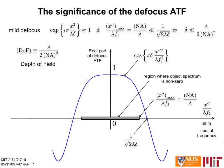
The significance of the defocus ATF 1 0 mild defocus
advertisement

The significance of the defocus ATF mild defocus Depth of Field Real part of defocus ATF 1 region where object spectrum is non-zero 0 spatial frequency MIT 2.71/2.710 05/11/09 wk14-a- 7 The significance of the defocus ATF strong defocus Depth of Field the oscillatory nature of the defocus kernel when δ>DoF results in strong blur on the image because of the suppression of spatial frequencies near the nulls and sign changes at the negative portions Real part of defocus ATF 1 0 spatial frequency MIT 2.71/2.710 05/11/09 wk14-a- 8 The significance of DoF in imaging a≡x”max Depth of Field: defocus tolerance to the axial location of the input transparency (NA)in object plane (NA)out pupil (Fourier) plane Depth of Focus: defocus tolerance to the axial location of the output sensor (CCD or film) image plane Even though our derivations were carried out for spatially coherent imaging, and results apply to the (more common) spatially incoherent case MIT 2.71/2.710 the same 05/11/09 wk14-a-arguments 9 The significance of DoF in imaging a≡x”max δ (NA)in 2½D object A portion of the object at distance δ from the focal plane is locally convolved with the defocus kernel The portion within the in-focus axially cut slice MIT 2.71/2.710 05/11/09 wk14-a- 10 (NA)out pupil (Fourier) plane in-focus slice The point-spread function (PSF) now becomes a function of δ(x); therefore, the imaging system is now shift variant. is imaged with maximum sharpness on a slice of thickness Numerical examples: the object 2 (DoF)s focal plane MIT 2.71/2.710 05/11/09 wk14-a- 11 4 (DoF)s Intensity image: noise-free “M” convolved with standard diffraction-limited PSF MIT 2.71/2.710 05/11/09 wk14-a- 12 “M” convolved with diffraction-limited PSF and defocus equivalent to 2 DoFs “M” convolved with diffraction-limited PSF and defocus equivalent to 4 DoFs Can the blur be undone computationally? • The effect of the optical system is expressed in the Fourier domain as a product of the object spectrum times the ATF: at best, diffraction-limited ATF; it may also include the effect of defocus and higher-order aberrations • Therefore, if we multiply the image spectrum by the inverse ATF, we should expect to recover the original object: this is referred to as “inverse filtering” or “deconvolution” • However, direct inversion never works because: – the ATF may be zero at certain locations, whence the inverse filter would blow up – the image intensity measurement always includes noise; the inverse filter typically amplifies the noise more than the true signal, leading to nasty artifacts in the reconstruction MIT 2.71/2.710 05/11/09 wk14-a- 13 Tikhonov-regularized inverse filter • The following inverse filter behaves better than the direct inversion: – for µ=0, it reduces to the direct filter (not a good idea) – the value of µ should be monotonically increasing with the amount of noise present in the intensity measurement • e.g. if the noise is vanishingly small then we expect direct inversion to be less problematic so a small value of µ is ok; however, the problem of zeros in the ATF remains so µ≠0 is still necessary • if the noise is strong, then a large value of µ should be chosen to mitigate noise amplification at high frequencies • in the special case when both signal and noise obey Gaussian statistics, it can be shown that the optimal value of µ (in the sense of minimum quadratic error) is 1/SNR; this special case of a Tikhonov regularizer is also known as a Wiener filter MIT 2.71/2.710 05/11/09 wk14-a- 14 Tikhonov regularized inverse filter, noise-free Deconvolution using Tikhonov regularized inverse filter Utilized a priori knowledge of depth of each digit (alternatively, needs depth-from defocus algorithm) Artifacts due primarily to numerical errors getting amplified by the inverse filter (despite regularization) MIT 2.71/2.710 05/11/09 wk14-a- 15 Intensity image: noisy SNR=10 MIT 2.71/2.710 05/11/09 wk14-a- 16 Tikhonov-regularized inverse filter with noise SNR=10 Deconvolution using Wiener filter (i.e. Tikhonov with µ=1/SNR=0.1) Noise is destructive away from focus (especially at 4DOFs) Utilized a priori knowledge of depth of each digit Artifacts due primarily to noise getting amplified by the inverse filter MIT 2.71/2.710 05/11/09 wk14-a- 17 Today • Polarization – the vector nature of electromagnetic waves revisited – basic polarizations: linear, circular – wave plates – polarization and interference • Effects of polarization on imaging – beyond scalar optics: high Numerical Aperture – engineering the focal spot with special polarization modes MIT 2.71/2.710 05/13/09 wk14-b- 1 Vector nature of EM fields Recall the vectorial nature of the EM wave equation: The polarization is given by the constitutive relationship: index of refraction The index of refraction (phase delay) depends on the polarization The index of refraction (phase delay) depends on the intensity MIT 2.71/2.710 05/13/09 wk14-b- 2 Linear polarization Fixed time (“snapshot”) t=0 x E(z=λ/2,t=0) E(z=0,t=0) y z E(z=λ,t=0) Φ=0 Φ=2π MIT 2.71/2.710 05/13/09 wk14-b- 3 Linear polarization Fixed space (“oscilloscope”) z=0 x E(z=0,t=λ/ω) E(z=0,t=0) y t E(z=0,t=2λ/ω) Φ=0 Φ=2π MIT 2.71/2.710 05/13/09 wk14-b- 4 Circular polarization Fixed time (“snapshot”) t=0 x E(z=λ/2,t=0) E(z=0,t=0) y z E(z=λ,t=0) Φ=0 Φ=2π MIT 2.71/2.710 05/13/09 wk14-b- 5 Circular polarization Decomposition into the two basic linear polarizations with π/2 phase delay Ex z + z Ey MIT 2.71/2.710 05/13/09 wk14-b- 6 Circular polarization Fixed space (“oscilloscope”) x rotation direction E(z=0,t=0) E(z=0,t=π/2ω) E(z=0,t=π/ω) MIT 2.71/2.710 05/13/09 wk14-b- 7 z E(z=0,t=3π/2ω) y λ/4 wave plate z birefringent λ/4 plate MIT 2.71/2.710 05/13/09 wk14-b- 8 λ/2 wave plate z birefringent λ/2 plate MIT 2.71/2.710 05/13/09 wk14-b- 9 λ MIT 2.71/2.710 05/13/09 wk14-b- 9 Polarization and interference I + φ -polarized waves interfere I + φ ⊥-polarized waves do not interfere MIT 2.71/2.710 05/13/09 wk14-b-11 Intensity in focal region Image removed due to copyright restrictions. Please see Fig. 2 in Linfoot, E. H., and Wolf. "Phase Distribution Near Focus in an Aberration-Free Diffraction Image." Proceedings of the Physical Society B 69 (August 1956): 823-832. Assumes: • Small angles (paraxial) • N large (Debye approximation) 12 McCutchen JOSA 54, 240-244 (1964) 3-D pupil 3-D point spread function h is 3-D Fourier transform of 3-D pupil (cap of spherical shell, Ewald sphere) 13 Two cases for Debye approximation (a) Aperture in plane of lens and N large N = a2/λz ~ a2/λf (b) Aperture in front focal plane of lens 14 Li and Wolf: finite Fresnel number Debye approximation not valid Diffraction of converging wave by an aperture (paraxial theory) Image removed due to copyright restrictions. Please see Fig. 4b in Li, Yajun, and Emil Wolf. "Three-dimensional intensity distribution near the focus in systems of different Fresnel numbers." Journal of the OSA A 1 (August 1984): 801-808. Maximum in intensity no longer at focus focal shift 15 Tight focusing of light • Microscopy • Laser micromachining and microprocessing • Optical data storage • Optical lithography • Laser trapping and cooling • Physics of light/atom interactions • Cavity QED 16 Focusing by high numerical aperture (NA) lens (Debye approximation) Front focal plane α f E(r) f E1(ρ, φ) Equivalent refractive locus (sphere for aplanatic system (sine condition)) 17 A plane polarized wave after focusing: Polarization on reference sphere direction of propagation • px (electric dipole along x axis) • my (magnetic dipole along y axis) • C is nearly linear polarization • Richards & Wolf polarization C 18 Richards and Wolf, 1959 Angular spectrum of plane waves Aplanatic factor I2: cross-polarization component I1: longitudinally-polarized component 19 Focal plane for aplanatic Image removed due to copyright restrictions. Please see Fig. 5 in Sheppard, C. J. R, A. Choudhury, and J. Gannaway. "Electromagnetic field near the focus of wide-angular lens and mirror systems." IEE Journal on Microwaves, Optics, and Acoustics 1 (July 1977): 129-132. 20 Not circularly symmetric Focus of an aplanatic lens Image removed due to copyright restrictions. Please see Fig. 6 in Sheppard, C. J. R., and P. Török. "Efficient calculation of electromagnetic diffraction in optical systems using a multipole expansion." Journal of Modern Optics 44 (1997): 803-818. 21 Bessel Beam Annular mask Axicon (McLeod, 1954) Diffractive axicon (Dyson, 1958) 22 Bessel beam J0 beam propagates without spreading: Image removed due to copyright restrictions. Please see Fig. 2 in Sheppard, C. J. R. "Electromagnetic field in the focal region of wide-angular annular lens and mirror systems." IEE Journal of Microwaves, Optics, and Acoustics 2 (September 1978): 163-166. Time-averaged electric energy density for plane polarized illumination 23 (e.g. with mirror) Annulus at high NA: circular polarization or TM0 (radial polarization) 0 (radial) •Paraxial: annulus narrower than Airy •High NA: circular polarized annulus is ~ same width as Airy •High NA: TM0 annulus is similar to paraxial 24 Polarization on reference sphere red: electric field direction of propagation 25 blue: magnetic field Radial polarization with phase mask Images removed due to copyright restrictions. Please see Fig. 2, 4, in Wang, Haifeng, et al. "Creation of a Needle of Longitudinally Polarized Light in Vacuum Using Binary Optics." Nature Photonics 2 (August 2008): 501-505. 26 Electric dipole wave: Ratio of focal intensity to power input TM0 (radial polarization) electric dipole ED ED is highest mixed dipole (plane polarized) 90o 180o C. J. R. Sheppard and P. Török, "Electromagnetic field in the focal region of an electric dipole wave," Optik 104, 27 175-177 (1997). Polarization of ED on reference sphere red: electric field blue: magnetic field Mixed = ED + MD direction of propagation 28 Bessel beams: TE1 polarization Mixed Mixed 30° ED 60° TE1 Mixed ED Mixed 150° 90° ED TE1 29 TE1 ED TE1 Polarization on reference sphere 30 90o plane polarized Polarization of input wave (azimuthally polarized) (radially polarized) 31 Area of focal spot NA = 0.89 32 NA = 0.91 Rotationally symmetric beams • TM0 = radial polarized input (longitudinal field in focus) • TE0 = azimuthal polarization • x polarized + i y polarized = circular polarized • TE1x + i TE1y = azimuthal polarization with a phase singularity (bright centre) • EDx + i EDy = elliptical polarization with a phase singularity (bright centre) • (TM1x + i TM1y = radial polarization with a phase singularity) • Same GT as for average over φ 33 Bessel beams: Transverse behaviour for rotationally symmetric (also average over φ) 30o 60o MD mixed mixed, TE1 narrowest mixed 34 90o Normalized width for rotationally symmetric TE narrowest for NA<0.98 TE annulus narrowest TE = azimuthal polarization with phase singularity (vortex) 35 Bessel beams for rotationally symmetric Side lobes Transverse gain TE1 is narrowest ED has weakest sidelobes Rad has weakest sidelobes NA=0.83 Eccentricity Rad 36 Conclusions • Focusing plane polarized light results in a wide focal spot • Focusing improved using radially polarized illumination • Strong longitudinal field on axis • Electric dipole polarization gives higher electric energy density at focus • Transverse electric (TE1) polarization gives smallest central lobe (smaller than radial for Bessel beam) • TE is asymmetric: symmetric version is azimuthal polarization with a phase singularity (vortex) 37 MIT OpenCourseWare http://ocw.mit.edu 2.71 / 2.710 Optics Spring 2009 For information about citing these materials or our Terms of Use, visit: http://ocw.mit.edu/terms.


