early-stage cancers. The linear echoendoscope allows the Abstract
advertisement

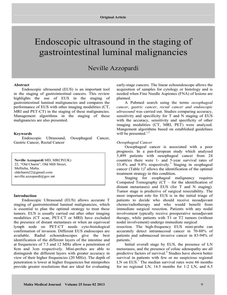
Original Article Endoscopic ultrasound in the staging of gastrointestinal luminal malignancies Neville Azzopardi Abstract Endoscopic ultrasound (EUS) is an important tool in the staging of gastrointestinal cancers. This review highlights the use of EUS in the staging of gastrointestinal luminal malignancies and compares the performance of EUS with other imaging modalities (CT, MRI and PET-CT) in the staging of these malignancies. Management algorithms in the staging of these malignancies are also presented. Keywords Endoscopic Ultrasound, Gastric Cancer, Rectal Cancer Oesophageal Cancer, Neville Azzopardi MD, MRCP(UK) 22, “Old Charm”, Old Mill Street, Mellieha, Malta. oldcharm22@gmail.com neville.azzopardi@gov.mt Introduction Endoscopic Ultrasound (EUS) allows accurate T staging of gastrointestinal luminal malignancies, which is essential to plan the optimal strategy to treat these tumors. EUS is usually carried out after other imaging modalities (CT scan, PET-CT or MRI) have excluded the presence of distant metastases or when an equivocal lymph node on PET-CT needs cyto-histological confirmation of invasion. Different EUS endoscopes are available. Radial echoendoscopes give the best identification of the different layers of the intestine and at frequencies of 7.5 and 12 MHz allow a penetration of 8cm and 3cm respectively. Mini-probes are able to distinguish the different layers with greater accuracy in view of their higher frequencies (20 MHz). The depth of penetration is lower at higher frequencies but miniprobes provide greater resolutions that are ideal for evaluating Malta Medical Journal early-stage cancers. The linear echoendoscope allows the acquisition of samples for cytology or histology and is needed when Fine Needle Aspirates (FNA) of lesions are planned. A Pubmed search using the terms oesophageal cancer, gastric cancer, rectal cancer and endoscopic ultrasound was carried out. Studies comparing accuracy, sensitivity and specificity for T and N staging of EUS with the accuracy, sensitivity and specificity of other imaging modalities (CT, MRI, PET) were analyzed. Mangement algorithms based on established guidelines will be presented.1,2 Volume 25 Issue 02 2013 Oesophageal Cancer Oesophageal cancer is associated with a poor prognosis. In a pan-European study which analysed 5,499 patients with oesophageal cancer from 24 countries there were 1- and 5-year survival rates of 33.4% and 9.8% respectively.3 Staging in oesphageal cancer (Table 1)4 allows the identification of the optimal treatment strategy in this condition. Staging for esophageal malignancy requires Computed Tomography (CT – for the identification of distant metastases) and EUS (for T and N staging). Tumor stage is predictive of surgical resectability. The most important role for EUS is in the initial triage of patients to decide who should receive neoadjuvant chemo/radiotherapy and who would benefit from immediate surgical resection. Patients with any nodal involvement typically receive preoperative neoadjuvant therapy, while patients with T1 or T2 tumors (without nodal involvement) undergo immediate surgical resection. The high-frequency EUS mini-probe can accurately detect intramucosal cancer in 70-88% of patients and submucosal invasive cancer in 83-94% of patients.5 Initial overall stage by EUS, the presence of LN metastases, and the presence of celiac adenopathy are all predictive factors of survival.6 Studies have shown better survival in patients with few or no suspicious regional LN on EUS.7 The median survival rates were 66 months for no regional LN, 14.5 months for 1-2 LN, and 6.5 9 Original Article months for >2 suspicious LN.7 Since the number of suspicious periesophageal LN detected by EUS is inversely associated with survival in patients with esophageal adenocarcinoma, N staging is very important before surgery. For this reason, the Surveillance, Epidemiology, and End Results database suggests that tumor length and number of LN should be routinely reported as part of the staging system.8 The management algorithm for esophageal cancer is described in Figure 1.9 Figure 1: Management of Oesophageal Cancer – from staging to treatment (PET- Positron emission tomography; CT Scan-Computed tomography scan; EUS +/- FNA-Endoscopic ultrasound with or without Fine Needle Aspiration; Neoadjuvant therapy – Chemo/Radiotherapy)7 patients undergoing CT scan showed 40-80% sensitivity and 14-97% specificity.12 N staging also appears to be superior with 60-97% sensitivity and 40-100% specificity for EUS versus 40-73% sensitivity and 2567% specificity for CT scan.12 In addition, FNA of suspicious LN allows cytological confirmation of malignancy. The sensitivity in staging LN increases from 71% with EUS to 83% with EUS-FNA.13 EUS also appears to be superior to PET in T staging and in detecting peritumoral and celiac LN.14 FDG-PET has a 15% false-positive rate in the detection of distant and hematogenous metastases.14 EUS FNA can be used to confirm positive findings on PET scans.15 The combination of CT, PET and EUS decreases the number of unnecessary operations by half when compared with CT alone.14 Detection of previously unidentified celiac axis metastases intraoperatively also decreased significantly when CT was combined with PET or EUS. EUS also detects occult liver metastases, though it visualizes only the medial two thirds of the liver. FNA can be performed for histological confirmation of liver metastasis.16 Gastric Cancer Gastric cancer is the second leading cause of cancer related deaths worldwide.17 Accurate staging of gastric cancer (Table 1) by determining tumor extent and nodal involvement is important in the treatment algorithm (Figure 2). Once passage with the echoendoscope through the stricture is possible, staging of esophageal cancer is done by inspecting the liver, celiac axis and gastrohepatic ligament for the presence of liver or LN metastasis. The esophageal lesion and the mediastinum are then examined to identify depth of invasion and peritumoral and mediastinal adenopathy.7 EUS is also important in restaging after patients receive chemo/radiotherapy. However, EUS is not accurate after neoadjuvant chemo/radiotherapy since it is unable to differentiate tumor from necrosis or inflammatory reaction.10 Other studies however indicate that EUS and EUS-guided FNA can be helpful in identifying residual tumor in the LN after preoperative chemo/radiotherapy in patients who may benefit from surgery.11 EUS is superior to CT scan in tumor (T) staging of esophageal cancer. Thirteen studies of patients with esophageal cancer undergoing EUS showed 71-100% sensitivity and 67-100% specificity while 5 studies in Malta Medical Journal Volume 25 Issue 02 2013 10 Original Article Table 1: American Joint Committee on Cancer TNM Classification for Esophageal and Gastric Cancers2 Oesophageal Cancer T Stage Tx T0 Tis T1 T2 T3 T4 N Stage Nx N0 N1 M Stage Mx M0 M1 Gastric Cancer T Stage Tx T0 Tis T1 T2 T3 T4 N Stage Nx N0 N1 N2 N3 M Stage Mx M0 M1 Tumor cannot be assessed No evidence of primary tumor Carcinoma in situ Invades lamina propria or submucosa Invades muscularis propria Invades adventitia Invades adjacent structures Regional nodes cannot be assessed No regional LN metastasis Regional LN metastasis Distant metastasis cannot be assessed No distant metastasis Distant metastasis Tumor cannot be assessed No evidence of primary tumor Carcinoma in situ Invades lamina propria or submucosa Invades muscularis propria or subserosa Penetrates serosa without invasion of adjacent structures Invades adjacent structures Regional nodes cannot be assessed No regional LN metastasis Metastasis in 1-6 regional LN Metastasis in 7-15 regional LN Metastasis in >15 regional LN Distant metastasis cannot be assessed No distant metastasis Distant metastasis Malta Medical Journal Volume 25 Issue 02 2013 Figure 2: Management of Gastric Cancer – from staging to treatment (CT Scan-Computed tomography scan; EUS +/- FNA-Endoscopic ultrasound with or without Fine Needle Aspiration; EMR-Endoscopic Mucosal Resection; Neoadjuvant therapy chemotherapy)27 Evidence shows that 5-year survival rate for early gastric cancer confined to the mucosa or submucosa is >75% following resection but < 30% with distant metastasis or with the involvement of >15 (N3) LN.18 Surgery is the recommended treatment of choice for localized gastric cancer with endoscopic mucosal resection (EMR) reserved for cancers limited to the mucosa. Neoadjuvant chemotherapy is recommended for patients with deeper invasion. CT is important to exclude distant metastases but lacks accuracy in T and N staging of gastric cancer19, while EUS has much greater accuracy in evaluating the depth of invasion of primary gastric cancer.19-21 In addition, EUS-guided FNA of LN adds to the accuracy of nodal staging.21 The overall sensitivity of EUS in determining T stages is best for T1 and T3 lesions, whereas EUS is least accurate for T2 and T4 lesions. In a meta-analysis of 54 studies carried out between 1988 and 2010, the pooled sensitivity was 83% for T1 lesions, 65% for T2, 86% for T3, and 66% for T4 lesions.22 This is believed to occur because identifying T1 and T3 invasion is facilitated by the hypoechogenicity of the muscularis mucosa and muscularis propria while identifying T4 invasion is made difficult by the limited depth of penetration of the ultrasound waves. The pooled sensitivity for N staging was 69% with a specificity of 84%.22 In situations where malignant LN can be difficult to distinguish from benign nodes, EUS-FNA cytology and biopsy may offer better diagnostic yields. A systematic review comparing the diagnostic T stage accuracy of EUS with CT revealed an accuracy of 65-92% with EUS and an accuracy varying between 11 Original Article 77.1-88.9% with CT.23 Another review comparing N stage sensitivities and specificities of EUS with CT showed that EUS had 71% sensitivity and 49% specificity while CT had 80% sensitivity and 78% specificity.24 CT is also superior in detecting distant metastasis and both techniques are complementary for overall staging. EUS is required in the absence of distant metastases since management decisions will then depend on the depth of invasion. Better surgical outcomes are obtained in patients with locally invasive gastric cancer who receive preoperative neoadjuvant chemotherapy. However, following chemotherapy, the accuracy of CT and EUS for both T and N staging drops significantly (57% and 47% for T staging and 37% and 39% for N staging).25 EUS is more sensitive than transabdominal ultrasound, CT and laparoscopy for the detection of intraperitoneal fluid, which is usually indicative of incurable disease. EUS-FNA of low-volume ascites can be performed safely.26 Visualisation of gastric lesions during EUS is facilitated by aspiration of air and instillation of deaerated water into the stomach to submerge the lesions completely, thus allowing better acoustic coupling. With the standard echoendoscope, five distinct layers are seen, with 3 hyperechoic alternating with 2 hypoechoic layers. The first 2 layers represent the mucosa, the third layer represents the submucosa, the fourth layer is the muscularis propria and the last layer is the serosa. Table 1 describes the T staging of gastric cancer. Tumors confined to the mucosa can be treated with EMR while those involving the submucosa carry a 20% risk of LN involvement and therefore require surgery.27 Perigastric and regional (celiac axis, gastrohepatic ligament) LN are next assessed (Figure 2). Suspicious LN features during endosonography include hypoechogenicity, sharp borders, round shape and size >10mm. All 4 features are present in only 25% of malignant LN, and FNA of nodes with any of these features is necessary to confirm or exclude malignancy.28 Distant metastasis (M) staging by checking the left lobe of the liver, the peritoneum, the pleural layers of the lung, and mediastinal LN is the final step. The finding of malignant ascites, pleural effusion and malignant mediastinal nodes is an indication for neoadjuvant or adjuvant treatment.29 Rectal cancer Rectal cancer represents 5% of malignant tumors and is the fifth commonest cancer in adults.30 Staging of rectal cancer identifies patients with locally invasive tumors who should be treated with neoadjuvant therapy before surgery (Figure 3). Malta Medical Journal Volume 25 Issue 02 2013 Figure 3: Management of Rectal Cancer – from staging to treatment (CT Scan: Computed tomography scan; EUS +/- FNA: Endoscopic ultrasound with or without Fine Needle Aspiration; EMR: Endoscopic Mucosal Resection; T1-Tumor invades lamina propria or submucosa; T2: Tmor invades muscularis propria; T3Tumor penetrates serosa; T4-Tumor invades adjacent structures; N0 – No regional lymph node metastases; N1 – Metastases in 1-3 regional nodes; N2 – Metastases in 4 or more regional nodes; M0 – No distant metastases; Neoadjuvant therapy – chemotherapy and 29 radiotherapy) The accuracy of EUS for T staging in rectal cancer is 85% while the accuracy for N staging is 75%. With the addition of FNA of LN, accuracy increases to 87%.31 During EUS staging of rectal cancers, the relationship of the tumor with adjacent organs such as the prostate, bladder, and seminal vesicles in men and the bladder, vagina, cervix and uterus in women is assessed. The perirectal area is studied for the presence of suspicious LN or involvement of the iliac vessels. Rectal cancer is seen as a hypoechoic lesion that disrupts the rectal wall pattern. A tumor limited to the submucosa is classified as T1, while if it invades the muscularis propria, it is classified as T2. T3 tumors penetrate into surrounding fat while T4 lesions invade into adjacent organs. Both radial and linear EUS scopes can be used for staging rectal lesions; however FNA of lymph nodes can only be done with the linear scope. High-frequency mini-probes using the water-filling technique may be a better option for small lesions (<1-2 cms). Alternatively, changing the patient position may allow better visualization of the lesion. EUS is superior to CT and MRI for T staging of rectal tumors. In a meta-analysis (5000 patients), CT accurately T staged the tumor in 73% of cases and had accurate N staging in 22-73%.32 MRI also has poor 12 Original Article accuracy in T staging of rectal cancer (52-54%).33 A meta-analysis (42 studies) evaluating EUS in rectal tumors revealed sensitivities and specificities of 88% and 98% for T1, 81% and 96% for T2, 91% and 96% for T3 and 95% and 98% for T4 tumors.34 This increased accuracy translates into improved patient management with more patients undergoing neoadjuvant treatment with chemotherapy and radiotherapy before surgery and a more cost-effective strategy when combining initial abdominal CT (to exclude distant metastases) and EUS (for local staging).35 N staging with EUS has not been shown to be superior to other imaging modalities. The pooled sensitivity and specificity of EUS in diagnosing nodal involvement in rectal cancers were 73% (95% confidence interval [CI] 70.6-75.6) and 76% (95% CI 73.5-78.0) with a pooled diagnostic odds ratio of 7.87 (meta-analysis of 35 studies), which is similar to the accuracy of CT and MRI.36 However, FNA of suspicious lymph nodes improves accuracy to 87%.37 Benign perirectal nodes are not usually visualized by EUS. Therefore, finding perirectal nodes during EUS is sufficient to warrant sampling by FNA. Conclusion With its high sensitivity and specificity in tumor (T) staging, endosonography has become an essential tool in the staging of gastrointestinal luminal malignancies. EUS is also important in lymph node (N) staging as it allows FNA cytological sampling of suspicious nodes. However, distant metastases (M staging) should be excluded by abdominal and thoracic computed tomography (with or without PET) before EUS is carried out. References 1. 2. 3. 4. 5. 6. 7. 8. 9. 10. 11. 12. 13. 14. 15. 16. 17. 18. Malta Medical Journal Volume 25 Issue 02 2013 Allum WH, Griffin SM, Watson D, Colin-Jones D. Guidelines for the management of oesophageal and gastric cancer. Gut 2002;50(Suppl V):v1-v23. NICE guidelines. Colorectal cancer: the diagnosis and management of colorectal cancer. Nov 2011. Gavin AT, Francisci S, Foschi R, Donnelly DW, Lemmens V, Brenner H, et al.. Oesophageal cancer survival in Europe: A EUROCARE-4 study. Cancer epidemiol. 2012 Dec;36(6):50551. Springer-Verlag. AJCC Cancer Staging Manual. 7th Edition. Springer-Verlag New York Inc. 2011:2:15-16. Yoshinaga S, Oda I, Nonaka S, Kushima R, Saito Y. Endoscopic ultrasound using ultrasound probes for the diagnosis of early esophageal and gastric cancers. World J Gastrointest Endosc. 2012 Jun 16;4(6):218-26. Eloubeidi MA, Wallace MB, Hoffman BJ, Leveen MB, Van Velse A, Hawes RH et al. Predictors of survival for esophageal cancer patients with and without celiac axis lymphadenopathy: impact of staging endosonography. Ann Thorac Surg 2001;72:212-219. Chen J, Xu R., Hunt GC, Krinsky ML, Savides TJ. Influence of the number of malignant regional lymph nodes detected by endoscopic ultrasonography on survival stratification in esophageal adenocarcinoma. Clin Gastroenterol Hepatol 2006;4(5):573-579. Eloubeidi MA, Desmond R, Arguedas M, Reed CE, Wilcox CM. Prognostic factors for the survival of patients with esophageal carcinoma in the US: the importance of tumor length and lymph node status. Cancer 2001;95:1434-1443. Eloubeidi MA. EUS in esophageal cancer. Endosonography. 2011;7:59-69. Griffin JM, Reed CE, Delinger CE. Utility of restaging endoscopic ultrasound after neoadjuvant therapy for esophageal cancer. Ann Thorac Surg. 2012 Jun;93(6):1855-9.Epub 2012 Apr 18. Agarwal B, Swisher S, Ajani J, Kelly K, Fanning C, Komaki RR et al. Endoscopic ultrasound after preoperative chemoradiation can help identify patients who benefit maximally after surgical esophageal reaction. Am J Gastroenterol 2004; 99(7):1258-1266. Kelly S, Harris KM, Berry E, Hutton J, Roderick P, Cullingworth J et al. A systematic review of the staging performance of endoscopic ultrasound in gastro-esophageal carcinoma. Gut. 2001;49(4):534-539. Vazquez-Sequeiros E, Wiersema MJ, Clain JE, Norton ID, levy MJ, Romero Y et al. Impact of lymph node staging on therapy of esophageal carcinoma. Gastroenterology. 2002;125:16261635. van Westreenen HL, Heeren PA, Jager PL, van Dullemen HM, Groen H, Plukker JT. Pitfalls of positive findings in staging esophageal cancer with F-18-fluorodeoxyglucose positron emission tomography. Ann Surg Oncol 2003;10(9):1100-1105. Eloubeidi M.A., Cerfolio R.J., Chen V.K., Desmond R, Syed S, Ojha B. Endoscopic ultrasound-guided fine needle aspiration of mediastinal lymph node in patients with suspected lung cancer after positron emission tomography and computed tomography scans. Ann Thorac Surg 2005; 79(1):263-268. Prasad P., Schmulewitz N., Patel A., Varadarajulu S, Wildi SM, Roberts S et al. Detection of occult liver metastases during EUS for staging of malignancies. Gastrointest Endosc 2004;59(1):49-53. Ferlay J, Bray F, Pisani P, Parkin DM: Globacan 2000. Cancer Incidence, Mortality and Prevalence Worldwide. Lyon, IARC Press, 2001. IARC Cancer Base No. 5. Agboola O. Adjuvant treatment in gastric cancer. Cancer Treat Rev 1994;20:217-240. 13 Original Article 19. Botet J, Lightdale C, Zauber A, Gerdes H, Winawer SJ, Urmacher C et al. Preoperative staging of gastric cancer: comparison of endoscopic US and dynamic CT. Radiology 1991;181:426-432. 20. Mouri R, Yoshida S, Tanaka S, Oka S, Yoshihara M, Chayama K. Usefulness of endoscopic ultrasonography in determining the depth of invasion and indication for endoscopic treatment of early gastric cancer. J Clin Gastroenterol 2009;43:318-322. 21. Jurgensen C, Brand J, Nothnagel M, Arlt A, Neser F, Habeck JO, et al. Prognostic relevance of gastric cancer staging by endoscopic ultrasound. Surg Endosc. 2012 Oct 6. [Epub ahead of print]. 22. Mocellin S, Marchet A, Nitti D. EUS for the staging of gastric cancer: a meta-analysis. Gastrointest Endosc. 2011 Jun;73(6):1122-34. 23. Kwee R., Kwee T. Imaging in local staging of gastric cancer: a systematic review. J Clin Oncol 2007; 25:2107-2116. 24. Kwee R., Kwee T. Imaging in assessing lymph node status in gastric cancer. Gastric Cancer 2009; 12:6-22. 25. Cunningham D, Allum W, Stenning S, Thompson JN, Van de Velde CJ, Nicolson M et al: Perioperative chemotherapy versus surgery alone for resectable gastroesophageal cancer. N Engl J Med 2006;355:11-20. 26. DeWitt J, LeBlanc J, McHenry L, McGreevy K, Sherman K. Endoscopic ultrasound-guided fine-needle aspiration of ascites. Clin Gastroenterol Hepatol 2007;5:609-615. 27. Hölscher A, Drebber U, Mönig S, Schukte C, Vallbohmer D, Bollschweiler E. Early gastric cancer: lymph node metastasis starts with deep mucosal infiltration. Ann Surg 2009;250:791797. 28. Bhutani M, Hawes R, Hoffman B. A comparison of the accuracy of echo features during endoscopic ultrasound (EUS) and EUS-guided fine-needle aspiration for diagnosis of malignant lymph node invasion. Gastrointest Endosc 1997;45:474-479. 29. Rosch T, Peter S, Varadarajulu S. EUS in the Evaluation of Gastric Tumors. Endosonography 2011;11:97-104. 30. Parkin DM, Bray F, Ferlay J, Pisani P. Global cancer statistics 2002. CA Cancer J Clin 2005;55:74-108. 31. Harewood GC. EUS in Rectal Cancer. Endosonography 2011;18:205-209. 32. Kwok H, Bissett IP, Hill GL. Preoperative staging of rectal cancer. Int J Colorectal Dis. 2000;15:9–20. 33. Chen CC, Lee RC, Lin JK, Wang LW, Yang SH. How accurate is magnetic resonance imaging in restaging rectal cancer in patients receiving preoperative combined chemoradiotherapy? Dis Colon Rectum. 2005;48:722–728. 34. Puli SR, Bechtold ML, Reddy JB, Choudhary A, Antillon MR, Brugge WR. How good is endoscopic ultrasound in differentiating various T stages of rectal cancer? Meta-analysis and systematic review. Ann Surg Oncol. 2009;16:254. 35. Harewood GC, Wiersema M.J. Cost-effectiveness of endoscopic ultrasonography in the evaluation of proximal rectal cancer. Am J Gastroenterol. 2002;97:874-882. 36. Puli SR, Reddy JB, Bechtold ML, Choudhary A, Antillon MR, Brugge WR. Accuracy of endoscopic ultrasound to diagnose nodal invasion by rectal cancers: a meta-analysis and systematic review. Ann Surg Oncol. 2009;16:1255. 37. Park HH, Nguyen PT, Tran Q. Endoscopic ultrasound-guided fine needle aspiration in the staging of rectal cancer. Gastrointest Endosc. 2000;51:AB171. Malta Medical Journal Volume 25 Issue 02 2013 14


