K
advertisement
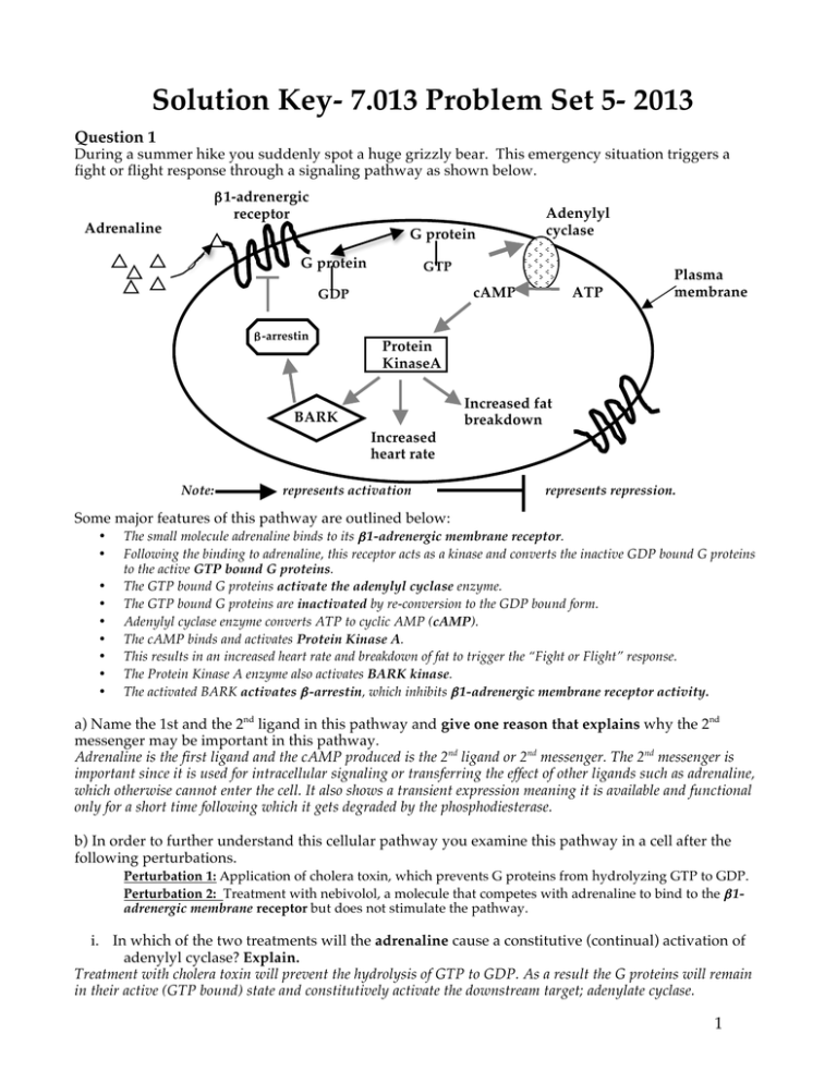
Solution Key- 7.013 Problem Set 5- 2013 Question 1 During a summer hike you suddenly spot a huge grizzly bear. This emergency situation triggers a fight or flight response through a signaling pathway as shown below. β1-adrenergic receptor Adrenaline G protein G protein GTP cAMP GDP β-arrestin Note: ATP Plasma membrane Protein KinaseA Increased fat breakdown BARK K Adenylyl cyclase Increased heart rate represents activation represents repression. Some major features of this pathway are outlined below: • • • • • • • • • The small molecule adrenaline binds to its β 1-adrenergic membrane receptor. Following the binding to adrenaline, this receptor acts as a kinase and converts the inactive GDP bound G proteins to the active GTP bound G proteins. The GTP bound G proteins activate the adenylyl cyclase enzyme. The GTP bound G proteins are inactivated by re-conversion to the GDP bound form. Adenylyl cyclase enzyme converts ATP to cyclic AMP (cAMP). The cAMP binds and activates Protein Kinase A. This results in an increased heart rate and breakdown of fat to trigger the “Fight or Flight” response. The Protein Kinase A enzyme also activates BARK kinase. The activated BARK activates β -arrestin, which inhibits β 1-adrenergic membrane receptor activity. a) Name the 1st and the 2nd ligand in this pathway and give one reason that explains why the 2nd messenger may be important in this pathway. Adrenaline is the first ligand and the cAMP produced is the 2nd ligand or 2nd messenger. The 2nd messenger is important since it is used for intracellular signaling or transferring the effect of other ligands such as adrenaline, which otherwise cannot enter the cell. It also shows a transient expression meaning it is available and functional only for a short time following which it gets degraded by the phosphodiesterase. b) In order to further understand this cellular pathway you examine this pathway in a cell after the following perturbations. Perturbation 1: Αpplication of cholera toxin, which prevents G proteins from hydrolyzing GTP to GDP. Perturbation 2: Treatment with nebivolol, a molecule that competes with adrenaline to bind to the β 1adrenergic membrane receptor but does not stimulate the pathway. i. In which of the two treatments will the adrenaline cause a constitutive (continual) activation of adenylyl cyclase? Explain. Treatment with cholera toxin will prevent the hydrolysis of GTP to GDP. As a result the G proteins will remain in their active (GTP bound) state and constitutively activate the downstream target; adenylate cyclase. 1 Question 1 continued ii. In which of the two treatments will you see very low or no activation of adenylyl cyclase in the presence of adrenaline? Explain. Treatment with Nebivolol will result in no or little activation of adenylate cyclase since this inhibitor will compete with adrenaline to bind to the β1- adrenergic receptor. c) Consider the following perturbations in different components of the above signaling pathway in a cell. • #1: BARK lacks its kinase domain. • #2: β arrestin is constitutively (always) active. • #3: β1-adrenergic membrane receptor lacks its ligand-binding domain. Complete the table for each of the following perturbations relative to unperturbed control cells in the presence of adrenaline. Note: Consider each perturbation independently while answering the questions. Perturbation Is cAMP produced? Is Protein Kinase A activated? Is β-arrestin activated? 1 Yes Yes No 2 No No N/A 3 No No No Effect on heart rate and fat breakdown relative to unperturbed control cells in the presence of adrenaline (choose from increased, decreased or unchanged)? Explain. Since mutant BARK is not activated by PKA, it will not activate arrestin, which is required for the feedback inhibition of β- adrenergic receptor. Therefore there will be an increased heart rate and fat breakdown relative to unperturbed control cells. Mutant arrestin will have a constitutive feedback inhibitory effect on β- adrenergic receptor. Therefore there will be no increase in the heart rate and fat breakdown relative to unperturbed control cells. Mutant β- adrenergic receptor will not bind to adrenaline. Accordingly, the pathway will not get activated and there will be no increase in heart rate and fat breakdown relative to unperturbed control cells. Question 2 The origin recognition complex (ORC) is a multi- subunit protein complex that binds to the ori site(s) and serves as a platform for the assembly of kinases like Cdk6 and Cdt1. During the G1 phase of the cell cycle in yeast, ORC forms a pre- replication complex by recruiting Cdk6 and Cdt1 that bind to both strands of DNA. These factors bind and inhibit the Mcm protein that functions as a helicase as is shown in the schematic below. Note: The activation is shown by an → and inhibition by ⊥ sign. 2 Question 2 continued a) Activation of the pre-replication complex occurs during the S phase and this requires its interaction with Cdk2 and Cyclin E proteins that degrade Cdk6 and Cdt1. This results in the replication of DNA. Draw a schematic, similar to the one on page 2, to show the regulatory interactions between ORC, Cdk6, Cdt1, Mcm, Cdk2 and Cyclin E proteins. Note: Please indicate the activation by an → and inhibition by ⊥ sign. Orc bound to Ori Cyclin E Cdk2 5’ 3’ 3’ 5’ Unwinding of DNA and replication Mcm (Active) Cdt1 (inactive) Cdk6 (inactive) Replication complex in S phase b) In a cell showing a Cdk2 loss-of–function mutation, in which phase (choose from G1, S, G2, M, all or none) would the cell arrest? Explain why you selected this option. In this mutant, since the Mcm protein is never activated the cell will not replicate its DNA and instead remain arrested in the G1 phase. c) If the cdk-2 gene encodes Cdk-2 protein, in which phase (choose from G1, S, G2, M, all or none) will the cdk-2 gene be expressed? Explain why you selected this option. The cdk-2 gene will be expressed in all the phases of cell cycle but the synthesized Cdk-2 protein will be activated only in G1/ S interphase by the Cyclin E protein. d) If the cyclin E gene encodes Cyclin E protein, in which phase (choose from G1, S, G2, M or all) will the cyclin E gene be optimally expressed? Explain why you selected this option. Each cyclin shows a transient/ cyclic expression in a specific phase of cell cycle. Therefore, the cyclin-E gene will be expressed only in the G1/ S interphase where it encodes Cyclin E protein, which can bind to and activate Cdk6 protein. e) One method for studying the essential components that regulate different steps of the cell cycle involves the use of conditional temperature-sensitive mutants. You isolate temperature-sensitive Cdk2 yeast mutants. These mutant cells grow normally at the permissive temperature (25°C). However these cells arrest at a specific point in the cell cycle when the temperature is shifted to 36°C (non-permissive temperature). i. Is the primary structure of the temperature-sensitive Cdk2 protein at 36°C same as or different from that at 25°C? The primary structure will be the same at both temperatures. ii. Is the three- dimensional conformation of the temperature-sensitive Cdk-2 protein at 36°C same as or different from that at 25°C? Explain why you selected this option. The three- dimensional structure of the protein that is predominantly stabilized by the non-covalent bonds be disrupted at higher temperature since these bonds are sensitive to temperature changes. f) You grow the mutant cells, described in part (e), at 36°C or non- permissive temperature. i. Predict the effect on G1 phase in the mutant cells grown at 36°C compared to those at 25°C. The cells will arrest in the G1 phase. ii. Predict the effect on the S phase in the mutant cells at 36°C compared to those at 25°C. The cells will arrest in the G1 phase. 3 Question 2 continued g) You further create two mutant cells each having a mutation in Cdk-2 genes as described below. • Mutant cell- type 1: The Cdk-2 protein lacks its kinase domain. • Mutant cell- type 2: The Cdk-2 lacks its Cyclin E binding domain. Predict what would happen to the cell cycle in… i. Mutant 1: The cells will arrest in the G1 phase. ii. Mutant 2: The cells will arrest in the G1 phase. h) The proper development and functioning of different tissues and organs involves a tight regulation of cell division and cell death. i. A cell can die by “apoptosis”. What does this term mean? Apoptosis refers to the programmed cell death ii. Apoptosis may occur either via extrinsic or intrinsic signaling pathways. Based on what you have learnt in 7.013, if a cell makes the decision to die when subjected to hypoxia (low oxygen concentration) would you regard this as an example of extrinsic or intrinsic signaling pathway? Explain why you selected this option. This is an intrinsic pathway since the hypoxia condition is being sensed by the cell. Note: please take a look at Slide 26 of Cell Biology-III lecture. iii. Apoptosis involves the activation of Caspases. Briefly explain the function of these enzymes. They act by causing the degradation of the proteins that ultimately results in DNA degradation. Question 3 Development of human and chicken hearts is thought to occur by very similar mechanisms. The heart develops from a layer of cells called the “mesoderm”, which lies on top of another layer of cells called “endoderm”. a) The mesoderm forms the heart, the kidney and the blood. i. What does the term “potency” mean? Potency refers to the number of fates a cell can acquire. ii. What term would describe the potency of the mesoderm (choose from totipotent, multipotent or unipotent)? Since the mesoderm can produce heart, the kidney and the blood, it is multi-potent. b) You want to know when the future heart cells decide to become such. You isolate a small piece of tissue (or explant), from the heart forming anterior mesoderm of a young embryo (stage 2), and also from a slightly older (stage 4) embryo (about 6 hours older than stage 2). You make sure to take the same relative region from both. You culture the tissues in tissue culture plates, examine them three days later and tabulate your results below. Explant Stage 2 anterior mesoderm explant Stage 4 anterior mesoderm explant Culture time 3 days in culture Observation No change from original cells 3 days in culture Beating heart 4 Question 3 continued i. At the time of isolation, are the stage 2 cells determined, differentiated or undetermined compared to Stage 4 cells? Explain. They are undetermined since after 3 days of culture they still remain unchanged and do not differentiate into the beating heart (perhaps in the absence specific inducers) ii. Are the beating heart cells determined, differentiated or neither compared to Stage 4 cells? Explain. They are defferentiated since they function as beating heart cells. c) You ask why the isolated Stage 2 cells did not become a heart when they were isolated. Since the anterior mesoderm cells lie on top of the “endoderm” layer, you therefore modify your experiment as shown in the table below and obtain the following results. Tissue Stage 2 anterior mesoderm alone- labeled with green fluorescent protein (GFP) Culture 3 days culture Result Same as original GFP labeled cells Stage 2 anterior mesoderm labeled with GFP) along with underlying endoderm that is unlabeled 3 days culture Beating heart comprised of cells that fluoresce green Endoderm alone that is unlabeled 3 days culture Same as original unlabeled cells Why does the stage 2 anterior mesoderm explant when cultured with endoderm make a heart, whereas the stage 2 anterior mesoderm explant alone does not? Endoderm, most likely, secretes an inducer (or a signaling factor), which binds to a specific receptor located on the surface of anterior mesoderm cells so that they become heart cells. d) You later observe that if Stage 2 cells are cultured for three days with a purified secreted protein, BMP4, they develop and form a beating heart. Based on all the data above, where in the embryo (choose from mesoderm, endoderm or both) would you expect BMP4 to be expressed if it directs normal heart formation? Explain why you selected this option. BMP4 is most likely expressed and secreted by endodermal cells since it is inducing the heart. On the mesoderm cells, it binds to a specific receptor so that they can transition into a beating heart. e) Posterior mesoderm does not form the heart. To test whether BMP4 is able to turn posterior mesoderm into beating heart you culture the cells of the posterior mesoderm in the presence and absence of BMP4 and obtain the following results. Explant Culture Result Stage 2 posterior mesoderm explant 3 day culture No Change Stage 2 posterior mesoderm explant + BMP4 3 day culture No change You know that BMP4 is a secreted protein that activates its receptor via phosphorylation. The activated receptor phosphorylates a transcription factor, Smad1. Phospho-Smad1 moves to the nucleus to change transcription of target genes. Using the above information, suggest why posterior mesoderm does not respond to BMP4. The posterior mesoderm either lacks the receptor specific for BMP4 or the transcription factor Smad1. Therefore BMP4 has no effect on posterior mesoderm. 5 Question 3 continued f) Anterior mesoderm cultured with BMP4 alone develops into a heart, while anterior mesoderm treated with BMP and fibroblast growth factor (FGF, another signaling protein) develops into kidney. You examine the combinatorial code required to determine heart or kidney, using tissue from these treatments, and obtain the following results. Treatment Regulatory genes expressed Anterior mesoderm alone A, B Anterior mesoderm + BMP A, C, D Anterior mesoderm + FGF A, E, F Anterior mesoderm +BMP + FGF A, C, D, E, G i. Complete the following table: Tissue Genes Present Genes expressed Heart Kidney A, B, C, D, E, F, G Same as above A, C, D A, C, D, E, G ii. Give a possible role of Gene A in heart and kidney formation? It encodes a regulatory protein that is ubiquitously involved in the formation of both heart and kidney cells i.e. it regulates a protein whose function is common to both cell types. iii. What role might Gene B play in heart and kidney formation? It encodes a protein that most likely functions as an inhibitor for heart and kidney formation. Alternatively, this protein may keep the cells undetermined. Question 4 Kidney is an organ that is comprised of 10 different cell types and has a specific 3- dimensional structure. Its formation involves the reciprocal interactions between the nephric duct (a tube) and the metanephric mesenchyme. First, the metanephric mesenchyme induces the duct to form a new tube called the ureteric bud. The ureteric bud induces the metanephric mesenchyme, which then forms the glomerulus, the principal filtration region of the kidney. Induction Nephric duct Induction Metanephric mesenchyme Ureteric bud Glomerulus tubule Kidney formation 6 Question 4 continued a) Briefly explain what is reciprocal induction and explain how it relates to the kidney development? Reciprocal induction is differing tissues causing changes in each other due to signals and receptors in each. The reciprocal induction between the metanephric mesenchyme and duct is critical for the formation of glomerulus (the principal filtration region) and ureteric bud that are the key elements of the urinary system. b) Formation of the ureteric bud, a tube, requires branching and lengthening of the existing nephric duct epithelium. List two processes that can lengthen an epithelium. Briefly explain what happens to cells during each process. Epiboly: The cells of the tube lengthen or stretch out. Convergent extension: Cells intercalate into the epithelial sheet forming the tube. c) The metanephric mesenchyme condenses and becomes an epithelial tube. i. Name the transition associated with this conversion? Mesenchymal to epithelial transition ii. Of the two cell states associated with mesenchyme and epithelium, which can migrate? iii. Give two possible changes that allow the mesenchymal cells to convert to a cell sheet? They form cell- cell junctions, and acquire apical – basal polarity and planer polarity. d) During ureteric bud formation, the cells elongate to form the tubule. Predict the effect of each of the following events on ureteric bud formation. i. Cells are treated with blebbistatin, a small molecule inhibitor of myosin function. Blebbistatin inhibits myosin function, which is required for contraction of actin filaments and cell shape change, so the ureteric bud will likely not form as the cells cannot elongate. ii. Cells express a defective cadherin protein (a protein involved in homotypic cell adhesion) that lacks its extracellular domain. Epithelial cells would not show the cadherin dependent homotypic cell– cell adhesion required to form the ureteric bud. The epithelium would be defective and the bud would be small, disorganized or non-existent. Question 5 Stem cells are found in almost all organs of multi-cellular organisms. Stem cells can undergo mitotic cell division to form cell types that can differentiate into diverse specialized cells. Stem cells are believed to have immense therapeutic potential. a) A stem cell is known to divide asymmetrically. When a stem cell divides asymmetrically, what are the possible fates of its two daughter cells? A stem cell undergoes asymmetric division to produce more daughter cells similar to the parental cell and also some other cell progenitor cells that differentiate to form specific cell type(s). b) Four human embryonic cell types, originally prepared from the SAME embryo, were tested for their potency in vitro. Based on the data below, complete the table by ranking the potency of these cell types. Cell types Cell types differentiated in vitro A motor Potency from 1-4 (1=most potent and 4= least potent). 4 B motor, sensory, lateral, hippocampal 1 C sensory, lateral, hippocampal 2 D motor, sensory 3 7 Question 5 continued c) Draw a lineage tree for the cell types A-D using the information in the table on Page 7. B C D A d) Do each of the above cell types have the same DNA (Yes/ No)? Explain. They have the same DNA sequence since they are originating from the same embryo. e) Induced pluripotent stem (iPS) cells hold great promise since they have the potential to differentiate into multiple cell types. i. Which cell types do you start with while making iPS cells? Adult differentiated cells ii. If you would like to generate new kidney tissue for a patient would you start with iPS cells or the embryonic stem cells? Provide a brief explanation for the choice that you made. Since you make the iPS cells from the adult differentiated cells of the patient the iPS cells are genotypically the same as the host, unlike the embryonic cells and they will not be rejected by the immune system of the patient. f) Stem cells exist in most organs including bone marrow. Describe one experiment that proved that stem cells exist in the bone marrow. Lethally irradiate a mouse to kill all existing stem cells in the bone marrow and then transplant bone marrow from a compatible healthy donor mouse. The re-constituted bone marrow in the recipient mouse must then be serially transplanted into a third lethally irradiated mouse. The reconstitution of the full hematopoietic lineage in the secondary recipient provides definitive proof of the existence of stem cells. Note: The second transplantation is required since it is possible that only progenitor/ transit amplifying cells, but not truly self-renewing, cells were transplanted in the first round.) g) The following schematic shows the cell lineage hierarchy in a small intestine crypt of a mouse. The epithelial surface of the intestine sloughs off continually and is renewed by fresh cells. Renewal occurs by movement of transit amplifying cells from invaginations called crypts. 8 Question 5 continued By doing a pulse chase experiment you determine that stem cells give rise to daughter stem cells and to transit amplifying cells. Transit amplifying cells can divide to produce daughter cells that ultimately differentiate into goblet cells and mature enterocytes. These differentiated cells die within 2-3 days and are shed from the epithelial surface. Briefly describe the underlying principle of a pulse chase experiment. You label the cells with BrdU, a thymidine analog, for a small time interval. Then you allow the cells to grow and divide in the presence of dTTP. The faster the cells turn over, the more will be the dilution of BrdU labeled cells. So by looking at the decrease in the number of BrdU labeled cells you can determine the rate of cell division i.e. turn over and also the migration of cells in an organ in vivo. h) In the diagram on the right on Page 8, i. Mark the region (choose from A, B or C) where you would find the transit amplifying cells? Regions B (which divide to form mature enterocytes and goblet cells) and C (which can divide to form the paneth cells). ii. Are the transit amplifying cells uncommitted, committed or differentiated relative to the Stem cells? Choose one, and explain your choice. They are somewhat more committed than the stem cells as they are further down the stem cell lineage. But they are not differentiated. iii. Accumulation of mutations in which cell type (choose from stem cells, transit amplifying cells, goblet cells, enterocytes and paneth cells) is potentially most harmful to the animal? Explain your choice. Stem cells persist through the life-time of an individual. They self renew and form the cells of the transit lineage pathway, which further divide to form the differentiated cells. Thus, the mutation in stem cells will be most harmful since they will persist throughout the lifetime and will also be transferred both to the progenitor and the differentiated cells. iv. Stem cells give rise to specific cell types based on the signals they receive from the "niche". Looking at the diagram of a crypt, and taking into account the information given, indicate which are likely to be the niche cells of the crypt. Explain your choice. Most likely, the cells that close to regions B or C of the crypt are the cells that make the niche since they are most likely to send the signals that can induce the proliferation of the stem cells to make the progenitor cells that can give rise to different differentiated cell types. Alternatively, one can also argue the apoptosis of the cells that form the intestinal lining may be signal that allows the stem cells in the crypt to divide. 9 MIT OpenCourseWare http://ocw.mit.edu 7.013 Introductory Biology Spring 2013 For information about citing these materials or our Terms of Use, visit: http://ocw.mit.edu/terms.
