Document 13541264
advertisement

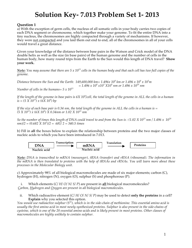
Solution Key- 7.013 Problem Set 1- 2013 Question 1 a) With the exception of germ cells, the nucleus of all somatic cells in your body carries two copies of each DNA segment or chromosome, which together make your genome. To fit the entire DNA into a tiny nucleus, the chromosomes are highly compacted through a variety of mechanisms. If however, they were not compacted and you laid them out end to end, all of the chromosomes in all of your cells would travel a great distance. Given your knowledge of the distance between base pairs in the Watson and Crick model of the DNA double helix as well as the size (in base pairs) of the human genome and the number of cells in the human body, how many round trips from the Earth to the Sun would this length of DNA travel? Show your work. Note: You may assume that there are 5 x 10¹³ cells in the human body and that each cell has two full copies of the genome. Distance between the Sun and the Earth: 149,600,000 km= 1.496x 10⁸ km or 1.496 x 108 x 103m = 1.496 x 108 x103 X109 nm or 1.496 x 10²⁰ nm 13 Number of cells in the humans= 5 x 10 If the length of the genome in base pairs is 6X 10⁹/cell, the total length of the genome in ALL the cells in a human is = (5 X 1013) x (6X 109) bp If the size of each base pair is 0.34 nm, the total length of the genome in ALL the cells in a human is = (5 X 1013) x (6X 109) X 0.34nm or 1.02 X 10²³ nm So the number of times this length of DNA could travel to and from the Sun is : (1.02 X 10²³ nm / 1.496 x 10²⁰ nm)/2 = (0.682 X 10³)/2 = 681/ 2 = 340.5 times b) Fill in all the boxes below to explain the relationship between proteins and the two major classes of nucleic acids to which you have been introduced in 7.013. DNA Nucleic acid Transcription mRNA Translation Proteins Nucleic acid Note: DNA is transcribed to mRNA (messenger), tRNA (transfer) and rRNA (ribosomal). The information in the mRNA is then translated to proteins with the help of tRNAs and rRNAs. You will learn more about these processes in the Molecular Biology unit. c) Approximately 98% of all biological macromolecules are made of six major elements; carbon (C), hydrogen (H), nitrogen (N), oxygen (O), sulphur (S) and phosphorous (P). i. Which elements (C/ H/ O/ N/ S/ P) are present in all biological macromolecules? Carbon, Hydrogen and Oxygen are present in all biological macromolecules. ii. Which radioactive element (C/ H/ O/ N/ S/ P) may be used to detect only the proteins in a cell? Explain why you selected this option. You would use radioactive sulphur (S35), which is in the side-chain of methionine. This essential amino acid is usually the first amino acid in most newly synthesized proteins. Sulphur is also present in the side-chains of cysteine, which is one of the 20 essential amino acids and is likely present in most proteins. Other classes of macromolecules are highly unlikely to contain sulphur. 1 Question 2 a) Consider the structure of the following polymer. H #1 i. Classify this polymer as a carbohydrate, lipid, protein or nucleic acid. It is a Carbohydrate ii. From the following, circle all the options that best characterize this polymer. Explain why you selected these options. Hydrophobic Polar Hydrophilic Nonpolar Charged Uncharged The presence of multiple hydroxyl (–OH) groups makes this molecule polar and hydrophilic. iii. Which of the above bonds (choose either bond 1 or bond 2) is an example of glycosidic bond? Explain why you selected this option. Bond 2 is an example of glycosidic bond that by definition is formed by a condensation/ dehydration reaction and joins the monomers to form polymers such as the one shown in the schematic above. In comparison, Bond 1 represents a covalent bond between two carbon atoms of the same monomer. iv. Circle the correct option(s). The breakdown of this polymer into individual monomers is an example of condensation, hydrolysis, dehydration or isomerization reaction. This is an example of hydrolysis reaction. v. From the following, circle the type of bond or interaction that occurs between two such polymers, when they are placed in an aqueous environment. Explain why you selected this option(s). Hydrogen bonds van der Waals forces (VDW) Covalent bond Ionic bond The presence of multiple hydroxyl (–OH) groups makes this molecule polar and hydrophilic and allows it to form hydrogen bonds with the –OH groups of the adjacent polymer as well as the surrounding water molecules. b) Below is a schematic of two different dimers, Dimer 1 and Dimer 2. CH2 CH2 Dimer 1 Dimer 2 If Enzyme X cleaves the bond that joins the two monomers of Dimer 1, will the same enzyme be able to cleave the bond that results in the formation of Dimer 2 (Yes or No)? Explain why you selected this option. The stereochemistry of these two dimers is different, which means that they have different 3D- shapes. Each enzyme has a binding pocket that is exquisitely specific for the 3D- shape of its substrate, thus the binding pocket of Enzyme X that cleaves the covalent bond circled in the schematic for dimer 1 will not be able to accommodate dimer 2. 2 Question 2 continued c) Consider the molecules below to answer the following questions. #2 #1 i. Which structure (choose either molecule #1 or #2) is most likely to assemble and form a lipid bilayer? What property of the molecule that you selected allows it to form a lipid bilayer? Molecule 2 has non-polar hydrocarbon fatty acid chains and polar, charged globular head due to the phosphate group. This makes it amphipathic. This allows them to form a lipid bilayer where the globular heads are exposed to the aqueous exterior and the inner cytosol and the non-polar fatty acid chains interact with each other to form the hydrophobic interior of the lipid bilayer. ii. Which bond or interaction (choose from covalent, hydrogen, hydrophobic or ionic) is most likely to stabilize the lipid bilayer? The hydrophobic interactions between the nonpolar, hydrophobic fatty acid side-chains of these molecules allow them to assemble into a stable lipid bilayer. List the option (choose either saturated or unsaturated) that best characterizes Molecule 2. Explain why you selected this option. The schematic shows a phospholipid molecule with saturated fatty acid chains; the carbon atoms are saturated with hydrogen and there are no double bonds. iii. iv. Draw two possible conformations that the molecule you selected in part (i) may assume when placed in an aqueous environment. Vesicle Lipid bilayer Micelle v. What bonds or interactions (choose from ionic, covalent, hydrophobic and hydrogen) are most likely to occur between the molecules and the surrounding aqueous environment when they acquire the conformations that you have drawn in part (iv) above? The polar, globular heads of these molecules can form hydrogen bonds with the surrounding water molecules. In addition, the negatively charged phosphate groups can potentially form ionic bonds with any positively charged ions in the surrounding aqueous environment. 3 Question 3 a) The following diagram represents a nucleotide that serves as a monomer for ribonucleic acid (RNA). 5’ 4’ 3’ i. 1’ 2’ Classify this nucleotide as purine or pyrimidine base. It is a purine base Besides serving as a monomer of RNA, what is the other major role of this nucleotide within a cell? It may serve as a source of energy. It may also participate in specific enzymes catalyzed reactions that are a part of different cell signaling cascades. ii. iii. Box the group or atom that you would remove, so that the nucleotide drawn above can serve as a monomer for DNA. You would remove the ‘O” from the 2’OH group so that it becomes a deoxyribonucleotide (dNTP) What type of bonds would hold two such adjacent nucleotides together in a growing nucleic acid chain? Circle the group(s) that would participate in the formation of this bond if the nucleotide shown above, was added to the growing nucleic acid chain. It is the phosphodiester bond that is formed in between the 3’OH end of a growing nucleic acid chain and 5’Phosphate of the incoming nucleotide. iv. Name the type of bonds that the above nucleotide will form with its complementary nucleotide. How many of these bonds would you expect between this nucleotide pair? The above schematic represents the GTP (Guanosine triphosphate), which can form three hydrogen bonds with the complentary CTP (Cytsoine triphosphate). v. The nucleotide (N) shown above is a part of the following nucleic acid sequence. vi. 5’GGCCANACCA3’ For the nucleic acid sequence that is given above… • Which nucleotide base (A/T/G/C/U) has a free phosphate group? G • Which nucleotide base (A/T/G/C/U) has a free hydroxyl group? A vii. If adenosine (A) is added to the above nucleic acid sequence in a cell, would it be added to the 5’ end or the 3’ end? 3’ end viii. The nucleic acid sequence shown above interacts with a specific protein. Of the following amino acids that are a part of this protein, circle those whose side-chains are most likely to interact with the phosphates of the sugar-phosphate backbone of nucleic acids. Explain why you selected these amino acids. Methionine Lysine Alanine Leucine Arginine Both Lysine and arginine are polar, hydrophilic amino acids with positively charged side-chains unlike methionine, alanine and leucine, which are non-polar, hydrophobic amino acids. So these positively charged amino acids, if placed in the right orientation, can form ionic bonds with the negatively charged phosphates of the sugar phosphate backbone. 4 Question 3 continued b) Consider the following amino acid sequence that is part of a protein. i. Circle all the peptide bonds in the above amino acid sequence. Peptide bond ii. Name the amino acid in this sequence that is closest to the N- terminus of the protein. Cysteine iii. In the sequence above…. • Name the amino acid(s) that is hydrophilic. Cysteine & tyrosine • Name the amino acid(s) that is hydrophobic. Alanine & phenylalanine • Name the amino acid(s) that can be phosphorylated. Tyrosine • Box the side-chain group (R group) of each amino acid in the sequence above. Question 4 The following is a schematic of a plasma membrane protein that functions as a growth factor (GF) receptor. When a specific growth factor binds to the extracellular domain of this protein, the receptor protein dimerizes i.e. two polypeptide chains, each comprised of many amino acid residues and shown by lines in the schematic, join together to make a dimerized protein. The GF receptor is active following dimerization as shown below. N N N N Growth factor Extracellular side Extracellular side Plasma membrane Plasma membrane Cytosolic side Cytosolic side C GF receptor protein C C Dimerized GF receptor protein C a) What is the highest order of this protein’s structure in its inactive state? Choose from primary, secondary, tertiary and quaternary given in an ascending order. Explain your choice. Tertiary, since the protein, in its inactive state, exists as a monomeric protein that is comprised of a single polypeptide chain. Quaternary structure is observed for proteins that have two or more than two polypeptide chains. 5 Question 4 continued b) Circle the amino acid(s) that is part of the extracellular domain of the GF receptor and has a sidechain that could form a hydrogen bond with the surrounding water molecules. Include all that apply and explain why you selected this option(s). Lysine Tyrosine Tryptophan Glutamic acid The underlined amino acids have polar, hydrophilic side-chains, which can hydrogen bond with water molecules. c) Circle the amino acid(s) in the transmembrane domain (amino acid sequence of the protein that spans the plasma membrane) of the GF receptor whose side-chains can most likely interact with the lipid bilayer. Include all that apply and explain why you selected this option(s). Lysine Tyrosine Tryptophan Glutamic acid The underlined amino acid can interact with hydrophobic lipid bilayer since it have nonpolar, hydrophobic sidechain. d) Interaction between specific amino acid residues found in two identical GF receptors is critical for their dimerization. In the table below, state the strongest interaction that likely occurs between the side-chains of listed amino acid pairs. Interacting amino acid pair. Note: In the column below Glutamic acid 155 means that the amino acid located at the 155th position in the peptide chain is Glutamic acid. Glutamic acid155 and Lysine68 Strongest bonding/ interaction (ionic/ hydrogen/ covalent/ hydrophobic)? Ionic bond Tyrosine140 and Glutamine62 Hydrogen bond Alanine133 and Methionine 54 Hydrophobic interaction e) Substitution of a single amino acid can influence the dimerization and function of the GF receptor protein. Predict whether the receptor will be able to dimerize, given the substitutions below. The original amino acid bonding pair, Glutamic acid155 and Lysine68, is changed to Glutamic acid155 and Arginine68. Explain your answer. The receptor will still be able to dimerize since arginine has a positively charged side-chain (similar to original lysine at this position) as a result of which it can still form the ionic bond with glutamic acid. i. The original amino acid bonding pair, Alanine133 and methionine54, is changed to Tryptophan133 and methionine54. Explain your answer. Both alanine and tryptophan have nonpolar, hydrophobic amino acid side-chains and hence may be able to undergo hydrophobic interaction with methionine. However, the side-chain of tryptophan is much larger than that of alanine. So replacing alanine by tryptophan may generate steric hindrance thus disrupting the interaction of the receptor with its ligand. ii. 6 Question 5 a) The enzyme E1 catalyzes the hydrolysis of GTP to GDP (Reaction 1) as shown below. E1 GTP GDP i. Draw the energy profile of the forward E1 catalyzed reaction on the axes below. Label the reactants and the products and indicate the overall free energy change. ii. Draw the energy profile of the forward reaction in the absence of E1 on the axes below. Label the reactants and the products and indicate the overall free energy change. Rtrans Profile EAC (ii) Rtrans Profile ∆G EAC (i) R P Reaction rate b) The following is a reaction (Reaction 2) catalyzed by a different enzyme, E2. S1 + S2 E2 P2 In a living cell, the coupling of Reaction 1 with Reaction 2 increases the rate of Reaction 2. Explain why this is so. The hydrolysis of GTP to GDP results in the release of energy that was stored in the phosphate bond. This energy can then be used drive Reaction 2. c) Enzymes enhance the rate of a reaction by decreasing the activation energy of the reactants. But they neither alter the free energy change (Δ G) of the reaction nor the reaction equilibrium (Keq). Briefly explain… i. How an enzyme may lower the activation of energy of the reactants. The enzymes lower the activation energy of reactants by promoting their transition state. ii. Why the enzymes have no effect on the Δ G and Keq of the reaction. The Keq is the ratio of the concentration of products and reactants, which is not influenced by the presence or absence of enzymes. Similarly ΔG represents the difference between the free energy of reactants and products of a reaction. It is the inherent property of the reactants and products and remains unchanged by the enzyme. 7 Question 5 continued d) You perform the coupled reactions in two separate test tubes (Tube 1 and Tube 2). Both these tubes contain GTP, substrates S1 and S2, enzymes E1 and E2 and either Drug 1 or Drug 2 as described below. • Tube 1 contains Drug 1 that is a manufactured chemical and inhibits the hydrolysis of GTP but it does not prevent the binding of GTP to the active site of E1. This drug has no effect on E2. Furthermore, you observe that the effect of this drug is irreversible. • In comparison, Tube 2 contains Drug 2 under appropriate reaction conditions. This drug is found naturally in a cell and it inhibits the binding of S1 to the active site of E2 but has no effect on E1. The inhibitory effect of this drug can be reversed by the excess amount of S1. You perform the reactions under optimal conditions and measure the amount of P2 formed after 30 minutes in both the tubes. i. Circle the option(s), from the choices below, that best characterizes Drug 1 and explain why you selected this option(s). Competitive inhibitor Allosteric inhibitor Non-competitive inhibitor Since the binding of this drug does not inhibit the binding of the substrate to an active site of the enzyme, this drug is not a competitive inhibitor. Instead it is either a non-competitive inhibitor or an allosteric inhibitor. It is worth noting that non-competitive inhibitors are mostly manufactured chemicals unlike allosteric inhibitors are mostly present in the biological system. ii. Circle the option(s), from the choices below, that best characterizes Drug 2 and explain why you selected this option(s). Competitive inhibitor Allosteric inhibitor Non-competitive inhibitor Since the inhibitory effect of this drug is reversed by the excess amount of substrate, this drug is competing with the substrate to bind to the active site of the enzyme. Hence this drug is a competitive inhibitor. e) The product P2 can be converted either to product ‘Z” or product “R” through a biochemical pathway where each step is catalyzed by a specific enzyme (E3 –E10) as shown below. The metabolites (products) at each step are indicated. Note: An arrow means activation and a “T” means inhibition of the reaction. P2 E3 B E4 C E9 H E5 D E6 F E7 G E8 Z E10 R What is the effect of Compound Z on Enzyme E4? Of what is this an example? Briefly explain why the cell may need such a regulatory mechanism. Compound Z shows feedback inhibitory effect on enzyme E4. This inhibitory mechanisms helps the cell to maintain homeostasis i.e. if there is excess of compound Z the cell decides to inhibit the biochemical cascade that makes compound Z. 8 Question 6 An eye lens is comprised of cells that are created when an eye is formed and are retained for its lifetime. These cells lack organelles and can be regarded as “sacs” that are filled with a loose uniform arrangement of water-soluble structural proteins called crystallins. The uniform distribution of crystallins in a cell prevents light scattering, maintains lens transparency and allows us to see. With age, the crystallins undergo different modifications that increase the opacity of a lens and may lead to the onset of cataracts. Biologists are currently using computer-modeling programs to view the structure and function of various proteins in both normal and diseased states. For this problem you will use a computer to view the structure of γ–crystallin. To begin, go to http://web.mit.edu/star/biochem, click on the “Start and allow the link to open. You may have to install JAVA to open this program. This can be downloaded free from the JAVA website. Click on “Samples-> Select from samples ->Amino acids/ Proteins ->1HK0 -> open”. You will see a schematic of γ- crystallin on the right and a menu on the left. a) Does the current view of 1HK0 show a monomeric (single polypeptide chain) or multimeric (more that one polypeptide chain) protein? It is a monomeric protein. b) What is the highest order of protein structure that you observe for 1HK0? Explain why you selected this level. Note: In the sequence window, click on “Structure-> Protein -> Quaternary”. Increase the “Size slide bar” in the “Surfaces” window. Each polypeptide chain in this program is shown by a different color. “Tertiary” is the highest order of protein structure for IHKO since IHKO is comprised of a single polypeptide chain. Quaternary structure is only observed for proteins that contain two or more than two polypeptide chains. c) The crystal structure of 1HK0 exhibits two homologous domains that are joined intra-molecularly by a six amino acid linker. What is the secondary structure of this amino acid linker (choose from helices, sheets and coils)? Note Click on “Reset - > Reset structure”. Then click on “Structure -> Protein-> Secondary”. Select helices, sheets and ribbons one at a time and look at the amino acid sequences that get highlighted in the “sequence window”. For better reviewing of protein structure you can increase the “Size slide bar” within in the “Secondary structure window”. It is the coils that connect the two homologous domains of 1HKO. d) Open another protein structure by going to “Samples-> Select from samples ->Amino acids/ Proteins ->1BLB -> open”. What is the overall difference between structure of 1HKO and 1BLB? 1BLB is multimer made of IHKO monomers. e) Based on what you have learned from this exercise, hypothesize how the change in crystallin structure, as seen in 1BLB, may lead to the development of cataract. Single amino acid substitutions within crystallins i.e. substitution of Gln54 and Arg79 polar amino acids by alanine that has a nonpolar, hydrophobic side-chain may disrupt the uniform distribution of crystallins in the lens cells. Furthermore, these substitutions may also promote the aggregation of crystallin monomers to form multimers. As a result there occurs an increase in the opacity of the lens leading to the onset of cataracts. 9 MIT OpenCourseWare http://ocw.mit.edu 7.013 Introductory Biology Spring 2013 For information about citing these materials or our Terms of Use, visit: http://ocw.mit.edu/terms.


