Picte eum quam aut et officatur, nonse-
advertisement
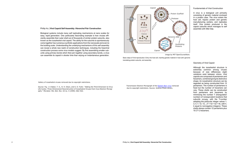
Cytoplasm Capsid Fundamentals of Viral Construction Protein Scaffold Protease Pentamers 3KLOOLS+X | Viral Capsid Self Assembly: Hierarchal Pair Construction Biological systems include many self replicating mechanisms at nano scales for easy, rapid generation. One particularly fascinating example is how viruses efficiently assemble their outer shell out of thousands of similar protein subunits, also known as the icosahedral viral capsid. The ability for the subunits to spontaneously come together has numerous synthetic applications from the nanoscale and even to the building scale. Understanding the underlying mechanisms of this self assembly can reveal a whole new realm of construction techniques, including the hiearchal construction process some virus models suggest. By first assembling smaller subunits using primary bonds which then join together using secondary bonds, a virus can assemble its capsid in shorter time than relying on instantaneous generation. Hexamers A virus is a biological unit primarily consisting of genetic material enclosed in a protein case. The virus enters the host cell, hijacks protein and genetic manufacturing centers, and replicates itself. One protein produced is the capsid subunits which manages to self assemble with little help. Triplexes Genomic DNA Protease Precursor Scaffolding Proteins Capsid Image by MIT OpenCourseWare. Basic steps of Viral reproduction: Entry into host cell, inserting genetic material in host cell’s genome translating protein subunits, and assembly Geometry of Viral Capsid Gallery of icosahedral viruses removed due to copyright restrictions. Source: Fig. 1 in Baker, T. S., N. H. Olson, and S. D. Fuller. "Adding the Third Dimension to Virus Life Cycles: Three-Dimensional Reconstruction of Icosahedral Viruses from Cryo-Electron Microgr aphs." Microbiol. Mol. Biol. Rev. 63 no. 4 (1999): 862–922. 2 Transmission Electron Micrograph of the Epstein-Barr virus removed due to copyright restrictions. Source: Science Photo Library. 3KLOOLS+X | Viral Capsid Self Assembly: Hierarchal Pair Construction Although the isosahedral structure is extremely common among viruses because of size differences slight variations exist between virions. Viral capsids are composed of pentamers and hexamers, combined giving its distinctive shape. An icosahedron structure can be regarded as being constructed from 12 pentamers. The number of pentamers is fixed but the number of hexamers can vary. These shells can be constructed from pentamers and hexamers by minimizing the number T (triangulation number) of nonequivalent locations that subunits occupy, with the T-number adopting the particular integer values 1, 3, 4, 7, 12, 13,...(T = h2 + k2 + hk, with h, k equal to nonnegative integers). These shells always contain 12 pentamers plus 10 (T-1) hexamers. B3 B4 o o 100 108 o 12 120 o 90 0 90 o o o 108 o 60 o 60 o 60 o 60 o 60 B5 Modeling the System Assembly Pathways Accurately simulating the complex, detailed capsomeres, the molecular protein subunits, is nearly impossible and incredibly time consuming, since observing the transient forms, which constantly bind and unbind, is currently not possible. Thus, most computation research have simplified the form into clearer abstract forms such as the sphere. Specifically, the model uses only a single spherical excluded volume per capsomer, and uses internal bond vectors to capture the effects of protein shape and complementarity as illustrated on the left [Hagan, Chandler]. By setting bond priorities, certain steps will accumulate parts, just like in an assembly line. Type I assembly favors the intercapsomer forward rate, creating numerous dimer units, which then through nucleation forms the actual subunit piece, either the square for octahedral or pentagon for isocahedral arrangements. Conversely in Type II, intracapsomer forward rate is higher, rapid accumulation of tetrameric pieces occurs, followed by their paring into the complete octahedral form. If rates are about equal, Type III, we achieve a hybrid of tetramer production with monomer attachments through elongation. These are just examples of how changing certain rates can result in varying pathways which could affect the speed and final yield of the assembly. Image by MIT OpenCourseWare. Illustration of possible bonding pair angle sites. Note that bonding angles are not coplanar Hierarchal Paths of Assembly Courtesy of Elsevier, Inc., http://www.sciencedirect.com. Used with permission. . Comparison of simple geometric model to actual, complex viral capsid Courtesy of Elsevier, Inc., http://www.sciencedirect.com. Used with permission. . Primary B1 B4 Secondary α β γ ε η υ 1 2 2 1 3 3 2 3 1 3 1 1 2 2 3 2 3 1 1 4 2 3 2 3 4 3 2 1 1 4 3 4 1 2 1 5 5 1 The next step is modelling bond strengths and kinetics. In the molecular scale, normal newtonian forces have much less affect, while covalent bonds and Van der Walhs forces greatly affect attractions, especially due to the large nature of protein chains. In terms of modelling, we can create a hierarchy of priorities in bonding. For example, according to Table 1 from Hagan’s discussion, (1,2) in 2 2 3 1 1 3 the B3 configuration is a primary B5 3 4 2 5 4 3 4 3 5 2 3 4 complementary pair while (3,3) is a 5 1 1 5 4 2 secondary pair, meaning there is a favourable interaction for (1,2), also Primary and secondary complementary pairs and associated angles for the three capsomer models requiring that bi be nearly coplanar with bj. This of course is an imbedded method Image by MIT OpenCourseWare. for quality assurance, favoring certain configurations over others. Because of these rigid attractions, we can create Courtesy of Elsevier, Inc., http://www.science such complex forms as the icosahedron direct.com. Used with permission. capsid. 4 2 After adjusting parameters, Sweeney discovered a balance between yield and speed of assembly. While concurrent low rates of assembly result in 100% yield, the assembly is extremely slow. But if the rates are too high, it is impossible to form complete capsids. Such a result has interesting implications on how a self replicating drawing following the rules do not always result in complete forms, implying the possibility for numerous kinds of forms, not limited . by the original isocahedral viral capsid. 4 Left: Pattern of Construction under normal conditions. Right: Pattern of Construction under abnormal conditions Courtesy of Elsevier, Inc., http://www.sciencedirect.com. Used with permission. 3KLOOLS+X | Viral Capsid Self Assembly: Hierarchal Pair Construction 5 Transmission Electron Micrograph of the smallpox virus removed due to copyright restrictions. Source: Science Photo Library. Abstract Logic Bibliography After observing the basic mechanisms behind viral capsid self assembly, we can start to develop a set of overarching rules, interior inputs and exterior inputs, which dictate the final form and assembly process. Hagan, Michael, and David Chandler. “Dynamic Pathways for Viral Capsid Assembly.” Biophysical Journal 91 (2006): 42-54. Controls Within one simulation, we are limited by the ‘natural’ bond vectors of the identical protein subunits. We also must initially choose the ‘ideal’ configuration, either the octahedral form or icosahedron. Multiple drawings can be made though, by adding more possible vector bond angles. Alternatively, it may be possible to include sites that repel, replicating nonpolar-polar intermolecular forces. Other details include bond lengths and our capsomere size. Sweeney, Blake , Tiequan Zhang, and Russell Schwartz. “Exploring the Parameter Space of Complex Self-Assembly through Virus Capsid Models.”Biophysical Journal 94 (2008): 772-783. Images from Wikipedia, Science Photo Library Variables By changing the rates of certain bonds, we can achieve aggregated or dispersed forms as illustrated above. By doing this, we can observe impossible transitional stages which could inform new construction forms. The rates we can vary must include a unbinding rate as well, as part of the quality control in self replication. 6 3KLOOLS+X | Viral Capsid Self Assembly: Hierarchal Pair Construction 7 Subunits Connections Subunits Connections Joinery Joinery (i) (i) (i) (i) (ii) (ii) (ii) (iii) (iii) (iii) (iii) (iv) (iv) (iv) (v) (v) (v) Strong (iv) Medium Weak Weak (v) 8 Dimer formation (ii) Strong Medium Hierarchy of Connections Hierarchy of Connections Free movement and Rotation Mechanism Dimer formation Free movement and Rotation Mechanism 3KLOOLS+X| Viral Capsid Self Assembly: Hierarchal Pair Construction 9 Growth Growth Hexagonal + Branching networks Hexagonal +Branching networks (i) 10 (i) (ii) (ii) (iii) (iii) 3KLOOLS+X | Viral Capsid Self Assembly: Hierarchal Pair Construction (iv) (iv) 11 12 3KLOOLS+X | Viral Capsid Self Assembly: Hierarchal Pair Construction 13 MIT OpenCourseWare http://ocw.mit.edu 4.112 Architecture Design Fundamentals I: Nano-Machines Fall 2012 For information about citing these materials or our Terms of Use, visit: http://ocw.mit.edu/terms.
