Document 13355304
advertisement

20.320 Problem Set 2
Question 1
In the last problem set, we discovered that Histidine 142 is conserved across many variants of
influenza hemagglutinins and thus may play a key role in mediating their pH-dependent
conformational change. Now, we will look at the effects the charge of His142 has on the
electrostatics of both hemagluttinin conformations.
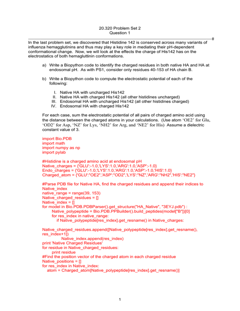
a) Write a Biopython code to identify the charged residues in both native HA and HA at
endosomal pH. As with PS1, consider only residues 40-153 of HA chain B.
b) Write a Biopython code to compute the electrostatic potential of each of the
following:
I. Native HA with uncharged His142
II. Native HA with charged His142 (all other histidines uncharged)
III. Endosomal HA with uncharged His142 (all other histidines charged)
IV. Endosomal HA with charged His142
For each case, sum the electrostatic potential of all pairs of charged amino acid using
the distance between the charged atoms in your calculations. (Use atom ‘OE2’ for Glu,
‘OD2’ for Asp, ‘NZ’ for Lys, ‘NH2’ for Arg, and ‘NE2’ for His) Assume a dielectric
constant value of 3.
import Bio.PDB import math
import numpy as np
import pylab #Histidine is a charged amino acid at endosomal pH Native_charges = {'GLU':-1.0,'LYS':1.0,'ARG':1.0,'ASP':-1.0}
Endo_charges = {'GLU':-1.0,'LYS':1.0,'ARG':1.0,'ASP':-1.0,'HIS':1.0}
Charged_atom = {'GLU':"OE2",'ASP':"OD2",'LYS':"NZ",'ARG':"NH2",'HIS':"NE2"}
#Parse PDB file for Native HA, find the charged residues and append their indices to Native_index
native_range = range(39, 153)
Native_charged_residues = []
Native_index = []
for model in Bio.PDB.PDBParser().get_structure("HA_Native", "3EYJ.pdb") : Native_polypeptide = Bio.PDB.PPBuilder().build_peptides(model["B"])[0]
for res_index in native_range:
if Native_polypeptide[res_index].get_resname() in Native_charges:
Native_charged_residues.append([Native_polypeptide[res_index].get_resname(),
res_index+1])
Native_index.append(res_index) print 'Native Charged Residues'
for residue in Native_charged_residues: print residue #Find the position vector of the charged atom in each charged residue Native_positions = [] for res_index in Native_index: atom = Charged_atom[Native_polypeptide[res_index].get_resname()]
1
#
20.320 Problem Set 2
Question 1
#
Native_positions.append([Native_polypeptide[res_index][atom].coord,Native_polypeptid
e[res_index].get_resname()]) #Parse PDB file for Endosomal HA, find the charged residues and append their indices
to Endo_index endo_range = range(0, 114) Endo_charged_residues = [] Endo_index = [] for model in Bio.PDB.PDBParser().get_structure("HA_Endo", "1HTM.pdb") : Endo_polypeptide = Bio.PDB.PPBuilder().build_peptides(model["B"])[0]
for res_index in endo_range: if Endo_polypeptide[res_index].get_resname() in Endo_charges: Endo_charged_residues.append([Endo_polypeptide[res_index].get_resname(),
res_index+40])
Endo_index.append(res_index)
print 'Endosomal Charged Residues'
for residue in Endo_charged_residues:
print residue
#Find the position vector of the charged atom in each charged residue
End_positions = []
for res_index in Endo_index:
atom = Charged_atom[Endo_polypeptide[res_index].get_resname()]
End_positions.append([Endo_polypeptide[res_index][atom].coord,Endo_polypeptide[res
_index].get_resname()])
#Electrostatics for uncharged HIS142 in Native HA
U_total = 0
pairs = 0
for i in range(0,len(Native_positions)):
for j in range(i+1,len(Native_positions)): pairs += 1
dist_vector = Native_positions[i][0] - Native_positions[j][0] distance = np.sqrt(np.sum(dist_vector**2)) U_total +=
(Native_charges[Native_positions[i][1]]*Native_charges[Native_positions[j][1]])/(3.0*dista
nce)
#Electrostatics for charged HIS142 in Native HA
#Add Histidine 142 to the list of charged residues
Native_positions.append([Native_polypeptide[141]["NE2"].coord,Native_polypeptide[141
].get_resname()])
ch_U_total = 0
ch_pairs = 0
for i in range(0,len(Native_positions)):
for j in range(i+1,len(Native_positions)): ch_pairs += 1
dist_vector = Native_positions[i][0] - Native_positions[j][0] distance = np.sqrt(np.sum(dist_vector**2)) 2
20.320 Problem Set 2
Question 1
ch_U_total +=
(Endo_charges[Native_positions[i][1]]*Endo_charges[Native_positions[j][1]])/(3.0*distanc
e)
#Electrostatics for charged HIS142 in Endosomal HA
End_U_total = 0
End_pairs = 0
for i in range(0,len(End_positions)):
for j in range(i+1,len(End_positions)): End_pairs += 1
dist_vector = End_positions[i][0] - End_positions[j][0]
distance = np.sqrt(np.sum(dist_vector**2)) End_U_total +=
(Endo_charges[End_positions[i][1]]*Endo_charges[End_positions[j][1]])/(3.0*distance)
#Electrostatics for uncharged HIS142 in Endosomal HA
#Remove Histidine 142 from the list of charged residues
del End_positions[37]
un_End_U_total = 0
un_End_pairs = 0
for i in range(0,len(End_positions)):
for j in range(i+1,len(End_positions)): un_End_pairs += 1
dist_vector = End_positions[i][0] - End_positions[j][0]
distance = np.sqrt(np.sum(dist_vector**2)) un_End_U_total +=
(Endo_charges[End_positions[i][1]]*Endo_charges[End_positions[j][1]])/(3.0*distance)
print 'Native HA with uncharged His142'
print 'Pairs considered: ' + str(pairs)
print 'U_total = ' + str(U_total)
print 'Native HA with charged His142'
print 'Pairs considered: ' + str(ch_pairs)
print 'U_total = ' + str(ch_U_total)
print 'Endosomal HA with uncharged His142'
print 'Pairs considered: ' + str(un_End_pairs)
print 'U_total = ' + str(un_End_U_total)
print 'Endosomal HA with charged His142'
print 'Pairs considered: ' + str(End_pairs)
print 'U_total = ' + str(End_U_total)
3
#
20.320 Problem Set 2
Question 1
#
Output:
Native Charged Residues
['ASP', 46] ['LYS', 51] ['ARG', 54]
['GLU', 57]
['LYS', 58] ['GLU', 61]
['LYS', 62] ['GLU', 67]
['LYS', 68] ['GLU', 69]
['GLU', 71]
['GLU', 74]
['ARG', 76]
['ASP', 79]
['GLU', 81]
['LYS', 82]
['GLU', 85]
['ASP', 86]
['LYS', 88]
['ASP', 90]
['GLU', 97]
['GLU', 103]
['ASP', 109]
['ASP', 112]
['GLU', 114]
['LYS', 117]
['GLU', 120]
['ARG', 121]
['ARG', 123]
['ARG', 124]
['ARG', 127]
['GLU', 128]
['GLU', 131]
['ASP', 132]
['GLU', 139]
['ASP', 145]
['GLU', 150]
['ARG', 153]
Endosomal Charged Residues
['ASP', 46] ['LYS', 51] ['ARG', 54]
['GLU', 57]
['LYS', 58] ['GLU', 61]
['LYS', 62] ['HIS', 64]
['GLU', 67]
['LYS', 68] ['GLU', 69]
['GLU', 72]
['GLU', 74]
['ARG', 76]
['ASP', 79]
['GLU', 81]
['LYS', 82]
['GLU', 85]
['ASP', 86]
['LYS', 88]
['ASP', 90]
['GLU', 97]
['GLU', 103]
['HIS', 106]
['ASP', 109]
['ASP', 112]
['GLU', 114]
['LYS', 117]
['GLU', 120]
['LYS', 121]
['ARG', 123]
['ARG', 124]
['ARG', 127]
['GLU', 128]
['GLU', 131]
['GLU', 132]
['LYS', 139]
['HIS', 142]
['LYS', 143]
['ASP', 145]
['GLU', 150]
['ARG', 153]
Native HA with uncharged His142
Pairs considered: 703
U_total = 0.0352466471996
Native HA with charged His142
Pairs considered: 741
U_total = -0.090493015543
Endosomal HA with uncharged His142
Pairs considered: 820
U_total = -0.371365158283
Endosomal HA with charged His142
Pairs considered: 861
U_total = -0.588778130675
4
20.320 Problem Set 2
Question 1
c) Do the calculated electrostatic potentials make sense? If not, why not? How could
we improve our energy calculation?
For endosomal HA, the electrostatic potential is lower when His142 is charged. This
makes sense because the protonation of histidine at the endosomal pH is important for
HA’s conformational change that allows the viral and cellular membranes to fuse. Thus
endosomal HA is at a lower energy when His142 is protenated.
For native HA, the electrostatic potentials don’t make sense because we know histidine
is uncharged in the native conformation and thus we would expect that to be the lower
energy conformation. One reason that our energy calculations may be off is that we did
not include the effect of water molecules.
One area for improvement in our energy calculation would be in choosing which charged
atoms we consider. Most of the charged side chains actually exist as resonance
structures where the charge is shared among the nitrogens/oxygens. For instance, in
glutamate and aspartate, the oxygens in the carboxylic acid groups are partially doublebound to carbon and carry a partial negative charge. Together, the carboxylic acid group
carries a charge of -1. For instance, we could assign a charge of -1/2 to each of the
oxygens in the side-chains of glutamate and aspartate and consider the position of both
in our electrostatic calculations.
5
#
20.320 Problem Set 2
Question 2
Once again we will be looking at the HA2 chain (chain ‘B’ in the PDB files) of Hemagglutinin in
its native state and at endosomal pH using the same PDB files from problem set 1.
#
The conformation potentials used by Chou and Fasman 1 appear in the chart below. These were
derived by examining 24 protein structures. The Pα/β value for each amino acid is proportional to
the frequency of that amino acid in alpha helices/beta sheets and has been normalized so that
they take on values between zero and two. The amino acids with Pα > 1 are assumed to have a
propensity for α-helices and similarly those with Pβ > 1 are assumed to have a propensity for βsheets. Thus Chou and Fasman classified amino acids as strong helix/sheet formers (Hα/β),
helix/sheet formers (hα/β), helix/sheet indifferent (Iα, iα/β), helix/sheet breakers (bα/β), and strong
helix/sheet breakers (Bα/β). These are also marked in the chart below. In order to understand the
Chou-Fasman algorithm, we will use the algorithm to predict alpha-helix propensity in an input
protein according to the following rules:
Criteria 1. Helix Nucleation. Locate clusters of four
helical residues (hα or Hα) out of six residues along
the polypeptide chain. Weak helical residues (Iα)
count as 0.5hα (i.e. three hα and two Iα residues out of
six could also nucleate a helix). Helix formation is
unfavorable if the segment contains 1/3 or more helix
breakers (bα or Bα).
Criteria 2. Helix Termination. Extend the helical
segment in both directions until terminated by
tetrapeptides with Pα,average < 1.00.
Criteria 3. Proline cannot occur in the alpha helix.
For this problem, you will need to download the
following files and put them in one folder, including
the pdb files for native HA (3EYJ.pdb) and
endosomal pH HA (1HTM.pdb):
CFalphaPredict.py
ChouFasman.py
a) Write the parsePDB function in ChouFasman.py to load chain ‘B’ of the proteins and
return a list of helical residues based on the criteria in PS1 question 1. You must also
complete the code for the function responsible for alpha-helix prediction - findAlpha(), in
the Python program CFalphaPredict.py according to the rules above. Attach a copy of
your code and output. Explain your results.
ChouFasman.py:
def parsePDB(fn): """
PDB file parsed to return:outputs:
seq == list of the protein's amino acids
1
Chou, P; Fasman, G.; “Empirical predictions of protein conformation,” Ann. Rev. Biochem. 47 (1978) 251-276.
6
20.320 Problem Set 2
Question 2
actual == list of seq indices to residues known to be in alpha helices """
seq = [] actual = [] for model in Bio.PDB.PDBParser().get_structure("File", fn) : polypeptides = Bio.PDB.PPBuilder().build_peptides(model["B"]) for poly_index, poly in enumerate(polypeptides) : phi_psi = np.float_(poly.get_phi_psi_list()) phi_psi_deg = phi_psi * 180 / math.pi
for res_index, res in enumerate(poly) : if res.id[1] >= 40 :
if res.id[1] <= 153 : seq.append(res.resname) if phi_psi_deg[res_index, 0] < -57 : if phi_psi_deg[res_index, 0] > -71 : if phi_psi_deg[res_index, 1] > -48 : if phi_psi_deg[res_index, 1] < -34 : actual.append(res.id[1])
print seq print actual
return seq, actual
CFalphaPredict.py
def P_average(window):
total = 0.0
for residue in window:
total += PA[residue]
return (total/float(len(window))) def findAlpha(seq,PA):
"""
Uses Chau-Fasman criteria to suggest alpha helical regions
but does not take beta sheets into account
Inputs:
seq == (list) the amino acids sequence of the protein
PA == dictionary whose keys are amino acids and values are the
CF <Palpha> parameters from the table in your problem set PA2 == dictionary of CF a-helix Classifaction for each amino acid
Outputs:
AHindices == (list) contains the residue indices of seq that are
predicted to form helices
""" AHindices=[]
#Search for helix nucleation region
for i in range(len(seq)-5): window = seq[i:i+6]
if not 'PRO' in window:
7
#
20.320 Problem Set 2
Question 2
helix_propensity = 0.0
breakers = 0
for aa in window:
if PA2[aa] == 'H' or PA2[aa] == 'h':
helix_propensity += 1.0
if PA2[aa] == 'I':
helix_propensity += 0.5
if PA2[aa] == 'b' or PA2[aa] == 'B':
breakers += 1 if helix_propensity >= 4.0 and breakers < 2:
begin = i end = i+5 helix = (begin,end) #Extend nucleation region while (begin-4) >= 0:
score = P_average(seq[begin-4:begin]) if ('PRO' in seq[begin-4:begin]) or (score < 1.0): break else:
begin -= 1 #Extend the nucleation region in the N-term direction
helix = (begin,end)
while (end+4) < len(seq):
score = P_average(seq[end+1:end+5])
if ('PRO' in seq[end+1:end+5]) or (score < 1.0): break
else:
end += 1 #Extend the nucleation region in the C-term direction
helix = (begin,end) #Store residues in AH indices for n in range(begin,end+1): if not n in AHindices:
AHindices.append(n) return AHindices b) E
xplain the reasons behind the occasional failure of Chou-Fasman alpha-helix
predictions.
Chou-Fasman algorithm is based off a very small data set, and therefore is not very
representative of all proteins. Also the algorithm is based off of alpha helices and does not
include beta sheets. If a beta sheet requirement was included then the algorithm would be more
stringent. It also considers only primary structures; therefore if we try to consider the proteins in
tertiary or quaternary structure, Chou-Fasman would not be very useful.
c) Using the PyMOL PDB viewer (zip file attached), look at the structures of these two
proteins. Attach print outs of the structures from the viewer. Focusing on the HIS 142
residue, explain the differences in the structures.
8
#
20.320 Problem Set 2
Question 2
#
3EYJ:
1HTM:
Note the major difference between the two images with the HIS 142 residue is that in native HA
(3EYJ) the HIS is tucked in between the alpha helices, whereas in the endosomal HA (1HTM)
the HIS 142 residue is exposed on the side. This explains the accessibility of the HIS 142
residue at different pHs.
9
20.320 Problem Set 2
Question 3
In bacteria, the lactose repressor (lacI) is involved with regulating the transcription of genes
involved in lactose metabolism. When lactose levels are low, lacI is bound to the lac operator,
preventing the expression of β-galactosidase, which cleaves lactose into its galactose and
glucose components. The following sequences were investigated for lacI binding in a 1987
paper mapping the recognition helix of lacI with the lac operator:
#
ACTTGTGAGC
ATTTGTGAGC
AAATGTGAGC
AATTGTGAGC
AACTGTGAGC
AATTGTGAGT
AATGGTGAGC
AAGTGTGAGC
AGTTGTGAGC
a) Calculate the log2(odds) matrix for these sequences. Use pseudocounts of 0.0025 for
zero frequencies.
In order to calculate the log2(odds) matrix, we first create a table with the number of occurrences
of each base at each position:
A
C
G
T
9
0
0
0
6
1
1
1
1
1
1
6
0
0
1
8
0
0
9
0
0
0
0
9
0
0
9
0
9
0
0
0
0
0
9
0
0
8
0
1
We can then determine the frequency of each occurrance by dividing the number of
occurrences by the total number of sequences – in this case, 9. For each zero frequency, we
insert a pseudocount of 0.25%. We must then subtract the sum of the pseudocounts for each
nonzero frequency in order to get a total frequency of 1 at each position. This results in the
following frequencies matrix:
A
0.9925
0.6667
0.1111
0.0025
0.0025
0.0025
0.0025
0.9925
0.0025
0.0025
C
0.0025
0.1111
0.1111
0.0025
0.0025
0.0025
0.0025
0.0025
0.0025
0.8844
G
T
0.0025
0.0025
0.1111
0.1111
0.1111
0.6667
0.1106
0.8844
0.9925
0.0025
0.0025
0.9925
0.9925
0.0025
0.0025
0.0025
0.9925
0.0025
0.0025
0.1106
In order to calculate the odds of an occurrence at each position, we assume that each base is
equally likely to occur at each position. Since there are four bases, we multiply each by 4. This
results in the following odds matrix:
A
3.9700
2.6667
0.4444
0.0100
0.0100
0.0100
0.0100
3.9700
0.0100
0.0100
C
0.0100
0.4444
0.4444
0.0100
0.0100
0.0100
0.0100
0.0100
0.0100
3.5378
G
0.0100
0.4444
0.4444
0.4422
3.9700
0.0100
3.9700
0.0100
3.9700
0.0100
T
0.0100
0.4444
2.6667
3.5378
0.0100
3.9700
0.0100
0.0100
0.0100
0.4422
10
20.320 Problem Set 2
Question 3
#
Taking the log2 of each odds in the above matrix yields:
A
C
1.9891
-6.6439
1.4150
-1.1699
-1.1699
-1.1699
-6.6439
-6.6439
-6.6439
-6.6439
-6.6439
-6.6439
-6.6439
-6.6439
1.9891
-6.6439
-6.6439
-6.6439
-6.6439
1.8228
G
-6.6439
-1.1699
-1.1699
-1.1772
1.9891
-6.6439
1.9891
-6.6439
1.9891
-6.6439
T
-6.6439
-1.1699
1.4150
1.8228
-6.6439
1.9891
-6.6439
-6.6439
-6.6439
-1.1772
You are interested in a sequence of DNA from a newly discovered organism with an apparently
functional lac regulation system. Based on your sequencing results, you predict that lacI binds
somewhere in the following sequence.
ATCTCATATAATTGTGAGCTCTAATAGAGTTCATGAGCAATG
b) Calculate the log2(odds) score for each hypothetical binding site in your sequence of
interest. Use these values to plot the log2(odds) score for each 10-base window as a
function of the window starting point. For instance, the first value in your plot should be
the log2(odds) score of the sequence ATCTCATATA.
c) Based on your results in Part B, determine the most likely lacI binding site in the given
sequence. Report the log2(odds) score of your choice.
The highest log2(odds) score is 18.41, occurring in a window that begins 9 bases after the
beginning of the sequence. Therefore, the lacI binding site is likely to be AATTGTGAGC.
11
MIT OpenCourseWare
http://ocw.mit.edu
20.320 Analysis of Biomolecular and Cellular Systems
Fall 2012
For information about citing these materials or our Terms of Use, visit: http://ocw.mit.edu/terms.


