Document 13309728
advertisement
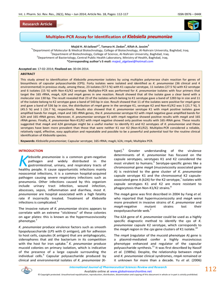
Int. J. Pharm. Sci. Rev. Res., 26(1), May – Jun 2014; Article No. 18, Pages: 112-117 ISSN 0976 – 044X Research Article Multiplex-PCR Assay for Identification of Klebsiella pneumoniae 1 *1 2 3 Majid H. Al-Jailawi* , Tamara H. Zedan , Kifah A. Jassim Department of Molecular & Medical Biotechnology, College of Biotechnology, Al-Nahrain University, Baghdad, Iraq. 2 Department of Biotechnology, College of Science, Al-Nahrain University, Baghdad, Iraq. 3 Department of Bacteriology, Central Public Health Laboratory, Ministry of Health, Baghdad, Iraq. *Corresponding author’s E-mail: majed_algelawi@hotmail.com Accepted on: 17-02-2014; Finalized on: 30-04-2014. ABSTRACT This study aimed to identification of Klebsiella pneumoniae isolates by using multiplex polymerase chain reaction for genes of biosynthesis of capsular polysaccharide (CPS). Forty isolates were isolated and identified as K. pneumoniae (36 clinical and 4 environmental) in previous study, among these, 23 isolates (57.5 %) with K1 capsular serotype, 11 isolates (27.5 %) with K2 serotype and 6 isolates (15 %) with Non-K1/K2 serotype. Multiplex-PCR was performed for K. pneumoniae isolates with four primers that target the 16S rRNA, magA, k2A and rmpA genes in one reaction. Result showed that all the isolate gave a clear band with a molecular size 130 bp. The result revealed that 23 of the isolates which belong to K1 serotype gave a band of 1283 bp in size and 11 of the isolate belong to K2 serotype gave a band of 543 bp in size. Result showed that 11 of the isolates were positive for rmpA gene and gave a band of 536 bp in size, the distribution of rmpA gene in the serotype K1, serotype K2 and Non-K1/K2 was 5 (21.7 %), 5 (45.5 %) and 1 (16.7 %), respectively. Results also showed that K. pneumoniae serotype K1 with rmpA positive isolates gave amplified bands for magA, rmpA and 16S rRNA genes, the K. pneumoniae serotype K2 with rmpA negative gave amplified bands for k2A and 16S rRNA genes. Moreover, K. pneumoniae serotype K1 with rmpA negative showed positive results with magA and 16S rRNA genes. Finally, K. pneumoniae Non-K1/K2 with rmpA negative showed only positive results with 16S rRNA gene. These results suggested that magA and k2A genotype might be a useful marker to identify K1 and K2 serotypes of K. pneumoniae and these serotypes have been more prevalent than those that were neither K1 nor K2 (Non-K1/K2). Multiplex-PCR considered a reliable, relatively rapid, effective, easy application and repeatable and possible to be a powerful and potential tool for the routine clinical identification of Klebsiella species. Keywords: Klebsiella pneumoniae; Capsular serotype; 16S rRNA; magA; k2A; rmpA; Multiplex PCR. INTRODUCTION K lebsiella pneumoniae is a common gram-negative pathogen and widely distributed in the gastrointestinal, urinary, and respiratory tracts of healthy people. It cause opportunistic infections mainly nosocomial infections, it is a common hospital-acquired pathogen causing severe respiratory infections such as pneumonia. Other infections caused by this organism include urinary tract infection, wound infection, abscesses, sepsis, inflammation and diarrhea, most K. pneumoniae are hospital associated with a high fatality rate if incorrectly treated. Treatment of Klebsiella infections is complicated.1 The invasive nature of K. pneumoniae strains appears to correlate with an extreme “stickiness” of these colonies on agar plates: this is known as the hypermucoviscosity phenotype.2 K. pneumoniae produce virulence factors such as smooth lipopolysaccharide (LPS with O antigen), pili for adhesion to host cells, capsules (K antigen) that are antiphagocytic, siderophores that aid the bacterium in its competition 3 with the host for iron uptake. K. pneumoniae produce mucoid colonies on primary isolation, which is indicative of the presence of a large capsule surrounding the individual cells.4 Capsular polysaccharide produced by clinical and environmental isolates of K. pneumoniae (K- type).5 Greater understanding of the virulence determinants of K. pneumoniae has focused on the capsule serotypes, serotypes K1 and K2 considered the most virulent to humans.6 Serotype-specific genes like a chromosomal gene magA (mucoviscosity associated gene A) is restricted to the gene cluster of K. pneumoniae capsule serotype K1 and the chromosomal K2 capsuleassociated gene A (k2A) for the K2 serotype,7 isolates with capsule serotypes K1 and K2 are more resistant to phagocytosis than Non-K1/K2 strains.8 The magA gene was first described in 2004 by Fang et al. who reported that hypermucoviscosity and magA were more prevalent in invasive strains of K. pneumoniae and magA-negative mutant strains lost their exopolysaccharide web.2 The k2A gene of K. pneumoniae could be used as a highly specific diagnostic method to identify the cps of K. pneumoniae capsule K2 serotype, which corresponds to 10 the magA region in the cps gene clusters of K1 isolate. The rmpA (regulator of the mucoid phenotype A) gene is a plasmid-mediated confer a highly mucoviscous phenotype enhanced and regulator of the capsular 11 polysaccharide synthesis. It was first described by Nassif et al. (1989a). Despite, the relationship between rmpA and K. pneumoniae clinical syndromes, rmpA remained or it unknown for more than a decade. Yu et al. (2006) International Journal of Pharmaceutical Sciences Review and Research Available online at www.globalresearchonline.net © Copyright protected. Unauthorised republication, reproduction, distribution, dissemination and copying of this document in whole or in part is strictly prohibited. 112 Int. J. Pharm. Sci. Rev. Res., 26(1), May – Jun 2014; Article No. 18, Pages: 112-117 demonstrated that rmpA-carrying strains were associated with the hypermucoviscosity phenotype, as well as with the invasive clinical syndrome. Nassif et al. (1989b) explained that remove of the rmpA gene can decrease virulence in mouse lethality tests by 1000-fold. Identification of the infectious agent of the diseases caused by K. pneumoniae is an important step in the choice of an effective therapy. Since, bacterial culture procedure and other routine invasive methods is costly, 15 time consuming, laborious, and sometimes inconclusive. Recent advances in molecular biology have generated culture independent diagnostic methods. The multiplexPCR is one such technique, which has been proved useful for the culture independent diagnosis of various microbial infections.16, 17 The aim of the present study was to evaluate of multiplex-PCR technique for the specific detection and identification of K. pneumoniae isolates utilizing gene clusters for biosynthesis of capsular polysaccharide (CPS). MATERIALS AND METHODS Bacterial isolates This study was carried out in Central Health Laboratory/Ministry of Health/Baghdad/Iraq, during the period from 1/11/2012 to 7/1/2013. K. pneumoniae isolates were isolated and identified as described previously by Zedan et al. (2013). DNA Extraction: The template DNA prepared from 1.5 ml of fresh cultures of bacterial isolates grown at 37˚C in Luria-Bertani ISSN 0976 – 044X 19 broth. DNA was extracted using genomic DNA extraction kit/Geneaid according to the manufacture protocol. The extracted DNA solution was stored at -20˚C. DNA concentration and purity measurement The concentration of DNA was measured by Nanodrop spectrophotometer according to the Nanodrop Optizen/Korea manual, DNA purity was measured depending on the ratio of sample absorbance at wave lengths 260 and 280 nm. A ratio of ~1.8 is considered as 19 “pure” DNA. Multiplex-PCR of K. pneumoniae The extracted DNA was subjected to amplification with a multiplex-PCR thermal cycler (Applied biosystems/ Singapore) and specific primers (Bioneer/Korea) (table 1) were used to amplify fragment from the 16S rRNA, magA, k2A and rmpA genes. PCR were carried out in 20 µl reaction mixture for amplification of 16S rRNA, magA, k2A and rmpA genes, contained 3 µl DNA template, forward and reverse primers 0.7 µl (10 pmol) for each primer, 12.5 µl of master mix (2x) (MgCl2 1.5 mM, Taq polymerase 1 U, each dNTPs 200 µM) and 11.4 μl DNase Free Water (Bioneer, Korea). The multiplex-PCR conditions for amplification of the 16S rRNA, magA, k2A and rmpA genes were as follows: 5 min. of initial denaturation at 94˚C, followed by 35 cycles of 30 s at 94˚C, 1.5 min at 60 C˚ and 1.5 min at 72˚C, with a final extension step at 72˚C for 10 min. The amplified DNA was visualized in a 2 % agarose gel containing ethidium bromide (0.5 µg/ml). DNA bands were visualized by UV illumination at 302 nm on a UV transilluminator. Table 1: PCR primers Target Gene 16S rRNA magA k2A rmpA Primer Sequence (5'-3') K 16S F ATT TGA AGA GGT TGC AAA CGA T K 16S R TTC ACT CTG AAG TTT TCT TGT GTT C magA F GGT GCT CTT TAC ATC ATT GC magA R GCA ATG GCC ATT TGC GTT TGC GTT AG k2A F CAACCATGGTGGTCGATTAG k2A R TGGTAGCCATATCCCTTTGG rmpA F ACT GGG CTA CCT CTG CTT CA rmpA R CTT GCA TGA GCC ATC TTT CA RESULTS AND DISCUSSION Product size(bp) Ref. 130 20 1283 21 543 22 536 20, 23 Amplification of specific and capsule biosynthesis genes for K. pneumoniae using Multiplex-PCR Bacterial isolates Forty isolates were isolated and identified as K. pneumoniae, Thirty six (90%) of these isolates were isolated from clinical sources (urine 21 (52.5 %), wound 1 (2.5 %), burn 2 (5 %), sputum 2 (5 %), ear swab 1 (2.5 %), blood 9 (22.5 %)) and 4 (10 %) isolates from hospital’s environment, as described in previous study.18 The multiplex-PCR was designed by using a primer pairs 16S rRNA-F and 16S rRNA-R, magA-F and magA-R, k2A-F and k2A-R, rmpA-F and rmpA-R specific for amplification of 16S rRNA magA, k2A and rmpA genes, respectively in one reaction. In order to molecular typing of K. pneumonia isolates, DNA was extracted from all isolates. Results showed that the recorded range of DNA International Journal of Pharmaceutical Sciences Review and Research Available online at www.globalresearchonline.net © Copyright protected. Unauthorised republication, reproduction, distribution, dissemination and copying of this document in whole or in part is strictly prohibited. 113 Int. J. Pharm. Sci. Rev. Res., 26(1), May – Jun 2014; Article No. 18, Pages: 112-117 concentration was 47.4-123.8 ng/µl and the DNA purity was 1.6-2.0. All isolates were subjected to molecular identification through multiplex-PCR amplification, Results in table (2) showed that All (40) isolates gave positive results (130 bp bands) (figure 1), and identified as K. pneumoniae. Results of PCR amplification confirmed that all isolates were K. pneumoniae. Amplification of the 16S rRNA gene represents a highly accurate and versatile method for the identification of bacteria to the species level, even when ISSN 0976 – 044X the species in question is notoriously difficult to identify by biochemical methods.20 Turton et al. (2010) reported that these findings were confirmed with a number of clinical isolates, the former having previously been identified by biochemical testing. They demonstrated that multiplex-PCR carried out on isolates of Klebsiella species by using primers for nine targets, 16S rRNA was used in this multiplex-PCR, result showed that all the isolate gave a clear band with a molecular size 130 bp. Figure 1: Gel electrophoresis for amplification of magA, k2A, 16S rRNA and rmpA genes of K. pneumoniae using multiplex-PCR. Electrophoresis was performed on 2 % agarose gel and run with a 70 volt/35 mAmp current for 2 hrs. Lane M is a (100 bp) ladder, Line: 1 – T1(clinical isolate K1), 2 – T8 (clinical isolate K1), 3 – T18 (clinical isolate K1), 4 – T105 (clinical isolate K1), 5 – T91 (environmental isolate Non-K1/K2), 6 – DNA-free negative control, 7 – DNA-free negative control, 8 – T57 (clinical isolate K2), 9 – T80 (clinical isolate K2), 10 – T120 (clinical isolate K2). 11– T121(clinical isolate K2). K. pneumoniae serotype K1 was diagnosed with multiplex-PCR by using a primer pair magA-F and magA-R specific for amplification magA gene. Result represented in Table 2 show that 23 isolate (57.5%) was positive for magA gene (gave a band 1283 bp in size) (figure 1). These results demonstrated that these pathogenic (23 isolates) have a K1 serotype. Chuang et al. (2006) demonstrated that magA is located within an operon that is specific to serotype K1 cps gene clusters regardless of their sources. Similarly, Struve et al. (2005) investigated 495 worldwide isolates and Yeh et al. (2006) screened 134 K. pneumoniae isolates and both studies found that magA is restricted to the gene cluster of K. pneumoniae capsule serotype K1 and that all the Non-K1 strains were magA negative. Thus, PCR analysis for magA is a rapid and accurate method to molecular diagnosis of K. pneumoniae serotype K1 isolates. K. pneumoniae serotype K2 was diagnosed with multiplex-PCR by amplified k2A using a specific primer pair k2A-F and k2A-R. All K. pneumoniae isolates were subjected to amplification using this primer. Figure (1) illustrated that PCR product was 543 bp in size. Table (2) revealed the k2A fragment of 543 bp was detected in 11 (27.5 %) of K. pneumoniae isolates. These results referred that these (pathogenic) isolates have a K2 serotype. That PCR analysis for the open reading frame (ORF)–9 region k2A of K. pneumoniae serotype K2, which corresponds to the magA region in the cps gene clusters of K1 isolates, could be used as a highly specific molecular diagnostic method to identify the K. pneumoniae capsule K2 serotype.24 Results pointed that all the K1 and K2 isolates were magA and k2A positive, respectively and all the Non-K1/K2 isolates (6 isolates) were negative to magA and k2A. NonK1/K2 strain is a less virulent and cross-react with K1 and K2 in serotyping but did not yield magA and k2A specific amplicon. The lack of such cross-reactions may be an advantage of developed assay when compared with a classical serotyping.25 Our results were in agreement with Victor et al. (2007) who reported that K. pneumoniae serotype K1 is dominant on the other serotypes in the different infections, and consistent with Fung et al. (2002) who reported that the prevalence of serotype K1 and serotype K2 was 52.3% and 22.7% respectively. International Journal of Pharmaceutical Sciences Review and Research Available online at www.globalresearchonline.net © Copyright protected. Unauthorised republication, reproduction, distribution, dissemination and copying of this document in whole or in part is strictly prohibited. 114 Int. J. Pharm. Sci. Rev. Res., 26(1), May – Jun 2014; Article No. 18, Pages: 112-117 Moreover, these results agreed with results of Doud et al. (2009) who reffered that K1 and K2 serotype of K. pneumoniae is the most common type of isolates. However, Chuang et al. (2006) reported that prevalence of K1 and K2 was 83.3% and 2.4% respectively. In addition our results disagreed with results elucidated by Lin et al. (2010) who noticed that serotypes K1, K2 and Non-K1/K2 accounted for 14.3 % (7/49), 38.8 % (19/49) and 46.9 % (23/49) of all K. pneumoniae isolates, respectively. The other virulence factor was studied including the ISSN 0976 – 044X extracapsular polysaccharide synthesis regulator gene (rmpA) related to the hypermucoviscosity phenotype.29 In the present study rmpA gene was amplified with multiplex-PCR by using a primer pair (rmpA-F and rmpAR) specific for amplification of this gene. The amplified DNA with the rmpA primer resulting in a PCR product with a band of a molecular size of about 536 bp, as shown in figure (1). Table 2 showed that 11 isolate (27.5 %) was positive for rmpA gene. Detection of this gene may 4 indicate the virulence potential of the isolates. Table 2: Prevalence of 16S rRNA, magA, k2A and rmpA genes within different serotypes of K. pneumoniae isolates No 1 2 3 4 Isolate symbol T1 T5 T8 T11 Isolate source Urine Urine Urine Blood 16S rRNA + + + + magA + + + – k2A – – – + rmpA + + – – Serotype K1 K1 K1 K2 5 6 7 T13 T18 Blood Urine + + – + + – + + K2 K1 T21 Urine + + – – K1 8 9 10 11 T22 T23 T24 T26 Blood Blood Blood Environ. + + + + + + + – – – – – – – – + K1 K1 K1 K2 12 13 14 15 16 T28 T31 T33 T37 T38 Blood Urine Urine Urine Urine + + + + + + + – – – – – + – – – – + – – K1 K1 K2 Non-K1/K2 Non-K1/K2 17 18 19 20 21 T39 T40 T48 T52 T57 Urine Burn Sputum Urine Blood + + + + + + – + – – – + – – + – + – – – K1 K2 K1 Non-K1/K2 K2 22 23 T58 T59 Urine Urine + + + + – – – – K1 K1 24 25 T63 T70 Urine Urine + + + – – – + – K1 Non-K1/K2 26 27 28 29 30 T73 T78 T80 T81 T88 Sputum Environ. Urine Urine Blood + + + + + – + – – + + – + – – + – – – – K2 K1 K2 Non-K1/K2 K1 31 32 33 34 35 T91 T92 T93 T98 T105 Environ. Wound Blood Ear swab Burn + + + + + – + + – + – – – + – – – – + + Non-K1/K2 K1 K1 K2 K1 36 37 T108 120 Environ. Urine + + + – – + – – K1 K2 38 39 121 122 Urine Urine + + – + + – – – K2 K1 40 123 Urine + + – – K1 International Journal of Pharmaceutical Sciences Review and Research Available online at www.globalresearchonline.net © Copyright protected. Unauthorised republication, reproduction, distribution, dissemination and copying of this document in whole or in part is strictly prohibited. 115 Int. J. Pharm. Sci. Rev. Res., 26(1), May – Jun 2014; Article No. 18, Pages: 112-117 A previous study has documented that the rmpA was located on a 180-Kb virulence plasmid. This plasmid is a multi-copy plasmid and responsible for expressing the 30 mucoid phenotype of K. pneumoniae. It was found that rmpA carrying plasmid of the K. pneumoniae isolates contained also many virulence-associated genes.31 Yu et al. (2006) revealed that prevelance of rmpA gene was in 72 from 151 isolates (48 %). Yu et al. (2008) studied the prevalence of various virulence attributed in the causative isolates, they detect rmpA gene in 96 % of the isolates. They found also that among 45 liver abscess isolates with positive hypermucoviscosity phenotype, the prevalence of rmpA gene was 97.8 %. The rmpA and magA are the most frequently occurring ones, the latter being associated with K. pneumoniae hypermucoviscosity and 33, 34 high virulence. Prevalence of rmpA within different serotypes of K. pneumoniae Results in table (2) showed that distribution of rmpA gene in the serotype K1, serotype K2 and Non-K1/K2 was 5 (21.7 %), 5 (45.5 %) and 1 (16.7 %), respectively. Yu et al. (2008) demonstrated that distribution of rmpA gene within serotype K1, serotype K2 and Non-K1/K2 were 100 %, 100 % and 86 %, respectively. Yeh et al. (2007) reported that rmpA plays a minor role in virulence with Non-K1/K2 isolates while plays a major factor with the serotype K1 or K2 isolates. Moreover, serotype K1 or K2, rather than magA and rmpA, correlated best with the virulence of K. pneumoniae isolates. Abdul Razzaq et al. (2013) referred that although, previous studies on rmpA gene were restricted with serotype K2 strains, rmpA gene also exists in serotype other than K2. They found that 21 isolates were positive for this gene, 16 among K2 serotypes, 4 in Non-K1/K2 isolates and only one in K1 serotypes isolates. They suggested that rmpA gene was more prevalent in K2 than K1 and in Non-K1/K2 isolates; this will enhance the severity of K. pneumoniae isolates. Also Fung et al. (2011) mentioned that the rmpA gene is present in serotype K1 and serotype K2. Yeh et al. (2007) revealed that all isolates (34) of serotype K1, all isolates (15) of serotype K2 and 66.7 % (16/24) of Non-K1/K2 isolates carried rmpA gene. This referred that rmpA exists in serotypes other than K2. Report by Aher et al. (2012) demonstrated that the rmpA-negative isolates are less phagocytosis resistant and/or less virulent than their rmpA positive counter parts of the same serotype. Yeh et al. (2007) reported that with an almost 90 % prevalence rate of rmpA in liver abscess strains, it was not surprising that all of K1 or K2 isolates and more than half of the Non-K1/K2 isolates carried this gene. The diverse occurrence and distribution of rmpA as a virulence factor which associated with different capsule K serotypes in K. pneumoniae might reflect the seroepidemiology of the organisms that caused the 13 infection. ISSN 0976 – 044X Turton et al. (2008) demonstrated that the multiplex-PCR based identification can be considered, a reliable, relatively rapid, cost-effective, easy application and repeatable and a powerful potential tool for the routine clinical identification of Klebsiella species. This study recommended the multiplex-PCR assay as a relatively cheap, reliable, easy application, powerful tool and reduce workload of K. pneumoniae K1 and K2 capsular types identification in routine diagnostic and epidemiological surveys. CONCLUSION The overall study revealed that Amplification of 16S rRNA gene of K pneumoniae confirmed the identification of this bacterium. K. pneumoniae serotype K1 was the most common found in clinical and environmental samples than K2 and Non-K1/K2 serotype. Based on this study, Molecular diagnosis of K. pneumoniae serotype K1 using magA gene is rapid and accurate while using k2A is a rapid and accurate method to molecular diagnosis of K. pneumoniae serotype K2. In addition, The distribution of kfu gene is more frequent than rmpA gene. In serotype K1 isolates kfu gene was more frequent than serotype K2 and Non-K1/K2 serotype, while distribution of rmpA gene were more frequent in serotype K1 and serotype K2 than Non-K1/K2 serotype. The Multiplex-PCR for K. pneumoniae considered, a reliable, relatively rapid, costeffective, easy application and repeatable. REFERENCES 1. Chiu S, Wu T, Chuang Y, Lin J, Fung C, Lu P, Wang J, Wang L, Siu K and Yeh K, National surveillance study on carbapenem Nonsusceptible Klebsiella pneumoniae in taiwan: the emergence and rapid dissemination of kpc-2 carbapenemase, Plos One, 8(7), 2013, 1-7. 2. Dubey d, Raza F, Sawhney A and Pandey A, Klebsiella pneumoniae renal abscess syndrome: a rare case with metastatic involvement of lungs, eye, and brain, Case Reports in Infect. Dis., 2013, 1-3. 3. Izquierdo L, Merino S, Regue´ M, Rodriguez F and Tomas J, Synthesis of a Klebsiella pneumoniae O antigen heteropolysaccharide (O12) requires an ABC 2 transporter, J. Bacteriol., 185, 2003, 1634-1641. 4. Aher T, Roy A and Kumar P, Molecular detection of virulence genes associated with pathogenicity of Klebsiella spp. isolated from the respiratory tract of apparently healthy as well as sick goats, J. Veterinary Med., 67(4), 2012, 249-252. 5. Ørskov I and Ørskov F, Serotyping of Klebsiella methods, J. Microbiol., l4, 1984, 143-164. 6. Zacharczuk K, Piekarska K, Szych J, Zawidzka E, Sulikowska A, Wardak S, Jagielski M and Gierczyński1 R, Emergence of Klebsiella pneumoniae coproducing kpc-2 and 16S rRNA methylase armA in Poland, Antimicrob. Agents and Chemotherapy, 55(1), 2011, 443446. 7. Doud M, Zeppegno R, Molina E, Miller N, Balachandar D, Schneper L, Poppiti R and Mathee K, A k2A-positive Klebsiella pneumoniae causes liver and brain abscess in a Saint Kitt’s man, Int. J. Med. Sci., 6(6), 2009, 301-304. 8. Wang L, Gu H and Lu X, A rapid low-cost real-time PCR for the detection of Klebsiella pneumoniae carbapenemase genes, Annals of Clin. Microbiol. and Antimicrob., 11(9), 2012, 1-6. 9. Fang C, chuang Y, Shun C, Chany S and wang J, A novel virulence gene in Klebsiella pneumoniae strain cansing primary liver abscess and septic metastatic amplication, J. Exp. med., 199(5), 2004, 697705. International Journal of Pharmaceutical Sciences Review and Research Available online at www.globalresearchonline.net © Copyright protected. Unauthorised republication, reproduction, distribution, dissemination and copying of this document in whole or in part is strictly prohibited. 116 Int. J. Pharm. Sci. Rev. Res., 26(1), May – Jun 2014; Article No. 18, Pages: 112-117 ISSN 0976 – 044X 10. Chuang Y, Fang C, Lai S, Chang S and Wang J, Genetic determinants of capsular serotype K1 of Klebsiella pneumoniae causing primary pyogenic liver abscess, J. Infect. Dis., 193, 2006, 645-654. 24. Chuang Y, Polymerase chain reaction analysis for detecting capsule serotypes K1 and K2 of Klebsiella pneumoniae causing abscesses of the liver and other sites, J. Infect. Dis., 195, 2007, 1235–1236. 11. Rivero A, Gomez E, Alland D, Huang D and Chiang T, K2 serotype Klebsiella pneumoniae causing a liver abscess associated with infective endocarditis, J. Clin. Microbiol., 48(2), 2010, 639-641. 25. Cheng L, Siu L and Chiang T, A case of carbapenem resistant NonK1/K2 serotype Klebsiella pneumoniae liver abscess, J. Medicine and Healthcare, 3(3), 2013, 219-222. 12. Nassif X, Honoré N, Vasselon T, Cole S and Sansonetti P, Positive control of colonic acid synthesis in Escherichia coli by rmpA and rmpB, two virulence-plasmid genes of Klebsiella pneumoniae, J. Mol. Microbiol., 3, 1989a, 1349-1359. 26. Victor L, Dennis S, Wen C, Asia S, Keith P, Anne V, Herman G, Marilyn M and Vicente J, Virulence Characteristics of Klebsiella and Clinical Manifestations of K. pneumoniae Bloodstream Infections, Emerging Infect. Dis., 13(7), 2007, 986-993. 13. Yu W, Ko W, Cheng K, Lee H, Ke D, Lee C, Fung C and Chuang Y, Association between rmpA and magA genes and clinical syndromes caused by Klebsiella pneumoniae in Taiwan, Clin. Infect. Dis., 42, 2006, 1351-1358. 27. Fung C, Chang F, Lee S, Hu B, Kuo B, Liu C and Siu L, A global emerging disease of Klebsiella pneumoniae liver abscess: is serotype K1 an important factor for complicated endophthalmitis, Gut, 50, 2002, 420–424. 14. Nassif X, Fournier J, Arondel J and Sansonetti P, Mucoid phenotype of Klebsiella pneumoniae is a plasmid-encoded virulence factor, J. Infect. Immun, 57, 1989b, 546-552. 28. Lin Y, Yuan Y, Chen T and Chang P, Bacteremic community-acquired pneumonia due to Klebsiella pneumoniae: Clinical and microbiological characteristics in Taiwan, 2001-2008, BMC Infect. Dis., 10, 2010, 307. 15. Al-Salihi S, Mahmood Y and Al-Jubouri A, Pathogenicity of Klebsiella pneumoniae isolated from diarrheal cases among children in Kirkuk city, Tikrit J. of Pure Science, 17(4), 2012, 17-25. 16. Anbazhagan D, Kathirvalu G, Mansor M, Yan G, Yusof M and Sekaran S, Multiplex polymerase chain reaction (PCR) assays for the detection of Enterobacteriaceae in clinical samples, African J. of Microbiol. Res., 4(11), 2010, 1186-1191. 17. Li J, Sheng Z, Deng M, Bi1 S, Hu F, Miao1 H, Ji1 Z, Sheng J and Li L, Epidemic of Klebsiella pneumoniae ST11 clone coproducing kpc-2 and 16S rRNA methylase rmtB in a Chinese university hospital, BMC Infect. Dis., 12(373), 2012, 1-8. 18. Zedan T, Al-Jailawi M and Jassim K, Determination of K1 and K2 capsular serotypes for Klebsiella pneumoniae using magA and k2A genes as specific molecular diagnosis tools, Int. J. of Biolog. and Pharma. Res., 4(12), 2013, 1283-1288. 19. Green M and Sambrook J, Molecular Cloning: A Laboratory Manual, th 4 ed, Cold Spring Harbor, N.Y. : Cold Spring Harbor Laboratory Press, 2012. 20. Song Y, Liu C, McTeague M and Finegold S, 16S ribosomal DNA sequence-based analysis of clinically significant gram-positive anaerobic cocci, J. Clin. Microbiol., 41(4), 2003, 1363-1369. 21. Turton J, Perry C, Elgohari S and Hampton C, PCR characterization and typing of Klebsiella pneumoniae using capsular type-specific, variable number tandem repeat and virulence gene targets, J. Med. Microbiol., 59 2010, 541-547. 22. Struve C, Bojer M, Nielsen F, Hansen D, Krogfelt K, Investigation of the putative virulence gene magA in a worldwide collection of 495 Klebsiella isolates: magA is restricted to the gene cluster of Klebsiella pneumoniae capsule serotype K1, J. Med. Microbiol., 54, 2005, 1111-1113. 23. Yeh K, Chang F, Fung C, Lin J and Siu L, MagA is not a specific virulence gene for Klebsiella pneumoniae strains causing liver abscess but is part of the capsular polysaccharide gene cluster of K. pneumoniae serotype K1, J. Med. Microbiol., 55, 2006, 803-804. 29. Yu W, Fung C, Ko W, Cheng K, Lee C, Chuang Y, Polymerase chain reaction analysis for detecting capsule serotypes k1 and k2 of Klebsiella pneumoniae causing abscesses of the liver and other sites, J. Infect. Dis., 195, 2007, 1235-1236. 30. Suescun A, Cubillos J and Zambrano M, Genes involucrados en la biogenesis de fimbrias afectan la formacion de biopeliculas por parte de Klebsiella pneumoniae, J. Biomedica, 26 (4), 2006, 528-537. 31. Yeh K, Kurup A, Siu L, Koh Y, Fung C, Lin J, Chen T, Chang F and Koh T, Capsular serotype K1 or K2, rather than magA and rmpA, is a major virulence determinant for Klebsiella pneumoniae liver abscess in singapore and Taiwan, J. Clin. Microbiol., 45(2), 2007, 466-471. 32. Yu W, Ko W, Cheng K, Lee C, Lai C and Chuang Y, Comparsion of prevalence of virulence factors for Klebsiella pneumoniae liver abscesses between isolates with capsular k1/k2 and non-k1/k2 serotypes, Diagnostic Microbiol. and Infect. Dis., 62, 2008, 1-6. 33. Kawai T, Hypermucoviscosity: an extremely sticky phenotype of Klebsiella pneumoniae associated with emerging destructive tissue abscess syndrome, J. Clin. Infect. Dis., 42, 2006, 1359-1361. 34. Chang L, Bastian I and Warner M, Survey of Klebsiella pneumoniae bacteraemia in two South Australian hospitals and detection of hypermucoviscous phenotype and magA/rmpA genotypes in K. pneumoniae isolates, J. Infection, 41(2), 2013, 559-563. 35. Abdul Razzaq M, Trad J and Al-Maamory E, Genotyping and detection of some virulence genes of Klebsiella pneumonia isolated from clinical cases, Med. J. of Babylon, 10 (2), 2013, 387-399. 36. Fung C, Chang F, Lin J, Ho D, Chen C, Chen J, Yeh K, Chen T, Lin Y and Siu L, Immune response and pathophysiological features of Klebsiella pneumoniae liver abscesses in an animal model, J. Lab. Invest., 91(7), 2011, 1029-1039. 37. Turton J, Baklan H, Siu L, Kaufmann M and Pitt T, Evaluation of a multiplex PCR for detection of serotypes k1, k2 and k5 in Klebsiella spp. and comparison of isolates within these serotypes, FEMS Microbiol. Lett., 284, 2008, 247-252. Source of Support: Nil, Conflict of Interest: None. International Journal of Pharmaceutical Sciences Review and Research Available online at www.globalresearchonline.net © Copyright protected. Unauthorised republication, reproduction, distribution, dissemination and copying of this document in whole or in part is strictly prohibited. 117
