Document 13309294
advertisement
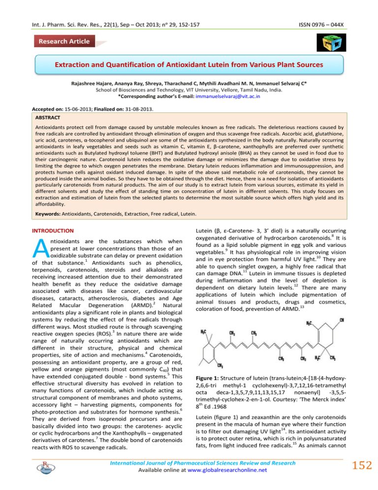
Int. J. Pharm. Sci. Rev. Res., 22(1), Sep – Oct 2013; nᵒ 29, 152-157 ISSN 0976 – 044X Research Article Extraction and Quantification of Antioxidant Lutein from Various Plant Sources Rajashree Hajare, Ananya Ray, Shreya, Tharachand C, Mythili Avadhani M. N, Immanuel Selvaraj C* School of Biosciences and Technology, VIT University, Vellore, Tamil Nadu, India. *Corresponding author’s E-mail: immanuelselvaraj@vit.ac.in Accepted on: 15-06-2013; Finalized on: 31-08-2013. ABSTRACT Antioxidants protect cell from damage caused by unstable molecules known as free radicals. The deleterious reactions caused by free radicals are controlled by antioxidant through elimination of oxygen and thus scavenge free radicals. Ascorbic acid, glutathione, uric acid, carotenes, α-tocopherol and ubiquinol are some of the antioxidants synthesized in the body naturally. Naturally occurring antioxidants in leafy vegetables and seeds such as vitamin C, vitamin E, β-carotene, xanthophylls are preferred over synthetic antioxidants such as Butylated hydroxyl toluene (BHT) and Butylated hydroxyl anisole (BHA) as they cannot be used in food due to their carcinogenic nature. Carotenoid lutein reduces the oxidative damage or minimizes the damage due to oxidative stress by limiting the degree to which oxygen penetrates the membrane. Dietary lutein reduces inflammation and immunosuppression, and protects human cells against oxidant induced damage. In spite of the above said metabolic role of carotenoids, they cannot be produced inside the animal bodies. So they have to be obtained through the diet. Hence, there is a need for isolation of antioxidants particularly carotenoids from natural products. The aim of our study is to extract lutein from various sources, estimate its yield in different solvents and study the effect of standing time on concentration of lutein in different solvents. This study focuses on extraction and estimation of lutein from the selected plants to determine the most suitable source which offers high yield and its affordability. Keywords: Antioxidants, Carotenoids, Extraction, Free radical, Lutein. INTRODUCTION A ntioxidants are the substances which when present at lower concentrations than those of an oxidizable substrate can delay or prevent oxidation of that substance.1 Antioxidants such as phenolics, terpenoids, carotenoids, steroids and alkaloids are receiving increased attention due to their demonstrated health benefit as they reduce the oxidative damage associated with diseases like cancer, cardiovascular diseases, cataracts, atherosclerosis, diabetes and Age Related Macular Degeneration (ARMD).2 Natural antioxidants play a significant role in plants and biological systems by reducing the effect of free radicals through different ways. Most studied route is through scavenging reactive oxygen species (ROS).3 In nature there are wide range of naturally occurring antioxidants which are different in their structure, physical and chemical properties, site of action and mechanisms.4 Carotenoids, possessing an antioxidant property, are a group of red, yellow and orange pigments (most commonly C40) that have extended conjugated double - bond systems.5 This effective structural diversity has evolved in relation to many functions of carotenoids, which include acting as structural component of membranes and photo systems, accessory light – harvesting pigments, components for 6 photo-protection and substrates for hormone synthesis. They are derived from isoprenoid precursors and are basically divided into two groups: the carotenes- acyclic or cyclic hydrocarbons and the Xanthophylls – oxygenated derivatives of carotenes.7 The double bond of carotenoids reacts with ROS to scavenge radicals. Lutein (β, ε-Carotene- 3, 3’ diol) is a naturally occurring oxygenated derivative of hydrocarbon carotenoids.8 It is found as a lipid soluble pigment in egg yolk and various vegetables.9 It has physiological role in improving vision and in eye protection from harmful UV light.10 They are able to quench singlet oxygen, a highly free radical that can damage DNA.11 Lutein in immune tissues is depleted during inflammation and the level of depletion is dependent on dietary lutein levels.12 There are many applications of lutein which include pigmentation of animal tissues and products, drugs and cosmetics, coloration of food, prevention of ARMD.13 Figure 1: Structure of lutein (trans-lutein;4-[18-(4-hydoxy2,6,6-tri methyl-1 cyclohexenyl)-3,7,12,16-tetramethyl octa deca-1,3,5,7,9,11,13,15,17 nonaenyl] -3,5,5trimethyl-cyclohex-2-en-1-ol. Courtesy: ‘The Merck index’ 8th Ed .1968 Lutein (figure 1) and zeaxanthin are the only carotenoids present in the macula of human eye where their function 14 is to filter out damaging UV light . Its antioxidant activity is to protect outer retina, which is rich in polyunsaturated fats, from light induced free radicals.15 As animals cannot International Journal of Pharmaceutical Sciences Review and Research Available online at www.globalresearchonline.net 152 Int. J. Pharm. Sci. Rev. Res., 22(1), Sep – Oct 2013; nᵒ 29, 152-157 16 synthesize lutein, they must obtain it from diet. A lot of information regarding the importance of lutein on human health has been gathered. Lutein can be obtained from various sources such as fruits and vegetables. To prevent ARMD a minimum of about 6mg of lutein is needed which we may not get from our diet, so we need an additional supplement of lutein.6 However, there is no much data available in the literature pertaining to lutein production such as its extraction, purification and phytochemical analysis. Hence we tried here to extract and quantify the lutein from various easily available low cost natural food sources. MATERIALS AND METHODS Chemicals used Acetone, ethanol, butanol, methanol, hexane, silica, sodium nitrite and sulphuric acid were purchased from Himedia chemicals. Standard lutein was obtained from Elnova pharma. Sample Preparation Flowers such as marigold, gerbera, fruits such as orange, grapes, papaya, vegetables such as spinach, cabbage, corn, and carrot were used in this study. They were purchased from the local market of Vellore, washed under tap water to remove all the dirt particles and again cleaned with sterile water and air dried at room temperature. The raw materials were chosen based on their local availability and also on the basis of information gathered during literature studies. The scientific and local names of the selected plants that were used in this study were mentioned in table 1. Marigold (Tagetes erecta L.) plants were authenticated and confirmed at Plant Biotechnology department, VIT University. Table 1: Scientific, family, common and local names of the plants investigated Scientific name Family name Common name Local name Tagetes erecta L. Asteraceae Marigold Samandhi Spinacia oleracea L. Amaranthaceae Spinach Keerai Carica papaya L. Caricaceae Papaya Papali Daucus carota Hoffm. Apiaceae Gerbera daisy Asteraceae Gerbera NA Vitis vinifera L. Vitaceae Grape Dratchai Citrus sinensis L. Rutaceae Orange Naaram Brassica oleracea L. Brassicaceae Cabbage Muttai gose Zea mays L. Poaceae Corn Cholam ISSN 0976 – 044X Extraction The samples (10 g each) were grinded with a mortar and pestle and mixed with solvent. Three different solvents (40 mL) i.e. acetone, ethanol and butanol were used. The solution was filtered using Whatman No. 1 filter paper. The filtrate was centrifuged at 10,000 rpm for 10 minutes. The aqueous phase was collected and stored at 4˚C.17 Measurement of Absorbance Absorbance was measured using spectrophotometer18 at th a wavelength of 446 nm at 0 hour and at intervals of every 3 hours standing time till 9 hours for both standard and samples and was recorded. Concentration of lutein was calculated using the following formula: A X V (ml) X dilution factor Concentration of lutein (µg/g of sample) = Where, A = Absorbance at 446 nm, V = Volume of extract in ml, €= Absorption coefficient (2589), W= Dry weight of sample. Test for carotenoids The colour of solution containing sample in solvent disappears after successive addition of 5% solution of sodium nitrite and 0.5M H2SO4.19 This test was performed for both standard and samples as well to confirm the presence of carotenoids. Detection Method To detect the lutein content in the extracted material, thin layer chromatography (TLC) was performed. The extracted samples were concentrated under vacuum using rotary evaporator. High temperature causes denature of lutein. Air dried samples were then used in TLC. TLC plates were made using silica slurry, mobile phase used was hexane. The samples were dissolved in methanol solvent and spot was placed on the TLC plate along with standard lutein.20 The plates were then kept in the beaker containing hexane as mobile phase and allowed to travel up to three-fourth of the length. The spots were detected under UV light and Rf value was calculated using the formula: Rf = Carrot Carrot € X W (g) Length of the spot travelled Length of mobile phase travelled RESULTS AND DISCUSSION The experiment was carried out to look for the most suitable raw material for the extraction of lutein. TLC was done to identify lutein in the extract by comparing Rf values of the standard with Rf value of the sample. Results obtained from different sources showed Rf values for acetone to be around 0.42, ethanol to be around 0.30 and butanol to be around 0.55 (Table 2). These values matched with the results obtained from the standard source of lutein, proving that the extract contained lutein. International Journal of Pharmaceutical Sciences Review and Research Available online at www.globalresearchonline.net 153 Int. J. Pharm. Sci. Rev. Res., 22(1), Sep – Oct 2013; nᵒ 29, 152-157 ISSN 0976 – 044X Table 2: Rf values for sample and standard lutein with different solvents on TLC Source Marigold Spinach Papaya Carrot Gerbera Grape Orange Cabbage Corn Standard / Sample Acetone Ethanol Butanol Standard 0.40 0.29 0.58 Sample 0.42 0.30 0.55 Standard 0.39 0.30 0.55 Sample 0.40 0.31 0.55 Standard 0.42 0.25 0.58 Sample 0.43 0.25 0.56 Standard 0.47 0.32 0.60 Sample 0.45 0.30 0.56 Standard 0.49 0.35 0.52 Sample 0.50 0.36 0.50 Standard 0.43 0.31 0.57 Sample 0.42 0.30 0.59 Standard 0.38 0.28 0.60 Sample 0.39 0.26 0.61 Standard 0.40 0.30 0.58 Sample Standard 0.42 0.45 0.32 0.28 0.54 0.56 Sample 0.47 0.28 0.56 Figure 2: Concentration of lutein at zeroth hour of standing time in three different solvents acetone, ethanol and butanol Figure 3: Concentration of lutein in various extracts after 3 hours of standing time International Journal of Pharmaceutical Sciences Review and Research Available online at www.globalresearchonline.net 154 Int. J. Pharm. Sci. Rev. Res., 22(1), Sep – Oct 2013; nᵒ 29, 152-157 ISSN 0976 – 044X Table 3: Absorbance and concentration of Lutein in various extracts Source Marigold Spinach Papaya Carrot Gerbera Grape Orange Cabbage Corn Standing time (hrs) Acetone Conc. (µg/g) 0.301 OD 0.301 Ethanol Conc. (µg/g) 1.163 OD 0.221 Butanol Conc. (µg/g) 0.854 0 OD 0.541 3 6 9 0 3 6 0.631 0.978 0.990 0.345 0.464 0.818 2.437 3.778 3.823 1.333 1.792 3.159 0.622 0.878 0.880 0.279 0.315 0.610 2.402 3.391 3.398 1.078 1.217 2.356 0.341 0.432 0.435 0.201 0.394 0.493 1.317 1.669 1.680 0.776 1.522 1.904 9 0 3 6 9 0.820 0.311 0.530 0.661 0.685 3.167 1.201 2.047 2.553 2.695 0.615 0.204 0.256 0.398 0.399 2.375 0.788 0.989 1.537 1.541 0.499 0.196 0.280 0.339 0.341 1.927 0.757 1.081 1.309 1.317 0 3 6 9 0 0.215 0.381 0.431 0.431 0.198 0.830 1.472 1.664 1.665 0.764 0.197 0.202 0.212 0.213 0.111 0.761 0.780 0.819 0.823 0.428 0.132 0.139 0.145 0.148 0.091 0.509 0.537 0.444 0.571 0.351 3 6 9 0 3 0.261 0.362 0.380 0.106 0.211 1.008 1.398 1.468 0.409 0.815 0.134 0.145 0.150 0.095 0.100 0.517 0.560 0.579 0.367 0.386 0.101 0.156 0.158 0.064 0.099 0.390 0.603 0.610 0.247 0.382 6 9 0 3 6 0.311 0.316 0.098 0.111 0.376 1.201 1.221 0.378 0.429 1.452 0.137 0.139 0.065 0.081 0.103 0.529 0.537 0.251 0.313 0.398 0.111 0.111 0.033 0.045 0.096 0.428 0.429 0.127 0.174 0.371 9 0 3 6 9 0.377 0.067 0.111 0.298 0.300 1.456 0.258 0.429 1.151 1.158 0.109 0.059 0.075 0.096 0.099 0.421 0.228 0.286 0.371 0.382 0.099 0.026 0.031 0.075 0.076 0.382 0.100 0.119 0.289 0.294 0 3 6 9 0.066 0.011 0.113 0.141 0.172 0.042 0.436 0.544 0.021 0.032 0.054 0.054 0.081 0.124 0.209 0.209 0.012 0.028 0.043 0.044 0.046 0.108 0.166 0.169 Figure 4: Concentration of lutein after 6 hours of standing time International Journal of Pharmaceutical Sciences Review and Research Available online at www.globalresearchonline.net 155 Int. J. Pharm. Sci. Rev. Res., 22(1), Sep – Oct 2013; nᵒ 29, 152-157 ISSN 0976 – 044X Figure 5: Concentration of lutein after 9 hours of standing time In TLC analysis, all the selected plant samples showed the presence of lutein in them. The relative amount of lutein was measured using UV Spectrophotometer at wavelength of 446 nm. The absorbance and concentration of lutein in different extracts at different standing times were mentioned in table 3. Figures 2, 3, 4 and 5 show the concentration of lutein at zeroth hour, after 3 hours, 6 hours and 9 hours of standing time respectively in the given three solvents. Within the range of standing time covered in this study, it was observed that the increment of standing time increased the amount of lutein extracted into the solvent. This is due to the longer duration that allowed for process to take place. Among all the samples selected marigold showed the highest yield and corn showed the lowest yield. In case of marigold the concentration of lutein in acetone increased from 0.541 µg/g of sample to 3.778 µg/g of sample after 6 hours of standing time. Comparing the performance of various solvents used in study, acetone extracted highest amount of lutein followed by ethanol and butanol. It is because acetone has lower polarity than that of ethanol and butanol. extraction was 6 hours. Moreover it is observed that the yield of the extraction depends on the solvent used also. Even though marigold is said to be highest in lutein content among the selected plants, it yield very low amount with butanol solvent at its optimum standing time whereas high yield is with acetone. Hence it is suggested that selection of the appropriate solvent for the extraction process is also important for further detailed study of this compound. Although, lutein is administered for macular degeneration, use of this carotenoid to reduce many systemic diseases and ease of purification needs further study. Acknowledgements: we would like to thank management of VIT University for providing the necessary facilities to carry-out this work. REFERENCES 1. Ratnam DV, Ankola DD, Bhardwaj V, Sahana DK, Ravi kumar MNV, Role of antioxidants in prophylaxis and therapy: A pharmaceutical perspective, Journal of Controlled release, 113, 2006, 189-207. 2. Xia F, Hu D, Jin H, Zhao Y, Liang J, Preparation of lutein proliposomes by supercritical anti-solvent technique, Food Hydrocolloids, 26, 2012, 456-463. 3. Junghans A, Sies H, Stahl W, Macular pigments lutein and zeaxanthin as blue light filters studied in lipososmes, Archives of Biochemistry and Biophysics, 391(2), 2001, 160-164. 4. Koh HH, Murray IJ, Nolan D, Carden D, Feather J, Beatty S, Plasma and macula responses to lutein supplements in subjects with and without age-related maculopathy: a pilot study, Experimental eye research, 79, 2004, 21-27. 5. Martinez AJM, Britton G, Vicario IM, Heredia FJ, HPLC analysis of geometrical isomers of lutein epoxide isolated from dandelion (Taraxacum officinale F. Weber ex Wiggers), Phytochemistry, 67, 2006, 771-777. 6. Quiros AR, Costa HS, Analysis of carotenoids in vegetable and plasma samples: A review, Journal of food composition and analysis, 19, 2006, 97-111. CONCLUSION Our study was carried out to determine the best source for extraction of lutein from locally available sources. Among all the sources used for extraction of lutein, marigold showed the highest concentration of lutein which was 3.82 µg/g of sample after letting it to stand for 9 hours in acetone. Also the literature studies shows that other carotenoids in marigold are less compared to lutein which is about 80% of total carotenoid content. The rate of extraction and yield on the basis of solubility and polarity of all the three solvents when evaluated showed that acetone yielded the highest concentration of lutein. As standing time was increased to 9 hours, there was increase in concentration of lutein as the solvent was in contact with the sample for a longer time period. However after certain time interval, the effect of standing time was constant. Therefore optimum time for International Journal of Pharmaceutical Sciences Review and Research Available online at www.globalresearchonline.net 156 Int. J. Pharm. Sci. Rev. Res., 22(1), Sep – Oct 2013; nᵒ 29, 152-157 ISSN 0976 – 044X 7. Tobias AV, Arnold FH, Biosynthesis of novel carotenoid families based on unnatural carbon backbones: A model for diversification of natural product pathways, Molecular and Cell biology of lipids, 1761(2), 2006, 235-246. 14. Batista AP, Raymundo A, Sousa I, Empis J, Rheological characterization of coloured oil-in-water food emulsions with lutein and phycocyanin added to the oil and aqueous phases, Food Hydrocolloids, 20, 2006, 44-52. 8. Tsao R, Yang R, Yang JC, Zhu H, Manolis T, Separation of geometric isomers of native lutein diesters in marigold (Tagetes erecta L.) by high-performance liquid chromatography-mass spectrometry, Journal of chromatography A, 1045, 2004, 65-70. 15. Sasaki M, Yuki K, Kurihara T, Miyake S, Noda K, Kobayashi S, Ishida S, Tsubota K, Ozawa Y, Biological role of lutein in the light-induced retinal degeneration, Journal of nutritional biochemistry, 23, 2012, 423-429. 9. Sujak A, Gabrielska J, Grudzinski W, Borc R, Mazurek P, Gruszecki WI, Lutein and zeaxanthin as protectors of lipid membranes against oxidative damage: the structural aspects, Archives of Biochemistry and biophysics, 371(2), 1999, 301-307. 10. Landrum JT, Bone RA, Lutein, Zeaxanthin and the Macular pigment, Archives of Biochemistry and biophysics, 385(1), 2001, 28-40. 11. Beatty S, Nolan J, Kavanagh H, Donovan O, Macular pigment optical density and its relationship with serum and dietary levels of lutein and zeaxanthin, Archives of Biochemistry and biophysics, 430, 2004, 70-76. 12. Koutsos EA, Lopez JCG, Klasing KC, Carotenoids from In ovo or dietary sources blunt systemic indices of the inflammatory response n growing chicks (Gallus gallus domesticus), The journal of nutrition, 136, 2006, 10271031. 13. Losso JN, Khachatryan A, Ogawa M, Godber JS, Shih F, Random centroid optimization of phosphatidylglycerol stabilized lutein-enriched oil-in-water emulsions at acidic pH, Food chemistry, 92, 2005, 737-744. 16. Zuorro A, Lavecchia R, New functional food products containing lutein and zeaxanthin from marigold(Tagetes erecta L.) flowers, Journal of Biotechnology, 150S, 2010, S296. 17. Murillo E, Martinez AJM, Portugal F, Screening of vegetables and fruits from Panama for rich sources of lutein and zeaxanthin, Food chemistry, 122, 2010, 167172. 18. Wang M, Tsao R, Zhang S, Dong Z, Yang R, Gong J, Pei Y, Antioxidant activity, mutagenicity/anti-mutagenecity, and clastogenecity/anti-clastogenecity of lutein from marigold flowers, Food and chemical toxicology, 44, 2006, 15221529. 19. Ajayi IA, Ajibade O, Oderinde RA, Preliminary phytochemical analysis of some plant seeds, Research Journal of Chemical Sciences, 1(3), 2011, 58-62. 20. Rodic Z, Simonovska B, Albreht A, Vovk I, Determination of lutein by high-performance thin-layer chromatography using densitometry and screening of major dietary carotenoids in food supplements, Journal of Chromatography A, 1231, 2012, 59-65. Source of Support: Nil, Conflict of Interest: None. International Journal of Pharmaceutical Sciences Review and Research Available online at www.globalresearchonline.net 157
