Document 13308902
advertisement
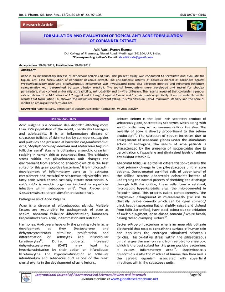
Int. J. Pharm. Sci. Rev. Res., 16(2), 2012; nᵒ 22, 97-103 ISSN 0976 – 044X Research Article FORMULATION AND EVALUATION OF TOPICAL ANTI ACNE FORMULATION OF CORIANDER EXTRACT * Aditi Vats , Pranav Sharma D.J. College of Pharmacy, Niwari Road, Modinagar-201204, U.P, India. *Corresponding author’s E-mail: sh.aditi.vats@gmail.com Accepted on: 29-08-2012; Finalized on: 29-09-2012. ABSTRACT Acne is an inflammatory disease of sebaceous follicles of skin. The present study was conducted to formulate and evaluate the topical anti acne formulation of coriander aqueous extract. The antibacterial activity of aqueous extract of coriander against Propionibacterium acne and Staphylococcus epidermidis was investigated using disc diffusion method and minimum inhibitory concentration was determined by agar dilution method. The topical formulations were developed and tested for physical parameters, drug content uniformity, spreadibility, extrudability and in-vitro diffusion. The results revealed that coriander aqueous extract showed the MIC values of 1.7 mg/ml and 2.1 mg/ml against P.acne and S. epidermidis respectively. It was revealed from the results that formulation Fa1 showed the maximum drug content (94%), in-vitro diffusion (93%), maximum stability and the zone of inhibition among all the formulations. Keywords: Acne vulgaris, antibacterial activity, coriander, topical gel, in-vitro activity. INTRODUCTION Acne vulgaris is a common skin disorder affecting more than 85% population of the world, specifically teenagers and adolescents. It is an inflammatory disease of sebaceous follicles of skin marked by comedones, papules and pustules and presence of bacterias Propionibacterium acne, Staphylococcus epidermidis and Malassezia furfur in follicular canal1. P.acne is obligatory anaerobic organism residing in human skin as cutaneous flora. The oxidative stress within the pilosebaceous unit changes the environment from aerobic to anaerobic which is the best suited for this gram positive bacterium.7 It is implicated in development of inflammatory acne as it activates complement and metabolize sebaceous triglycerides into fatty acids which chemo tactically attract neutrophills. S. epidermidis is aerobic organism involved in superficial infection within sebaceous unit1. Thus P.acne and S.epidermidis are target sites for anti acne drugs. Sebum: Sebum is the lipid- rich secretion product of sebaceous gland, secreted by sebocytes which along with keratinocytes may act as immune cells of the skin. The severity of acne is directly proportional to the sebum production16. The secretion of sebum increases due to enlargement of sebaceous glands under the stimulatory action of androgens. The sebum of acne patients is characterized by the presence of lipoperoxides due to peroxidation o f squalene and diminished levels of sebum antioxidant vitamin E. Acne is a disease of pilosebaceous glands. Multiple factors are responsible for pathogenesis of acne as sebum, abnormal follicular differentiation, hormones, Propionibacterium acne, inflammation and nutrition. Abnormal follicular epithelial differentiation-It marks the most primary change in the pilosebaceous unit in acne patients. Desquamated cornified cells of upper canal of the follicle become abnormally adherent; instead of undergoing the normal process of shedding and discharge through follicular orifice, these cells form a retained, microscopic hyperkeratotic plug (the microcomedo) in follicular canal. This process called comedogenesis. The progressive enlargement of microcomedo give rise to clinically visible comedo which can be open comedo/ black heads (appearing flat or slightly raised and distend from follicular orifice), have black colour due to oxidation of melanin pigment, or as closed comedo / white heads, having closed overlying surface17. Harmones: Androgens have only the priming role in acne development as they (testosterone and dehyrotestosterone) stimulate proliferation and differentiation of sebocytes and infundibular 14 keratinocytes . During puberty, increased dehyrotestosterone (DHT) may lead to hyperkeratinisation by their action on infundibular keratinocytes. The hyperkeratinisation in follicular infundibulum and sebaceous duct is one of the most crucial events in the development of acne lesions. Bacteria-Propionibacterium acne is an anaerobic obligate diptheroid that resides beneath the surface of human skin and populates the androgen stimulated sebaceous follicles. The oxidative stress within the pilosebaceous unit changes the environment from aerobic to anaerobic which is the best suited for this gram positive bacterium. It causes inflammatory acne18. Staphylococcus epidermidis is also the resident of human skin flora and is the aerobic organism associated with superficial infections within the sebaceous units. Pathogenesis of Acne Vulgaris International Journal of Pharmaceutical Sciences Review and Research Available online at www.globalresearchonline.net Page 97 Int. J. Pharm. Sci. Rev. Res., 16(2), 2012; nᵒ 22, 97-103 Inflammation: Inflammation is the direct or indirect result of proliferation of P. acne. The bacteria produce the extracellular lipase that hydrolyses sebum triglycerides to glycerol, used by organism as growth substrate, and fatty acids, which have proinflammatory and comedogenic properties. Further, P.acne may activate keratinocytes and sebocytes via TLR, CD14 and CD1 molecules. TLR2 is expressed on the surface of macrophages surrounding the pilosebaceous follicles in acne lesions. Activation of TLR2 leads to triggering of transcription factor nuclear factor and thus the production of cytokines which along with IL8 and IL 12, released from TLR2 positive monocytes, produce the inflammatory lesions of acne. Inflammatory acne comprises of pustules, papules and nodules14. Nutrition: Acne is also driven by growth factors (particularly insulin-like growth factor [IGF-1]) acting on sebaceous glands and keratinocytes lining the pilary canal. Dairy products contain 5α-reduced steroid harmones and other steroid precursors of DHT that drive sebaceous gland function. Drinking milk causes a direct rise in IGF-1 through a disproportionate elevation in blood sugar and serum insulin level. High glycemic load foods also cause IGF-1 mediated elevations in DHT. IGF-1 levels during teenage years closely parallel acne activity and are likely synergistic with the steroid harmones14.Through population based studies it is being observed that as the diet westernize, acne prevalence increases. The studies show the associations between acne and selected food dietary patterns15. Due to increased instances of resistance of acne inducing bacterias towards the antibiotics18, the alternate system of medicine for the treatment of acne have been investigated and adopted. Among the alternate systems of medicine the topical therapeutic agents are more convenient for application11. It is rightly being said that “for every disease there is a plant on every continent”. The herbs as ingredients in topical acne treatment are occupying the upper position as they are safe, dilute, patient familiar, economic, easily available and 20 multifunctional . Figures from the World Health Organization suggest that 4 billion people, who make nearly 70% of the world’s population, are users of herbal medicine for some purpose of primary health care. Medicinal herbs have always been in usage in some form or the other in indigenous systems of medicines including Ayurveda, Sidha and Unani in India. An increasing number of companies are making forays into the herbal market segment. An example is Ranbaxy, which launched herbal medicines in India. According to the company the global market size for herbals is estimated at US$ 20 Billion with a growth rate of 10-15 percent per year. The growth rate 19 for the Indian market is pegged at 12% per annum . Coriandrum sativum is medicinally proved to have 2 therapeutic activities like hypoglycemic , anti3 4 inflammatory , hypolipidemic, antioxidant , anti scarring ISSN 0976 – 044X 10 property due to the presence of salicylic acid microbial activity against bacteria and fungi5. and anti MATERIALS AND METHODS The coriander leaves and dried seeds were purchased from the local markets of Modinagar. The plant material was authenticated by taxonomist at Modinagar and the specimens were deposited at Botanical section of M. M. PG College, Modinagar. The test organisms, P.acne (MTCC 1951) and S. epidermidis (MTCC 931), were obtained from Microbial culture collection and Gene bank, Chandigarh, India. All media were purchased from Hi-Media. All reagents used were of analytical grade. Obtainment of Coriander extract The green leaves were dried in the shade and were powdered. 200gm of this powder was macerated with distilled water for seven days at room temperature (25±2ᵒC). Frequent agitation and circulation of solvent was maintained in order to decrease the boundary layer phenomenon and enhance the efficiency of extraction process. After seven days, the extracted solution was filtered and marc was pressed. Both the strained liquid and the expressed liquid were combined together. Then the enriched extract, called miscella, was concentrated in a rotary vacuum flash evaporator under reduced pressure. The procedure was adopted to prepare the hydroethanolic and the ethereal extract with a slight modification in the type of solvent used as distilled waterethanol (60:40) was used for hydroethanolic extract whereas ether was used for ethereal extract. Determination of antimicrobial activity Determination of antibacterial activity The antibacterial activity was determined by disc diffusion method. This experiment was performed by following the method of Hayes and Markovic (2002) with some modifications. Propionibacterium acne (P. acne) was incubated in ASLA agar medium for 48 hrs under anaerobic conditions and adjusted to yield approximately 1x10⁸ CFU/ml. The agar plates were swabbed with inoculums. The sterile filter paper disc of diameter 6mm were aseptically placed on the inoculated plates and were impregnated with the test material (100mg/ml of extracts). The plates were left at ambient temperature for 30 min to allow exceed pre diffusion prior to incubation at 37ᵒC for 72 hrs under anaerobic conditions in a anaerobic bag (Hi-Media) with gas pack and indicator tablets and the bag was kept in an incubator for 72 hrs at 37 ± 1ᵒC. Gas packs containing citric acid, sodium carbonate and sodium borohydride were used to maintain and check the anaerobiosis. The indicator tablet of methylene blue changed from dark pink-blue-light pink finally, which indicated the achievement of anaerobic condition. The culture of Staphylococcus epidermidis (S.epidermidis) was prepared in nutrient agar medium at 24 hrs under aerobic conditions. Test samples of this aerobic bacterium were incubated at 37ᵒC for 24 hrs under aerobic conditions. The anti bacterial activity was International Journal of Pharmaceutical Sciences Review and Research Available online at www.globalresearchonline.net Page 98 Int. J. Pharm. Sci. Rev. Res., 16(2), 2012; nᵒ 22, 97-103 ISSN 0976 – 044X estimated by measuring the diameter of the zone of inhibition. All disc diffusion tests were performed in three separate experiments and antibacterial activity was expressed as the mean ± standard deviation. Clindamycin phosphate was used as standard and control was used to determine the growth capacity of medium6. Physical parameters Determination of Minimum inhibitory Concentration Consistency- The consistency was checked by applying on skin. The minimum inhibitory concentration (MIC) values were determined by agar dilution method. The test material was added aseptically to 20ml aliquots of sterile molten agar at appropriate range of test material (0.05mg/ml5mg/ml). The resulting agar solutions were vortexed at high speed for 15 secs or until completely dispersed, immediately poured into sterile petri plates then allowed to set for 30 min. The plates were then inoculated with the P.acne. The inoculated plates were left until the inoculums had set and then incubated under anaerobic 0 conditions at 37 C for 72 hrs in gas bag (Hi-Media) with gas pack and indicator tablets and the bag was kept in an incubator for specified duration at specified temperature. Gas packs containing citric acid, sodium carbonate and sodium borohydride were used to maintain and check the anaerobiosis. The indicator tablet of methylene blue changed from dark pink-blue-light pink finally, which indicated the achievement of anaerobic condition. The test samples of S.epidermidis were prepared in nutrient agar medium and incubated for 24 hrs at 37ᵒC under aerobic conditions. Following the incubation period, the plates were observed and recorded for the presence or absence of growth. From the results, the MIC was recorded as the lowest concentration of test substance where the absence of growth was observed. Minimum bactericidal concentration (MBC) was determined by subculturing the samples on to the sterile agar plates from the three test plates, with each of bacterium, which had shown no growth during the determination of MIC. The plates for each of bacterium were incubated following the same procedure as described in MIC determination. The minimum bactericidal concentration values were interpreted as the highest dilution (lowest concentration) of sample which 12, 8, 9 showed no growth on agar plates. Preparation of gel The weighed amount of methyl paraben was dissolved in 5ml of hot water and propyl paraben was added on slight cooling of water. To this beaker carbopol 934 was dispersed with continous stirring for 20 min after addition of 50 ml of distilled water. This dispersion was kept overnight for soaking. In another beaker the required quantity of propylene glycol and polyethylene glycol (PEG 400) were added. This mixture along with concentration of aqueous extract corresponding to its MIC was incorporated to carbopol beaker with stirring. The volume was made up with distilled water and stirring was done vigorously. Triethanolamine was added form the gel by 23,24 adjusting pH to 6.8 (Table [2]) Physical appearance- The physical appearance of the formulation was checked visually which comprised of: Colour- The colour of the formulations was checked out against white background. Greasiness- The greasiness was assessed by the application on to the skin. Odour- The odour of the gels was checked by mixing the gel in water and taking the smell. pH About 20mg of the formulation was taken in a beaker and was subjected to the pH measurement using a digital pH meter within 24 hrs of manufacture.25 Viscosity Viscosities of formulated gels were determined using Brookfield viscometer spindle # 7 at 50 rmps and 25ᵒC. The corresponding dial reading on the viscometer was noted. Then the spindle was lowered successively. The dial reading was multiplied by the factor mentioned in catalog25. Extrudability Extrudability is defined as the weight in grams required for extruding 0.5cm long ribbon of formulation in 10 secs. The gel formulation was filled in a standard capped collapsible alluminium tubes and sealed by crimping to the end. The tubes were placed between two slides and were clamped. 500g weight was placed over the slides and then the cap was removed. The length of the ribbon of the formulation that came out in 10 secs was recorded.21 Spreadibility Spreadibility denotes the extent of area to which a gel readily spreads on the application to the skin or affected part. The bioavailability efficiency of the gel also depends on Spreadibility value. Spreadibility is defined in terms of time in secs required taken by the upper slide to slip off the gel placed between the two slides, under certain load. The lesser the time taken for the separation of two slides, the better the spreadibility. About 500mg of the formulation was sand witched between the two slides, each with dimensions of 6x2cm. A weight of 100g was placed upon the upper slide so that the formulation between the two slides get pressured uniformly to form a thin layer. The weight was removed and the excess of the formulation adhering to the slides was scrapped off. The lower slide was fixed on the board of apparatus and the upper slide was held to the non-flexible string to which 20g load was applied with the help of a simple pulley which was in horizontal level with the fixed slide. The time taken by the upper slide to slip off the lower slide 24 was noted . International Journal of Pharmaceutical Sciences Review and Research Available online at www.globalresearchonline.net Page 99 Int. J. Pharm. Sci. Rev. Res., 16(2), 2012; nᵒ 22, 97-103 ISSN 0976 – 044X Spreadibility= m x l/t Drug Content Where, m= weight tied to upper slide, l= length of the glass slide (6cm), t= time in secs. The drug content of the formulations was determined by dissolving an accurately weighed quantity of gel 1gm in 100ml of solvent (phosphate buffer pH 6.8 + ethanol in ratio 40:60). The solutions were kept for shaking for 4 hrs and then kept for 6 hrs for complete dissolution of the formulations. Then the solutions were filtered through 0.45mm membrane filters and proper dilutions were made and solution was subjected to the Spectrophotometric analysis. The drug content was calculated from the linear regression equation obtained from the calibration data. Antimicrobial studies of the formulation The solutions of the gels were prepared using 100mg of gel in 10ml of dimethyl sulfoxide. The anti bacterial activity was tested by well diffusion method. P. acne was incubated in ASLA agar medium for 48 hrs under anaerobic conditions and adjusted to yield approximately 8 1x10 CFU/ml. The solidified agar plates were swabbed with inoculums on the surface. The equidistance wells were cut in the plates with help of 8mm borer. In each of these wells the gel solutions in DMSO were placed and the plates were left at ambient temperature for 30 min to allow pre diffusion prior to incubation at 37ᵒC for 72 hrs under anaerobic conditions in a anaerobic bag (Hi-Media) with gas pack and indicator tablets and the bag was kept in an incubator for 72 hrs at 37 ± 1ᵒC. Gas packs containing citric acid, sodium carbonate and sodium borohydride were used to maintain and check the anaerobiosis. The indicator tablet of methylene blue changed from dark pink-blue-light pink finally, which indicated the achievement of anaerobic condition. The culture of S. epidermidis was prepared in nutrient agar medium at 24 hrs under aerobic conditions. Test samples of this aerobic bacterium were incubated at 37ᵒC for 24 hrs under aerobic conditions. The anti bacterial activity was estimated by measuring the diameter of the zone of inhibition. All well diffusion tests were performed in three separate experiments and antibacterial activity was expressed as the mean ± standard deviation22. Stability studies In-vitro diffusion studies The in-vitro diffusion studies for all formulations (Fa1-Fa4) were carried out using the Franz-diffusion cell. The diffusion cell apparatus was fabricated locally as an openended cylindrical tube with an area of 3.7994 cm2 and 2 100m height, having a diffusion area of 3.8 cm . A weighed quantity of formulation equivalent to 1gm of the drug was placed onto the dialysis membrane-70 (HiMedia) and was immersed slightly in 100ml of receptor medium (phosphate buffer pH 6.8+ ethanol in ratio 40:60) which was continuously stirred and the temperature was maintained at 37ᵒC±1ᵒC. Aliquots of 1ml were withdrawn from each of the system at time intervals of 5, 10, 15, 30, 60, 120, 240 and 360 min were analyzed for drug content using UV spectrophotometer.21 Drug release kinetics To study the release kinetics of the optimized formulation, the data obtained from invitro release studies were plotted in various kinetic models.28 The stability of the formulations was assessed according to the guide lines issued by International Conference on Harmonisation (ICH) on October 27, 1993.26,27 Table 1: Antibacterial activity of coriander aqueous extract. Zone of inhibitionb (mm), mean ±SD MIC Test sample P.acne S.epidermidis P.acne S.epidermidis Aqueous extract 21.5±1.4 20.6±1.09 1.7 mg/ml 2.1 mg/ml Hydroethanolic extract 19.1±1.0 18.1±1.9 2.5 mg/ml 3 mg/ml Ethereal extract 11.6±1.4 10.6±1.6 4.3 mg/ml 5 mg/ml b = diameter of zone of inhibition including disc diameter of 4mm. Table 2: Composition of topical formulations Mass (mg) Ingredients Fa1 Fa2 Fa3 Carbopol 934 0.5 1 1.5 PEG 400 5 5 5 Propylene glycol 15 15 15 Methyl paraben 0.15 0.15 0.15 Propyl paraben 0.03 0.03 0.03 Triethanolamine q.s q.s q.s Distilled water q.s 100 100 100 Fa4 2 5 15 0.15 0.03 q.s 100 International Journal of Pharmaceutical Sciences Review and Research Available online at www.globalresearchonline.net Page 100 Int. J. Pharm. Sci. Rev. Res., 16(2), 2012; nᵒ 22, 97-103 ISSN 0976 – 044X Table 3: Physicochemical properties of semisolid formulations of coriander aqueous extract. a a a a a Formulation pH Consistency Spreadability (g/sec) Extrudability (g) Viscosity (cps) Fa1 6.8±.01 *** 41.5±.7 542.9±.2 34.5±.8 Fa2 6.9±.04 ** 43±.2 545.6±.8 35.6±.6 Fa3 6.8±.07 ** 45±.9 549.4±.7 37.1±.1 Fa4 7.1±.04 ** 46.5±.4 552.8±.5 38.4±.4 a = mean ± standard deviation, *** = very good, ** = good.; All experiments were performed in triplicate. Table 4: Drug content and in-vitro release Formulation Drug content (%, m/m) % cumulative release Fa1 94.5 93 Fa2 93.4 92 Fa3 90 88 Fa4 88.5 87 All experiments were performed in triplicate Table 5: Antibacterial activity of formulations Zone of inhibition (mm), mean±SD Formulation P.acne S.epidermidis Fa1 21.5±1.2 20.6±1.06 Fa2 21.4±.1.0 20.5±1.02 Fa3 21.3±0.9 20.4±1.0 Fa4 21.3±.1.2 20.4±0.8 MH 20.4±0.6 20.4±0.9 Clin 31.8±2.6 30.9±2.4 All experiments were performed in triplicate. MH = marketed formulation, Clin = Clindamycin phosphate. Table 6: Release Kinetic parameters for optimized formulation of coriander extract. Formulation Zero order model First order model Higuchi model R2 K0 (min-1 ) R2 K1(min-1 ) R2 KH(min-1/2 ) Fa1 0.9686 0.684 0.9776 0.0036 0.9995 9.489 A: Fa1 First order release kinetic model B: Fa1 Zero order release kinetic model C: Fa1 Higuchi model. Figure 1: Release Kinetic model for optimized formulation of coriander extract. RESULTS AND DISCUSSION The aqueous extract of coriander showed the zone of inhibition of 21.5±1.4 and 20.6±1.09 mm as well as the MIC values of 1.7 mg/ml and 2.1 mg/ml against P.acne and S. epidermidis respectively. This activity of the aqueous extract was more than that of the hydroethanolic and ethereal extract. (Table [1]). The formulations were developed with Coriander aqueous extract using carbopol 934 as gelling agent in the concentration of 0.5% (Fa1),1% (Fa2),1.5% (Fa3) and 2% (Fa4) w/w. All the formulations were brown in colour and had characteristic odor of coriander. All formulations were glossy and translucent. Formulation Fa1 was found to have excellent consistency and Fa2 had good consistency. As indicated in the table 3, the pH of the formulation ranged from 6.8-7.1, which may be suitable for topical application without discomfort. The apparent viscosity of the formulations ranged from 34.5±.8 to 38.4±.4cps. The viscosity of formulation increased with increase in the concentration of carbopol content. The spreadability and extrudability of the formulations were found to range from 41.5±.7 to 46.5±.4g/sec and from 542.9±.2 to 552.8±.5g respectively. The viscosity was observed to increase with decrease in the spreadability and viceversa. International Journal of Pharmaceutical Sciences Review and Research Available online at www.globalresearchonline.net Page 101 Int. J. Pharm. Sci. Rev. Res., 16(2), 2012; nᵒ 22, 97-103 ISSN 0976 – 044X The drug concentration in dermatological bases was found to range from 88.5 to 94.5% (Table [4]). This proved that the method was suitable for the preparation of topical dosage forms. As indicated by the value of the drug content, there was no degradation of drug during the preparation process. Cumulative drug diffusion after 6hrs was found to be 93% for Fa1 which is highest among all the formulations. This is ascribed to the increased drug diffusion from the gel due to its low viscosity, soft nature and the nature of gelling agent used. Thus the formulation with the 0.5%w/w carbopol content (Fa1) was found to have the most optimized results. The results from the stability studies showed that formulations showed no changes in the ph, viscosity, spreadability, consistency, extrudability and drug content and hence are found to be stable. 2. Gallarher A.M., Flatt. P.R., Duffy. G. The effect of traditional antidiabetic plants on in vitro glucose diffusion. Nutrition Research 23:2003; 413-424. 3. Chithra V., Leelamma S. Coriandrum sativum changes the levels of lipid peroxides and activity of antioxidant enzymes in experimental animals. Indian J Biochemistry and Biophysics 36:1999; 59-61 4. Melo A De Enayde, Filho M Jorge. Characterisation of antioxidant compounds in aqueous coriander extract. Lebensm Wiss u Technol 38:2005; 15-19. 5. Matasyoh J.C.,Maiyo Z.C. Chemical composition and antimicrobial activity of the essential oil of coriandrum sativum. Food Chemistry 113:1999; 526-529. 6. Luangnarumitchai. S., Lamlertthon.S., Tiyaboonchai. W. Antimicrobial activity of essential oils against five strains of Propionibacterium acnes,J Pharma. Sci. 34: 2007; 60-64. When formulations were subjected to antimicrobial investigation, then it was found that all had inhibitory against P. acne and S. epidermidis. Among all the formulations, the formulation Fa1 had shown almost the same zone of inhibition as that shown by the aqueous extract (Table [5]). It was observed that Fa1 had shown the zone of inhibition greater than that of herbal marketed formulation, hence it is more effective than the marketed formulation. Clindamycin phosphate was found to have greater activity than the developed formulations. Thus Fa1 was found to be the most optimized formulation. 7. Bowe P Whitney, Logan C Alan. Clinical implication of lipid peroxidation in acne vulgaris: old wine in new bottles. Lipids in Health and Disease 9:2010; 140. 8. Determination of minimum inhibitory concentrations (MICs) of antimicrobial agents by agar dilution. EUCAST Definitive Document E. Def 3.1. 2000. 9. Andrews M. Jennifer. Determination of minimum inhibitory concentrations. J Antimicrobial Chemotherapy 48:2001; 516. The invitro diffusion data of the optimized formulation of coriander extract, Fa1, was subjected to kinetic models as zero order, first order, higuchi and hixon-crowell model. The selection criteria (R2) and the equations best describing the kinetics of invitro drug release are given in Tables [6]. The release of the drug from the optimized formulation was found to follow the first order release kinetics. The results are in agreement with the previous investigation performed by Dua et.al (2011). Higher correlation, as indicated by R2, was observed for the higuchi matrix release kinetics in the optimized formulation (Fig 1), suggesting diffusion as a probable prominent mechanism of drug release. In diffusion, the rate of drug dissolution within the matrix must be much higher than that of the diffusion rate of the drug leaving the matrix. This may be attributed to the nature of gelling agent used. CONCLUSION The coriander aqueous extract was found to have potency against acne inducing bacteria. The formulations developed from it also showed the same activity. So it can be further developed and sued commercially for treatment of acne. REFERENCES 1. Chomnawong Mullika Traidej, Surassmo Suvimol. Antimicrobial effects of Thai medicinal plants against acne inducing bacteria. J Ethanopharmacology 101:2005; 330333. 10. Kanlayavattanakul. M., Lourith N. Therapeutic agents and herbs in topical application for acne treatment. Int. J Cosmetic Sci. 33:2011; 289-29 11. Ghosh K. V., Nagore H. D., Kadbhune P. K. Different approaches of alternate medicines in acne vulgaris treatment. Orient Pharm Exp Med 11:2011; 1-9. 12. Viyoch J., Pisutthanan N., Faikreua A., Nupangta. K., Wangtorpol K., Ngokkuen J. Evaluation of in-vitro antimicrobial activity of thai basil oil and their microemulsion formulas against Propionibacterium acnes. Int J Cosmetic Sci. 28: 2006; 125-133. 13. McCall, C., Lawley T. Eczema, Psoriasis, Cutaneous Infections, Acne and other Common Skin Disorders. th Harrison`s Principles of Internal Medicines.,17 Ed,53.McGraw-Hill. 14. Kurokawa Ichiro, Danby William.W. New developments in our understanding of acne pathogenesis and treatment. Exp. Dermatol. 18: 2009; 821-832. 15. Spencer H. Elsa. Diet and acne: a review of evidence. Int. J Dermatol 48:2009; 339-347. 16. Brown K Sonya, Shalita R Alan. Acne vulgaris, Seminar. The Lancet 351:1998; 1871-1876.. 17. Blair C, Lewis CA. The pigment of comedones.Br J Dermatol 82:1970; 572-583. 18. Leyden J James. The evolving role of Propionibacterium acnes in acne. Seminar Cutaneous Medicine Surgery 3:2001; 139-143. 19. Robinson M Molly,Zhang Xiaorui. World medicine situation 2011. 3:2011; 1-12. 20. Aburjai Talal, Natsheh M Feda. Plants used in cosmetics. Phytother. Res. 17: 2003; 987-1000 International Journal of Pharmaceutical Sciences Review and Research Available online at www.globalresearchonline.net Page 102 Int. J. Pharm. Sci. Rev. Res., 16(2), 2012; nᵒ 22, 97-103 ISSN 0976 – 044X 21. Dua Kamal, Pabreja Kavita, Ramana V. M. Aceclofenac topical dosage forms: In vitro and in vivo characterization. Acta Pharmaceutica, 60:2010; 467–478. 25. Panigarhi L, John T, Shariff A, Shobarani R.S. Formulation and evaluation of Lincomycin HCL gels. Indian J Pharma. Sci. 59: 1997; 610-613. 22. Vijayalakshmi A., Tripura A., Ravichandiran V. Development and evaluation of anti acne products from Terminalia arjuna Bark. Int J Pharmtech Research 3:2011; 320-327. 26. ICH guidelines. Stability testing of new drug substances and products. 27 Oct 1993. 23. Sawarkar A H, Khadabadi SS, Mankar M.D. Development and biological evaluation of herbal anti acne gel. Int. J PharmTech Res. 2:2010; 2028-2031 24. Das Kuntal, Dang Raman, Machale U Manjunath. Formulation and evaluation of a novel herbal gel of Stevia extract. Iranian J Dermatol. 12:2009; 117-122. 27. Carl AB, Edward RA, David EB, Tietz NW (Ed).Textbook of th clinical chemistry and molecular diagnoistics, 4 rev ed. WB Saunders Philadelphia.2001. 28. Hamzah M. Maswadeh, Sereen H Mohmmad, Abdulhalim A. Abdulatif. In vitro dissolution kinetic study of theophylline from hydrophilic and hydrophobic matrices. Acta Poloniae Pharmaceutica- Drug research 63(1):2006, 63-67. ********************** International Journal of Pharmaceutical Sciences Review and Research Available online at www.globalresearchonline.net Page 103
