Computational analysis of kinetochore fibre organisation Supervisor:
advertisement
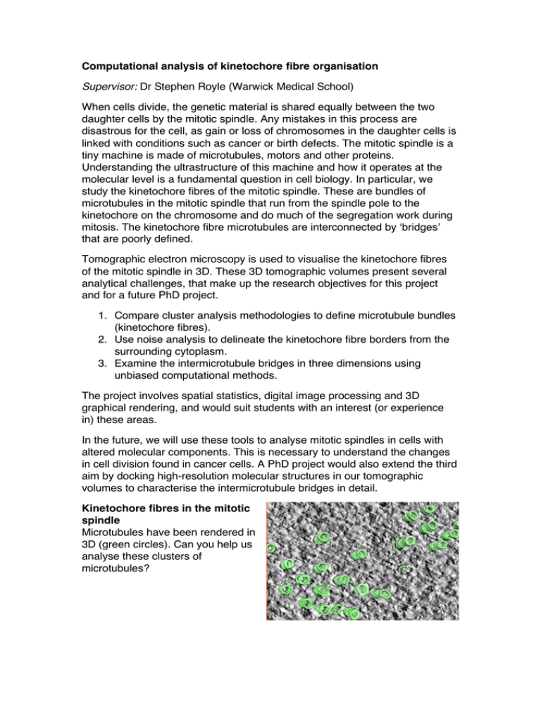
Computational analysis of kinetochore fibre organisation Supervisor: Dr Stephen Royle (Warwick Medical School) When cells divide, the genetic material is shared equally between the two daughter cells by the mitotic spindle. Any mistakes in this process are disastrous for the cell, as gain or loss of chromosomes in the daughter cells is linked with conditions such as cancer or birth defects. The mitotic spindle is a tiny machine is made of microtubules, motors and other proteins. Understanding the ultrastructure of this machine and how it operates at the molecular level is a fundamental question in cell biology. In particular, we study the kinetochore fibres of the mitotic spindle. These are bundles of microtubules in the mitotic spindle that run from the spindle pole to the kinetochore on the chromosome and do much of the segregation work during mitosis. The kinetochore fibre microtubules are interconnected by ‘bridges’ that are poorly defined. Tomographic electron microscopy is used to visualise the kinetochore fibres of the mitotic spindle in 3D. These 3D tomographic volumes present several analytical challenges, that make up the research objectives for this project and for a future PhD project. 1. Compare cluster analysis methodologies to define microtubule bundles (kinetochore fibres). 2. Use noise analysis to delineate the kinetochore fibre borders from the surrounding cytoplasm. 3. Examine the intermicrotubule bridges in three dimensions using unbiased computational methods. The project involves spatial statistics, digital image processing and 3D graphical rendering, and would suit students with an interest (or experience in) these areas. In the future, we will use these tools to analyse mitotic spindles in cells with altered molecular components. This is necessary to understand the changes in cell division found in cancer cells. A PhD project would also extend the third aim by docking high-resolution molecular structures in our tomographic volumes to characterise the intermicrotubule bridges in detail. Kinetochore fibres in the mitotic spindle Microtubules have been rendered in 3D (green circles). Can you help us analyse these clusters of microtubules?
![This article was downloaded by: [Simon Fraser University]](http://s2.studylib.net/store/data/013912039_1-4a9e0fe8c55b2150e18b853ce825775f-300x300.png)