Joining the dots: reconstructing complex microtubule networks WMS).
advertisement
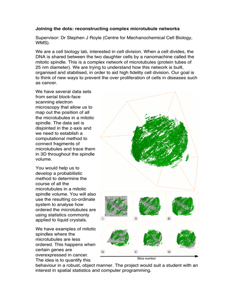
Joining the dots: reconstructing complex microtubule networks Supervisor: Dr Stephen J Royle (Centre for Mechanochemical Cell Biology, WMS). We are a cell biology lab, interested in cell division. When a cell divides, the DNA is shared between the two daughter cells by a nanomachine called the mitotic spindle. This is a complex network of microtubules (protein tubes of 25 nm diameter). We are trying to understand how this network is built, organised and stabilised, in order to aid high fidelity cell division. Our goal is to think of new ways to prevent the over proliferation of cells in diseases such as cancer. We have several data sets from serial block-face scanning electron microscopy that allow us to map out the position of all the microtubules in a mitotic spindle. The data set is disjointed in the z-axis and we need to establish a computational method to connect fragments of microtubules and trace them in 3D throughout the spindle volume. You would help us to develop a probabilistic method to determine the course of all the microtubules in a mitotic spindle volume. You will also use the resulting co-ordinate system to analyse how ordered the microtubules are using statistics commonly applied to liquid crystals. We have examples of mitotic spindles where the microtubules are less ordered. This happens when certain genes are overexpressed in cancer. The idea is to quantify this behaviour in a robust, object manner. The project would suit a student with an interest in spatial statistics and computer programming.
