Document 13294354
advertisement
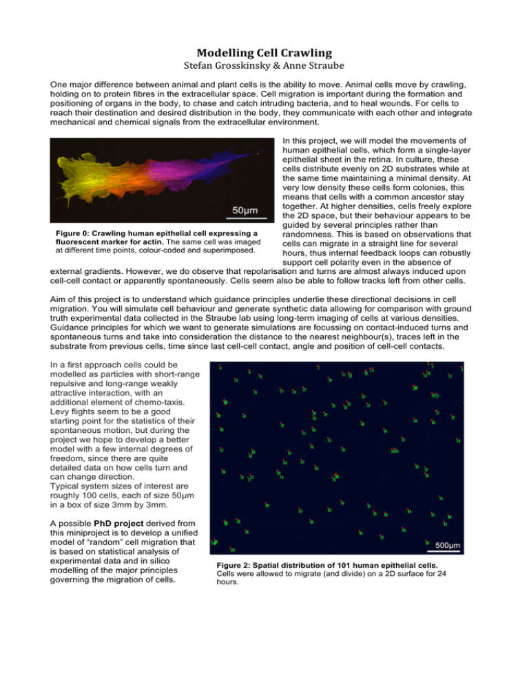
Modelling Cell Crawling Stefan Grosskinsky & Anne Straube One major difference between animal and plant cells is the ability to move. Animal cells move by crawling, holding on to protein fibres in the extracellular space. Cell migration is important during the formation and positioning of organs in the body, to chase and catch intruding bacteria, and to heal wounds. For cells to reach their destination and desired distribution in the body, they communicate with each other and integrate mechanical and chemical signals from the extracellular environment. In this project, we will model the movements of human epithelial cells, which form a single-layer epithelial sheet in the retina. In culture, these cells distribute evenly on 2D substrates while at the same time maintaining a minimal density. At very low density these cells form colonies, this means that cells with a common ancestor stay together. At higher densities, cells freely explore the 2D space, but their behaviour appears to be guided by several principles rather than Figure 0: Crawling human epithelial cell expressing a randomness. This is based on observations that fluorescent marker for actin. The same cell was imaged cells can migrate in a straight line for several at different time points, colour-coded and superimposed. hours, thus internal feedback loops can robustly support cell polarity even in the absence of external gradients. However, we do observe that repolarisation and turns are almost always induced upon cell-cell contact or apparently spontaneously. Cells seem also be able to follow tracks left from other cells. Aim of this project is to understand which guidance principles underlie these directional decisions in cell migration. You will simulate cell behaviour and generate synthetic data allowing for comparison with ground truth experimental data collected in the Straube lab using long-term imaging of cells at various densities. Guidance principles for which we want to generate simulations are focussing on contact-induced turns and spontaneous turns and take into consideration the distance to the nearest neighbour(s), traces left in the substrate from previous cells, time since last cell-cell contact, angle and position of cell-cell contacts. In a first approach cells could be modelled as particles with short-range repulsive and long-range weakly attractive interaction, with an additional element of chemo-taxis. Levy flights seem to be a good starting point for the statistics of their spontaneous motion, but during the project we hope to develop a better model with a few internal degrees of freedom, since there are quite detailed data on how cells turn and can change direction. Typical system sizes of interest are roughly 100 cells, each of size 50µm in a box of size 3mm by 3mm. A possible PhD project derived from this miniproject is to develop a unified model of “random” cell migration that is based on statistical analysis of experimental data and in silico modelling of the major principles governing the migration of cells. Figure 2: Spatial distribution of 101 human epithelial cells. Cells were allowed to migrate (and divide) on a 2D surface for 24 hours.
