Vol 459|14 May 2009
NEWS & VIEWS
ORIGINS OF LIFE
Systems chemistry on early Earth
Jack W. Szostak
Understanding how life emerged on Earth is one of the greatest challenges facing modern chemistry.
A new way of looking at the synthesis of RNA sidesteps a thorny problem in the field.
It is well established that the evolution of life
passed through an early stage in which RNA
played central roles in both inheritance and
catalysis1 — roles that are currently played by
DNA and protein enzymes, respectively. But
where did the RNA come from?
Experiments reported by Powner et al.2
(page 239 of this issue) provide fresh insight
into the chemical processes that might have
led to the emergence of information-coding
nucleic acids on early Earth.
For 40 years, efforts to understand the
prebiotic synthesis of the ribonucleotide building blocks of RNA have been based on the
assumption that they must have assembled from
their three molecular components: a nucleobase (which can be adenine, guanine, cytosine
or uracil), a ribose sugar and phosphate. Of the
many difficulties encountered by those in the
field, the most frustrating has been the failure to
find any way of properly joining the pyrimidine
nucleobases — cytosine and uracil — to ribose3
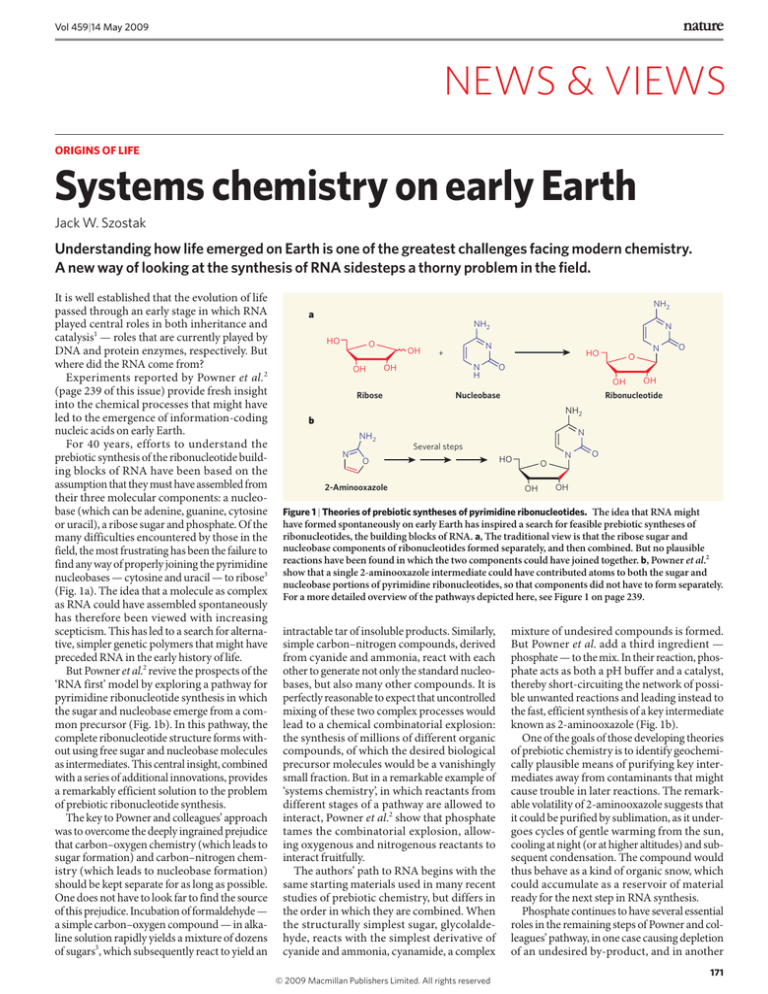
(Fig. 1a). The idea that a molecule as complex
as RNA could have assembled spontaneously
has therefore been viewed with increasing
scepticism. This has led to a search for alternative, simpler genetic polymers that might have
preceded RNA in the early history of life.
But Powner et al.2 revive the prospects of the
‘RNA first’ model by exploring a pathway for
pyrimidine ribonucleotide synthesis in which
the sugar and nucleobase emerge from a common precursor (Fig. 1b). In this pathway, the
complete ribonucleotide structure forms without using free sugar and nucleobase molecules
as intermediates. This central insight, combined
with a series of additional innovations, provides
a remarkably efficient solution to the problem
of prebiotic ribonucleotide synthesis.
The key to Powner and colleagues’ approach
was to overcome the deeply ingrained prejudice
that carbon–oxygen chemistry (which leads to
sugar formation) and carbon–nitrogen chemistry (which leads to nucleobase formation)
should be kept separate for as long as possible.
One does not have to look far to find the source
of this prejudice. Incubation of formaldehyde —
a simple carbon–oxygen compound — in alkaline solution rapidly yields a mixture of dozens
of sugars3, which subsequently react to yield an
NH2
a
NH2
HO
O
OH
OH
N
+
N
H
OH
Ribose
N
N
HO
O
O
O
OH
Nucleobase
OH
Ribonucleotide
NH2
b
N
NH2
Several steps
N
O
2-Aminooxazole
N
HO
O
O
OH
OH
Figure 1 | Theories of prebiotic syntheses of pyrimidine ribonucleotides. The idea that RNA might
have formed spontaneously on early Earth has inspired a search for feasible prebiotic syntheses of
ribonucleotides, the building blocks of RNA. a, The traditional view is that the ribose sugar and
nucleobase components of ribonucleotides formed separately, and then combined. But no plausible
reactions have been found in which the two components could have joined together. b, Powner et al.2
show that a single 2-aminooxazole intermediate could have contributed atoms to both the sugar and
nucleobase portions of pyrimidine ribonucleotides, so that components did not have to form separately.
For a more detailed overview of the pathways depicted here, see Figure 1 on page 239.
intractable tar of insoluble products. Similarly,
simple carbon–nitrogen compounds, derived
from cyanide and ammonia, react with each
other to generate not only the standard nucleobases, but also many other compounds. It is
perfectly reasonable to expect that uncontrolled
mixing of these two complex processes would
lead to a chemical combinatorial explosion:
the synthesis of millions of different organic
compounds, of which the desired biological
precursor molecules would be a vanishingly
small fraction. But in a remarkable example of
‘systems chemistry’, in which reactants from
different stages of a pathway are allowed to
interact, Powner et al.2 show that phosphate
tames the combinatorial explosion, allowing oxygenous and nitrogenous reactants to
interact fruitfully.
The authors’ path to RNA begins with the
same starting materials used in many recent
studies of prebiotic chemistry, but differs in
the order in which they are combined. When
the structurally simplest sugar, glycolaldehyde, reacts with the simplest derivative of
cyanide and ammonia, cyanamide, a complex
© 2009 Macmillan Publishers Limited. All rights reserved
mixture of undesired compounds is formed.
But Powner et al. add a third ingredient —
phosphate — to the mix. In their reaction, phosphate acts as both a pH buffer and a catalyst,
thereby short-circuiting the network of possible unwanted reactions and leading instead to
the fast, efficient synthesis of a key intermediate
known as 2-aminooxazole (Fig. 1b).
One of the goals of those developing theories
of prebiotic chemistry is to identify geochemically plausible means of purifying key intermediates away from contaminants that might
cause trouble in later reactions. The remarkable volatility of 2-aminooxazole suggests that
it could be purified by sublimation, as it undergoes cycles of gentle warming from the sun,
cooling at night (or at higher altitudes) and subsequent condensation. The compound would
thus behave as a kind of organic snow, which
could accumulate as a reservoir of material
ready for the next step in RNA synthesis.
Phosphate continues to have several essential
roles in the remaining steps of Powner and colleagues’ pathway, in one case causing depletion
of an undesired by-product, and in another
171
NEWS & VIEWS
saving a critical intermediate from degradation.
The penultimate reaction of the sequence, in
which the phosphate is attached to the nucleoside, is another beautiful example of the
influence of systems chemistry in this set 2 of
interlinked reactions. The phosphorylation is
facilitated by the presence of urea4; the urea
comes from the phosphate-catalysed hydrolysis
of a by-product from an earlier reaction in
the sequence.
The authors wrap up their synthetic tour de
force by using ultraviolet light to clean up
the reaction mixture. They report that ultraviolet irradiation destroys side products while
simultaneously converting some of the desired
ribocytidine product to ribouridine (the
second pyrimidine component of RNA). The
development of this complex photochemistry
required remarkable mechanistic insight from
NATURE|Vol 459|14 May 2009
Powner and colleagues, who not only correctly
predicted that ultraviolet irradiation would
destroy the majority of the by-products, but
also that the desired ribonucleotides would
withstand such treatment.
The authors’ careful study 2 of every potentially relevant reaction and side reaction in
their sequence is a model of how to develop the
fundamental chemical understanding required
for a reasoned approach to prebiotic chemistry. By working out a sequence of efficient
reactions, they have set the stage for a more
fruitful investigation of geochemical scenarios
compatible with the origin of life.
Of course, much remains to be done. We
must now try to determine how the various
starting materials could have accumulated in a
relatively pure and concentrated form in local
environments on early Earth. Furthermore,
MOLECULAR MICROBIOLOGY
A key event in survival
Dave Barry and Richard McCulloch
The parasitic microorganism Trypanosoma brucei evades recognition by its
host’s immune system by repeatedly changing its surface coat. The switch
in coat follows a risky route, though: DNA break and repair.
Like many other single-celled pathogens,
the protozoan Trypanosoma brucei, which
causes African sleeping sickness in humans,
undergoes antigenic variation — that is, it
periodically switches its variant surface glycoprotein (VSG), the molecule targeted by host
antibodies. But how switching is triggered
has remained largely elusive. On page 278 of
this issue, Boothroyd et al.1 show that a DNA
double-strand break (DSB) upstream of the
T. brucei VSG gene is the likely primary event in
this process. Their results add to the few, albeit
crucial, cases in which DSBs trigger developmental processes: these include mating-type
switching in yeast, rearrangements of immunesystem genes in humans and meiotic cell
division to produce sex germ cells2.
Antigenic switching can occur through
several genetic strategies, the most common
being the differential activation of an archive of
silent genes and pseudogenes. Although only
one gene is transcribed, from a specialized
expression site, switching occurs when silent
genes, or their fragments, are duplicated in the
expression site by a gene-conversion process,
replacing all or part of the expressed gene. In
some pathogens, the expressed gene can be
constructed as a mosaic from several archival
pseudogenes; such a combinatorial strategy
expands the scale of variation enormously,
with, for example, five pseudogenes giving rise
to hundreds of combinations3.
Trypanosoma brucei has evolved an even
more staggeringly complex system. It, too,
172
transcribes a single VSG gene, but the sources
of sequences that contribute to switching are
large and diverse. It has several inactive expression sites, and its archive contains up to 200
although Powner and colleagues’ synthetic
sequence yields the pyrimidine ribonucleotides,
it cannot explain how purine ribonucleotides
(which incorporate guanine and adenine)
might have formed. But it is precisely because
this work opens up so many new directions for
research that it will stand for years as one of the
great advances in prebiotic chemistry.
■
Jack W. Szostak is in the Howard Hughes Medical
Institute and Department of Molecular Biology,
Massachusetts General Hospital, Boston,
Massachusetts 02114, USA.
e-mail: szostak@molbio.mgh.harvard.edu
1. Joyce, G. F. & Orgel, L. E. in The RNA World (eds Gesteland,
R. F., Cech, T. R. & Atkins, J. F.) 23–56 (Cold Spring Harbor
Laboratory Press, 2006).
2. Powner, M. W., Gerland, B. & Sutherland, J. D. Nature 459,
239–242 (2009).
3. Orgel, L. E. Crit. Rev. Biochem. Mol. Biol. 39, 99–123 (2004).
4. Lohrmann, R. & Orgel, L. E. Science 171, 490–494 (1971).
VSG genes that lie at the ends (telomeres) of a
set of mini-chromosomes, as well as a further
1,600 silent genes — of which two-thirds are
pseudogenes — on the main chromosomes4.
The potential for mosaic variation therefore
seems beyond estimation. Intact archival
genes are duplicated starting from an upstream
set of repeat sequences each 70 base pairs (bp)
long 5, all the way to sequences at the downstream end of the coding sequence, or, in the
case of silent telomeric genes, perhaps to the
nearby end of the chromosome. As gene conversion in other organisms is initiated by a DSB
in the conversion site, such a break has been
proposed also to occur in the T. brucei VSG
Endonuclease
Gene promoter
Transcribed VSG gene
70-bp
repeats
DSB
Switch by repair
83%
a
Expression sites 5–15 VSG genes
17%
b
Mini-chromosomes <200 VSG genes
Not detected
c
Arrays ~1,600 VSG genes
1
Figure 1 | Antigenic switching and sources. Boothroyd et al. used an endonuclease enzyme to induce
a DNA double-strand break (DSB) adjacent to the 70-bp-repeat region of the active VSG gene in
Trypanosoma brucei. Consequently, the region from the DSB site to the end of the VSG gene was deleted.
The protozoan filled this gap by a repair process, using silent VSG loci on other chromosomes as template.
Locations of donor sequences included: (a) expression sites (of which there are 5–15 per strain) at
the telomeres of the main chromosomes; (b) telomeres of some 100 mini-chromosomes found in the
T. brucei genome; and (c) tandemly arrayed VSG genes in the main chromosomes. The copied regions
stretched from the 70-bp-repeat regions to the telomere, or, for intact genes, to the end of the VSG. The
frequencies of conversions the authors detected (shown as percentages) differ from those observed during
infections with natural strains of T. brucei, in which mini-chromosomes dominate as donors. Brackets
denote the duplicated region, with dashed sections indicating uncertainty over where the duplication
ends. Broad arrows indicate genes; narrow arrows, repetitive DNA sequences (70-bp repeats are shown
in black and white). Coloured arrows are different VSG genes; grey arrows, genes other than VSG.
© 2009 Macmillan Publishers Limited. All rights reserved


