An Unusual Presentation of Mycoplasma Pneumoniae
advertisement
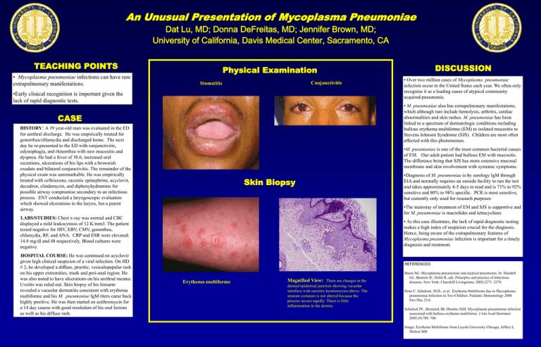
An Unusual Presentation of Mycoplasma Pneumoniae Dat Lu, MD; Donna DeFreitas, MD; Jennifer Brown, MD; University of California, Davis Medical Center, Sacramento, CA TEACHING POINTS • Mycoplasma pneumoniae infections can have rare extrapulmonary manifestations. Physical Examination Conjunctivitis Stomatitis •Early clinical recognition is important given the lack of rapid diagnostic tests. • Over two million cases of Mycoplasma pneumoniae infection occur in the United States each year. We often only recognize it as a leading cause of atypical community acquired pneumonia. • M. pneumoniae also has extrapulmonary manifestations, which although rare include hemolysis, arthritis, cardiac abnormalities and skin rashes. M. pneumoniae has been linked to a spectrum of dermatologic conditions including bullous erythema multiforme (EM) to isolated mucositis to Stevens Johnson Syndrome (SJS). Children are most often affected with this phenomenon. CASE HISTORY: A 19 year-old man was evaluated in the ED for urethral discharge. He was empirically treated for gonorrhea/chlamydia and discharged home. The next day he re-presented to the ED with conjunctivitis, odynophagia, and rhinorrhea with new mucositis and dyspnea. He had a fever of 38.6, increased oral secretions, ulcerations of his lips with a brownish exudate and bilateral conjunctivitis. The remainder of the physical exam was unremarkable. He was empirically treated with ceftriaxone, racemic epinephrine, acyclovir, decadron, clindamycin, and diphenyhydramine for possible airway compromise secondary to an infectious process. ENT conducted a laryngoscopic evaluation which showed ulcerations in the larynx, but a patent airway. •M. pneumoniae is one of the most common bacterial causes of EM. Our adult patient had bullous EM with mucositis. The difference being that SJS has more extensive mucosal membrane and skin involvement with systemic symptoms. Skin Biopsy •Diagnosis of M. pneumoniae is by serology IgM through EIA and normally requires an outside facility to run the test and takes approximately 4-5 days to read and is 71% to 92% sensitive and 80% to 98% specific. PCR is most sensitive, but currently only used for research purposes •The mainstay of treatment of EM and SJS is supportive and for M. pneumoniae is macrolides and tetracyclines LABS/STUDIES: Chest x-ray was normal and CBC displayed a mild leukocytosis of 12 K/mm3. The patient tested negative for HIV, EBV, CMV, gonorrhea, chlamydia, RF, and ANA. CRP and ESR were elevated; 14.8 mg/dl and 48 respectively. Blood cultures were negative. HOSPITAL COURSE: He was continued on acyclovir given high clinical suspicion of a viral infection. On HD # 2, he developed a diffuse, pruritic, vesiculopapular rash on his upper extremities, trunk and peri-anal region. He was also noted to have ulcerations on his urethral meatus. Uveitis was ruled out. Skin biopsy of his forearm revealed a vacuolar dermatitis consistent with erythema multiforme and his M. pneumoniae IgM titers came back highly positive. He was then started on azithromycin for a 14 day course with good resolution of his oral lesions as well as his diffuse rash. DISCUSSION • As this case illustrates, the lack of rapid diagnostic testing † makes a high index of suspicion crucial for the diagnosis. Hence, being aware of the extrapulmonary features of Mycoplasma pneumoniae infection is important for a timely diagnosis and treatment. REFERENCES: Erythema multiforme Magnified View: There are changes at the dermal/epidermal junction showing vacuolar interface with necrotic keratinocytes above. The stratum corneum is not altered because the process occurs rapidly. There is little inflammation in the dermis. Baum SG. Mycoplasma pneumoniae and atypical pneumonia. In: Mandell GL, Bennett JE, Dolin R, eds. Principles and practice of infectious diseases. New York: Churchill Livingstone, 2005;2271–2278. Peter C. Schalock, M.D., et al.. Erythema Multiforme due to Mycoplasma pneumoniae Infection in Two Children. Pediatric Dermatology 2006 Nov/Dec 23:6 Schalock PC, Brennick JB, Dinulos JGH. Mycoplasma pneumoniae infection associated with bullous erythema multiforme. J Am Acad Dermatol 2005;54:705–706 Image: Erythema Multiforme from Loyola University Chicago, Jeffery L. Melton MD
