Behavioral Analysis In vitro
advertisement
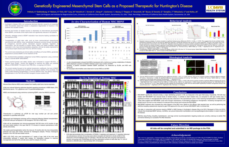
Genetically Engineered Mesenchymal Stem Cells as a Proposed Therapeutic for Huntington’s Disease Pollock, K 1 Dahlenburg, H 1 Nelson, H 1 Fink, KD 1 Cary, W 1 Hendrix K 1, Torrest A 1, Deng P 1, Gutierrez J 1, Nacey, C 1 Pepper, K 1 Gruenloh, W 1 Bauer, G1 Annett, G 1 Tempkin, T 2 Wheelock, V 2 and Nolta, JA1 1 Stem Cell Program and Institute for Regenerative Cures, University of California Davis Health System, Sacramento, CA, USA; 2 Dept. Neurology, University of California Davis Health System, Sacramento, CA, USA Introduction In vitro Characterization of Human MSC/BDNF • Huntington’s disease (HD) is an autosomal dominant disorder caused by an expanded CAG trinucleotide repeat that causes a progressive degeneration of neurons in the putamen and caudate nucleus. Behavioral Analysis B. A. • Survival and function of striatal neurons is dependent on brain-derived neurotrophic factor (BDNF), and levels of this trophic factor are significantly reduced in HD patients. • Recently, strategies aimed at BDNF restoration have become leading candidates for the treatment of HD. • Transplantation of adult stem cells, such as bone marrow-derived mesenchymal stem/stromal cells (MSCs), show considerable therapeutic promise through stimulation of endogenous neuronal growth, decreased neuronal apoptosis, regulation of inflammation, and the secretion of trophic factors. MSCs are readily available, easily expanded in vitro, have immunomodulatory properties, and can be easily engineered to over-produce trophic factors (see Fink et al. 2015 for review) A. Repeated measures analysis of variance revealed no significant between-group differences [F(3,51) = 0.438, p = 0.727]. This suggests that all mice exhibited similar spontaneous exploration of the box and that any differences observed in anxiety-like behaviors was not due to a motor impairment. The time in the center zone of the box was measured to test anxiety in the YAC128 mice. Repeated measures analysis of variance revealed a significant between-group difference [F(3,50) = 4.305, p = 0.009]. LSD post hoc analysis revealed a significant overall between group difference between WT mice and Tg + Normosol (p =0.007), and Tg + MSC (p = 0.002). LSD post hoc analysis also revealed an overall between group trend between Tg + Normosol and tg + BDNF hMSC (p = 0.169). * = Significant to WT; # = Significant to Tg + Normosol ; † = Significant to Tg + MSC; n = 16-17/group. B. Transplantation of MSC or MSC/BDNF increased the lifespan of R6/2 (CAG 120) mice. N= 7-9 mice per group, Kaplan-Meier analysis: 10% increase for WT MSC, 7.7% increase for MSC BDNF MOI 10, 15.5% increase for MSC BDNF MOI 20 • The current study tested the pre-clinical safety and efficacy of human MSCs, genetically engineered to produce BDNF, for the treatment of HD. • Our developmental candidate is allogeneic human MSCs engineered, using a lentiviral vector, to secrete BDNF (MSC/BDNF). Histological Analysis • These proof-of-concept and safety studies were conducted to support HD-CELL, a planned future Phase I clinical trial designed to examine the safety and potential efficacy of MSC/BDNF. The lead-in trial, PRE-CELL, is ongoing at UC Davis • • The current studies focused on the optimal transduction coefficients, the level of BDNF produced by our developmental candidate, the karyotypic stability of the candidate, the safety profile following transplantation in immune deficient mice, and efficacy following transplantation in the YAC128 and R6/2 transgenic mouse models of HD. • • A. B. In vitro characterization showed that MSCs transduced with a lentivirus at varying multiplicities of infection (MOI) displayed a positive correlation between vector copy number and MOI. Similarly, a positive correlation between BDNF production, as measured by ELISA, and MOI was observed. No karyotypic abnormalities were observed in human MSCs at all MOI In Vivo Retention of Human MSC Methods •Human Bone Marrow MSCs were isolated from healthy adult bone marrow aspirates. A. One-way ANOVA revealed no overall between-group differences [F(3,59) = 1.631, p = 0.193]. However, LSD post hoc analysis revealed a significant difference between WT and tg + Normosol (p = 0.038). No significant difference was observed between WT and tg + WT hMSC (p = 0.129) or WT and tg + BDNF hMSC (p = 0.252), suggesting an intermediate effect following transplantation. * = Significant to WT. B. One-way ANOVA revealed an overall between-group differences [F(3,26) = 5.196, p = 0.007]. LSD post hoc analysis revealed a significant difference between WT and Tg + MSC BDNF MOI 10 (p = 0.032), WT and Tg + MSC (p = 0.002), and Tg + MSC to Tg + Normosol (p = 0.009). A trend was observed between Tg + MSC BDNF MOI 10 and Tg + Normosol (p = 0.121). Taken together this data suggests that transplantation of MSC with and without BDNF significantly increases neurogenesis activity in the subventricular zone. * = Significant to WT, # = Significant to Tg + Normosol. n = 6-7/group. •Cells are cultured following optimized standard operating procedures in MEM Alpha, 20% fetal bovine serum, 1% L-glutamine, and 1% penicillin/streptomycin Conclusions •MSCs are transduced using a lentivirus containing BDNF under the MNDU3 promoter. • MSC/BDNF significantly reduced anxiety, a major debilitating symptom in HD, as measured in the open field assay. Importantly, YAC128 mice treated with MSC/BDNF had reduced levels of striatal atrophy, in contrast to sham treated mice, as compared to wild type control mice. A significant increase in neurogenesis-like activity was also observed in the subventricular zone in mice receiving transplantations of MSC/BDNF. These data suggest that MSC/BDNF could work through mechanisms of stimulating endogenous neurogenesis. Increasing neurogenesis and striatal neuron survival is a key endpoint of a planned future clinical trial involving the MSC/BDNF. • MSC/BDNF treatment also increased the mean lifespan of the R6/2 mice. Based on the efficacy data reported here, we will be performing the definitive biosafety assessments required to gain FDA approval for the use of human MSC/BDNF cells in a clinical trial. • This data, in conjunction with previous reports of BDNF or MSC therapy in Huntington’s disease (Dey et al 2010, Reviewed in Fink et al 2015), indicate that intrastriatal BDNF delivery, via MSCs, can prevent behavioral manifestation and neuropathological abnormalities in rodent models of the disease. •Transduction is confirmed via q-PCR for viral copy number per cell and protein expression is quantified with ELISA • Definitive dose-finding, biosafety, biodistribution, and large animal neurotransplantation targeting studies are currently underway to satisfy FDA comments following our successful Pre-IND interaction. •Behavioral and histological outcomes will be measured following bilateral transplantation of 500,000 cells per hemisphere in HD transgenic YAC128 or R6/2 mice. •Cells will be transplanted into immune-suppressed YAC128 mice at 8.5 months of age and were tested weekly in the open field for 10 minutes to measure spontaneous motor activity and anxiety. •Six weeks post-transplantation (when the mice are 10 months old), the mice intracardially perfused and have their brains removed for histological analysis. Primary histological analysis in the YAC128 was striatal volume. •R6/2 (120 CAG) mice were transplanted at 7 weeks of age. Half of the mice were intracardially perfused 3 weeks post surgery for histological analysis to examine neurogenesis and half of the animals were used for analysis of survival. Future Direction All data will be compiled and submitted in an IND package to the FDA. • IVIS data demonstrates that a combination of FK506 (1 mg/g/day) and rapamycin (1 mg/g/day) delivered via subcutaneous osmotic mini-pumps were able to increase cell retention to levels similar to that observed in immune-deficient mice (21-28 days). YAC128 mice that were not given subcutaneous osmotic mini-pumps with FK506/Rapa did not have detectable numbers of cells after day 14. Limit of detection is 250,000 cells or approximately 50% of transplant. Acknowledgements This work was supported by a National Institute of Health National Research Service Award Postdoctoral Fellowship [F32NS090722] (Fink), Graduate Fellowships [NSF GRFP 2011116000, NIH T32-GM008799, NSF GROW 201111600, T32-HL086350] (Anderson), National Institute of Health Director’s transformative award [R01GM099688] (Nolta), California Institute for Regenerative Medicine [CIRM DR2-05415] (Wheelock/Nolta), Dake Foundation (Fink), and philanthropic donors from the HD community, including the Roberson family and TeamKJ


